

Paper Menu >>
Journal Menu >>
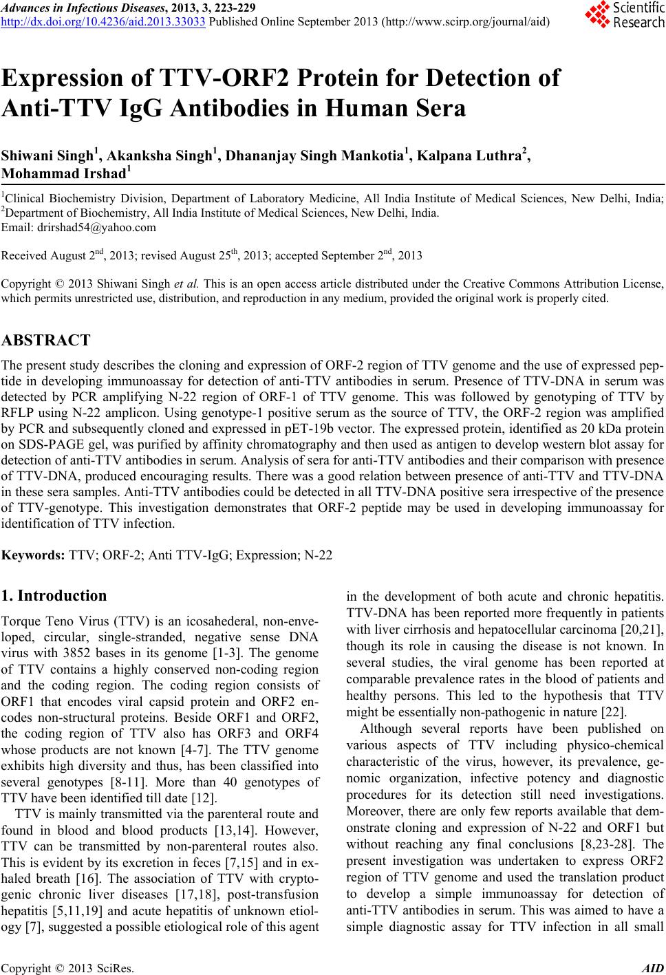 Advances in Infectious Diseases, 2013, 3, 223-229 http://dx.doi.org/10.4236/aid.2013.33033 Published Online September 2013 (http://www.scirp.org/journal/aid) 223 Expression of TTV-ORF2 Protein for Detection of Anti-TTV IgG Antibodies in Human Sera Shiwani Singh1, Akanksha Singh1, Dhananjay Singh Mankotia1, Kalpana Luthra2, Mohammad Irshad1 1Clinical Biochemistry Division, Department of Laboratory Medicine, All India Institute of Medical Sciences, New Delhi, India; 2Department of Biochemistry, All India Institute of Medical Sciences, New Delhi, India. Email: [email protected] Received August 2nd, 2013; revised August 25th, 2013; accepted September 2nd, 2013 Copyright © 2013 Shiwani Singh et al. This is an open access article distributed under the Creative Commons Attribution License, which permits unrestricted use, distribution, and reproduction in any medium, provided the original work is properly cited. ABSTRACT The present study describes the cloning and expression of ORF-2 region of TTV genome and the use of expressed pep- tide in developing immunoassay for detection of anti-TTV antibodies in serum. Presence of TTV-DNA in serum was detected by PCR amplifying N-22 region of ORF-1 of TTV genome. This was followed by genotyping of TTV by RFLP using N-22 amplicon. Using genotype-1 positive serum as the source of TTV, the ORF-2 region was amplified by PCR and subsequently cloned and expressed in pET-19b vector. The expressed protein, identified as 20 kDa protein on SDS-PAGE gel, was purified by affinity chromatography and then used as antigen to develop western blot assay for detection of anti-TTV antibodies in serum. Analysis of sera for anti-TTV antibodies and their comparison with presence of TTV-DNA, produced encouraging results. There was a good relation between presence of anti-TTV and TTV-DNA in these sera samples. Anti-TTV antibodies could be detected in all TTV-DNA positive sera irrespective of the presence of TTV-genotype. This investigation demonstrates that ORF-2 peptide may be used in developing immunoassay for identification of TTV infection. Keywords: TTV; ORF-2; Anti TTV-IgG; Expression; N-22 1. Introduction Torque Teno Virus (TTV) is an icosahederal, non-enve- loped, circular, single-stranded, negative sense DNA virus with 3852 bases in its genome [1-3]. The genome of TTV contains a highly conserved non-coding region and the coding region. The coding region consists of ORF1 that encodes viral capsid protein and ORF2 en- codes non-structural proteins. Beside ORF1 and ORF2, the coding region of TTV also has ORF3 and ORF4 whose products are not known [4-7]. The TTV genome exhibits high diversity and thus, has been classified into several genotypes [8-11]. More than 40 genotypes of TTV have been identified till date [12]. TTV is mainly transmitted via the parenteral route and found in blood and blood products [13,14]. However, TTV can be transmitted by non-parenteral routes also. This is evident by its excretion in feces [7,15] and in ex- haled breath [16]. The association of TTV with crypto- genic chronic liver diseases [17,18], post-transfusion hepatitis [5,11,19] and acute hepatitis of unknown etiol- ogy [7], suggested a possible etiological role of this agent in the development of both acute and chronic hepatitis. TTV-DNA has been reported more frequently in patients with liver cirrhosis and hepatocellular carcinoma [20,21], though its role in causing the disease is not known. In several studies, the viral genome has been reported at comparable prevalence rates in the blood of patients and healthy persons. This led to the hypothesis that TTV might be essentially non-pathogenic in nature [22]. Although several reports have been published on various aspects of TTV including physico-chemical characteristic of the virus, however, its prevalence, ge- nomic organization, infective potency and diagnostic procedures for its detection still need investigations. Moreover, there are only few reports available that dem- onstrate cloning and expression of N-22 and ORF1 but without reaching any final conclusions [8,23-28]. The present investigation was undertaken to express ORF2 region of TTV genome and used the translation product to develop a simple immunoassay for detection of anti-TTV antibodies in serum. This was aimed to have a simple diagnostic assay for TTV infection in all small Copyright © 2013 SciRes. AID 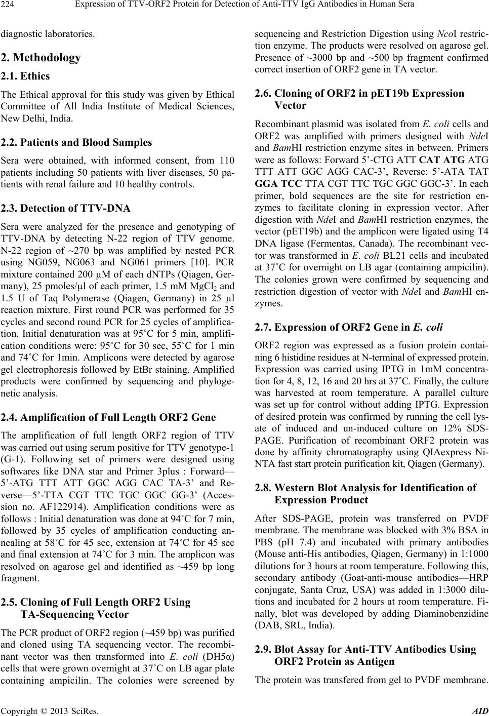 Expression of TTV-ORF2 Protein for Detection of Anti-TTV IgG Antibodies in Human Sera 224 diagnostic laboratories. 2. Methodology 2.1. Ethics The Ethical approval for this study was given by Ethical Committee of All India Institute of Medical Sciences, New Delhi, India. 2.2. Patients and Blood Samples Sera were obtained, with informed consent, from 110 patients including 50 patients with liver diseases, 50 pa- tients with renal failure and 10 healthy controls. 2.3. Detection of TTV-DNA Sera were analyzed for the presence and genotyping of TTV-DNA by detecting N-22 region of TTV genome. N-22 region of ~270 bp was amplified by nested PCR using NG059, NG063 and NG061 primers [10]. PCR mixture contained 200 µM of each dNTPs (Qiagen, Ger- many), 25 pmoles/µl of each primer, 1.5 mM MgCl2 and 1.5 U of Taq Polymerase (Qiagen, Germany) in 25 µl reaction mixture. First round PCR was performed for 35 cycles and second round PCR for 25 cycles of amplifica- tion. Initial denaturation was at 95˚C for 5 min, amplifi- cation conditions were: 95˚C for 30 sec, 55˚C for 1 min and 74˚C for 1min. Amplicons were detected by agarose gel electrophoresis followed by EtBr staining. Amplified products were confirmed by sequencing and phyloge- netic analysis. 2.4. Amplification of Full Length ORF2 Gene The amplification of full length ORF2 region of TTV was carried out using serum positive for TTV genotype-1 (G-1). Following set of primers were designed using softwares like DNA star and Primer 3plus : Forward— 5’-ATG TTT ATT GGC AGG CAC TA-3’ and Re- verse—5’-TTA CGT TTC TGC GGC GG-3’ (Acces- sion no. AF122914). Amplification conditions were as follows : Initial denaturation was done at 94˚C for 7 min, followed by 35 cycles of amplification conducting an- nealing at 58˚C for 45 sec, extension at 74˚C for 45 sec and final extension at 74˚C for 3 min. The amplicon was resolved on agarose gel and identified as ~459 bp long fragment. 2.5. Cloning of Full Length ORF2 Using TA-Sequencing Vector The PCR product of ORF2 region (~459 bp) was purified and cloned using TA sequencing vector. The recombi- nant vector was then transformed into E. coli (DH5α) cells that were grown overnight at 37˚C on LB agar plate containing ampicilin. The colonies were screened by sequencing and Restriction Digestion using NcoI restric- tion enzyme. The products were resolved on agarose gel. Presence of ~3000 bp and ~500 bp fragment confirmed correct insertion of ORF2 gene in TA vector. 2.6. Cloning of ORF2 in pET19b Expression Vector Recombinant plasmid was isolated from E. coli cells and ORF2 was amplified with primers designed with Nde I and BamHI restriction enzyme sites in between. Primers were as follows: Forward 5’-CTG ATT CAT ATG ATG TTT ATT GGC AGG CAC-3’, Reverse: 5’-ATA TAT GGA TCC TTA CGT TTC TGC GGC GGC-3’. In each primer, bold sequences are the site for restriction en- zymes to facilitate cloning in expression vector. After digestion with NdeI and BamHI restriction enzymes, the vector (pET19b) and the amplicon were ligated using T4 DNA ligase (Fermentas, Canada). The recombinant vec- tor was transformed in E. coli BL21 cells and incubated at 37˚C for overnight on LB agar (containing ampicilin). The colonies grown were confirmed by sequencing and restriction digestion of vector with NdeI and BamHI en- zymes. 2.7. Expression of ORF2 Gene in E. coli ORF2 region was expressed as a fusion protein contai- ning 6 histidine residues at N-terminal of expressed protein. Expression was carried using IPTG in 1mM concentra- tion for 4, 8, 12, 16 and 20 hrs at 37˚C. Finally, the culture was harvested at room temperature. A parallel culture was set up for control without adding IPTG. Expression of desired protein was confirmed by running the cell lys- ate of induced and un-induced culture on 12% SDS- PAGE. Purification of recombinant ORF2 protein was done by affinity chromatography using QIAexpress Ni- NTA fast start protein purification kit, Qiagen (Germany). 2.8. Western Blot Analysis for Identification of Expression Product After SDS-PAGE, protein was transferred on PVDF membrane. The membrane was blocked with 3% BSA in PBS (pH 7.4) and incubated with primary antibodies (Mouse anti-His antibodies, Qiagen, Germany) in 1:1000 dilutions for 3 hours at room temperature. Following this, secondary antibody (Goat-anti-mouse antibodies—HRP conjugate, Santa Cruz, USA) was added in 1:3000 dilu- tions and incubated for 2 hours at room temperature. Fi- nally, blot was developed by adding Diaminobenzidine (DAB, SRL, India). 2.9. Blot Assay for Anti-TTV Antibodies Using ORF2 Protein as Antigen The protein was transfered from gel to PVDF membrane. Copyright © 2013 SciRes. AID 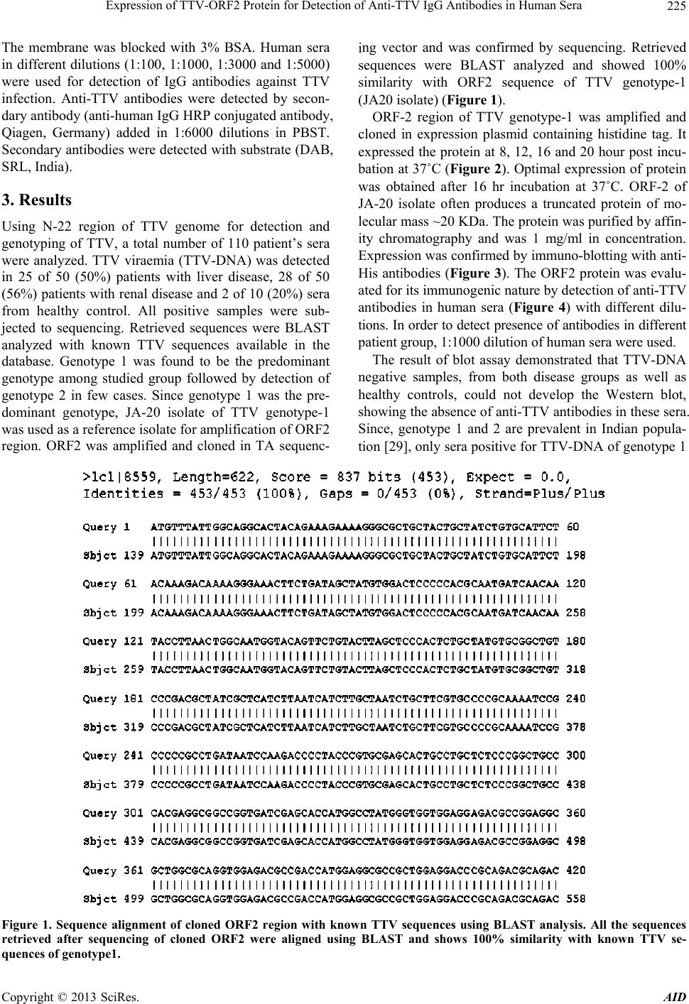 Expression of TTV-ORF2 Protein for Detection of Anti-TTV IgG Antibodies in Human Sera Copyright © 2013 SciRes. AID 225 The membrane was blocked with 3% BSA. Human sera in different dilutions (1:100, 1:1000, 1:3000 and 1:5000) were used for detection of IgG antibodies against TTV infection. Anti-TTV antibodies were detected by secon- dary antibody (anti-human IgG HRP conjugated antibody, Qiagen, Germany) added in 1:6000 dilutions in PBST. Secondary antibodies were detected with substrate (DAB, SRL, India). ing vector and was confirmed by sequencing. Retrieved sequences were BLAST analyzed and showed 100% similarity with ORF2 sequence of TTV genotype-1 (JA20 isolate) (Figure 1). ORF-2 region of TTV genotype-1 was amplified and cloned in expression plasmid containing histidine tag. It expressed the protein at 8, 12, 16 and 20 hour post incu- bation at 37˚C (Figure 2). Optimal expression of protein was obtained after 16 hr incubation at 37˚C. ORF-2 of JA-20 isolate often produces a truncated protein of mo- lecular mass ~20 KDa. The protein was purified by affin- ity chromatography and was 1 mg/ml in concentration. Expression was confirmed by immuno-blotting with anti- His antibodies (Figure 3). The ORF2 protein was evalu- ated for its immunogenic nature by detection of anti-TTV antibodies in human sera (Figure 4) with different dilu- tions. In order to detect presence of antibodies in different patient group, 1:1000 dilution of human sera were used. 3. Results Using N-22 region of TTV genome for detection and genotyping of TTV, a total number of 110 patient’s sera were analyzed. TTV viraemia (TTV-DNA) was detected in 25 of 50 (50%) patients with liver disease, 28 of 50 (56%) patients with renal disease and 2 of 10 (20%) sera from healthy control. All positive samples were sub- jected to sequencing. Retrieved sequences were BLAST analyzed with known TTV sequences available in the database. Genotype 1 was found to be the predominant genotype among studied group followed by detection of genotype 2 in few cases. Since genotype 1 was the pre- dominant genotype, JA-20 isolate of TTV genotype-1 was used as a reference isolate for amplification of ORF2 region. ORF2 was amplified and cloned in TA sequenc- The result of blot assay demonstrated that TTV-DNA negative samples, from both disease groups as well as healthy controls, could not develop the Western blot, showing the absence of anti-TTV antibodies in these sera. Since, genotype 1 and 2 are prevalent in Indian popula- tion [29], only sera positive for TTV-DNA of genotype 1 Figure 1. Sequence alignment of cloned ORF2 region with known TTV sequences using BLAST analysis. All the sequences retrieved after sequencing of cloned ORF2 were aligned using BLAST and shows 100% similarity with known TTV se- uences of genoty pe 1. q 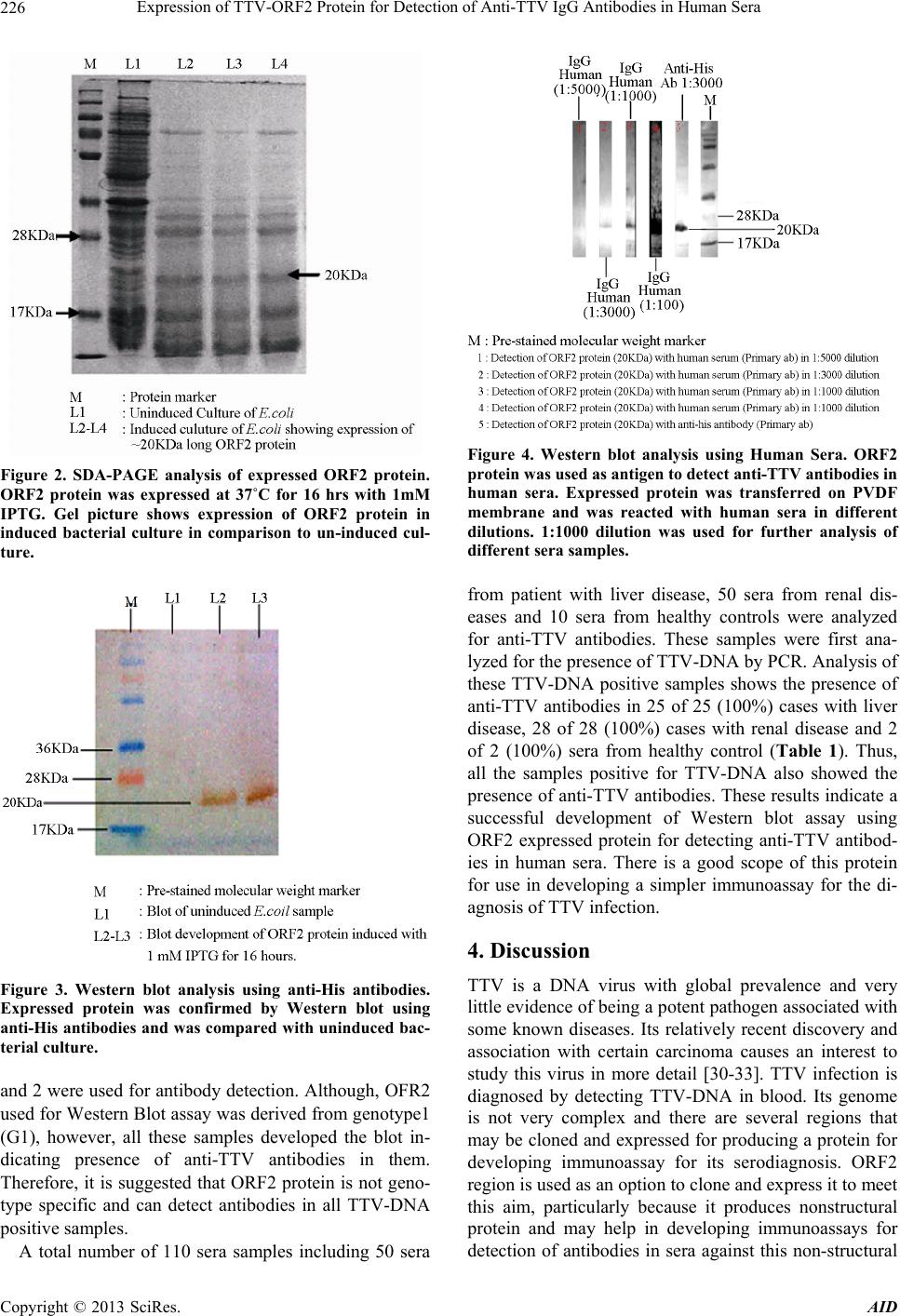 Expression of TTV-ORF2 Protein for Detection of Anti-TTV IgG Antibodies in Human Sera 226 Figure 2. SDA-PAGE analysis of expressed ORF2 protein. ORF2 protein was expressed at 37˚C for 16 hrs with 1mM IPTG. Gel picture shows expression of ORF2 protein in induced bacterial culture in comparison to un-induced cul- ture. Figure 3. Western blot analysis using anti-His antibodies. Expressed protein was confirmed by Western blot using anti-His antibodies and was compared with uninduced bac- terial culture. and 2 were used for antibody detection. Although, OFR2 used for Western Blot assay was derived from genotype1 (G1), however, all these samples developed the blot in- dicating presence of anti-TTV antibodies in them. Therefore, it is suggested that ORF2 protein is not geno- type specific and can detect antibodies in all TTV-DNA positive samples. A total number of 110 sera samples including 50 sera Figure 4. Western blot analysis using Human Sera. ORF2 protein was used as antigen to dete c t anti-TTV antibodies in human sera. Expressed protein was transferred on PVDF membrane and was reacted with human sera in different dilutions. 1:1000 dilution was used for further analysis of different sera samples. from patient with liver disease, 50 sera from renal dis- eases and 10 sera from healthy controls were analyzed for anti-TTV antibodies. These samples were first ana- lyzed for the presence of TTV-DNA by PCR. Analysis of these TTV-DNA positive samples shows the presence of anti-TTV antibodies in 25 of 25 (100%) cases with liver disease, 28 of 28 (100%) cases with renal disease and 2 of 2 (100%) sera from healthy control (Table 1). Thus, all the samples positive for TTV-DNA also showed the presence of anti-TTV antibodies. These results indicate a successful development of Western blot assay using ORF2 expressed protein for detecting anti-TTV antibod- ies in human sera. There is a good scope of this protein for use in developing a simpler immunoassay for the di- agnosis of TTV infection. 4. Discussion TTV is a DNA virus with global prevalence and very little evidence of being a potent pathogen associated with some known diseases. Its relatively recent discovery and association with certain carcinoma causes an interest to study this virus in more detail [30-33]. TTV infection is diagnosed by detecting TTV-DNA in blood. Its genome is not very complex and there are several regions that may be cloned and expressed for producing a protein for developing immunoassay for its serodiagnosis. ORF2 region is used as an option to clone and express it to meet this aim, particularly because it produces nonstructural protein and may help in developing immunoassays for detection of antibodies in sera against this non-structural Copyright © 2013 SciRes. AID 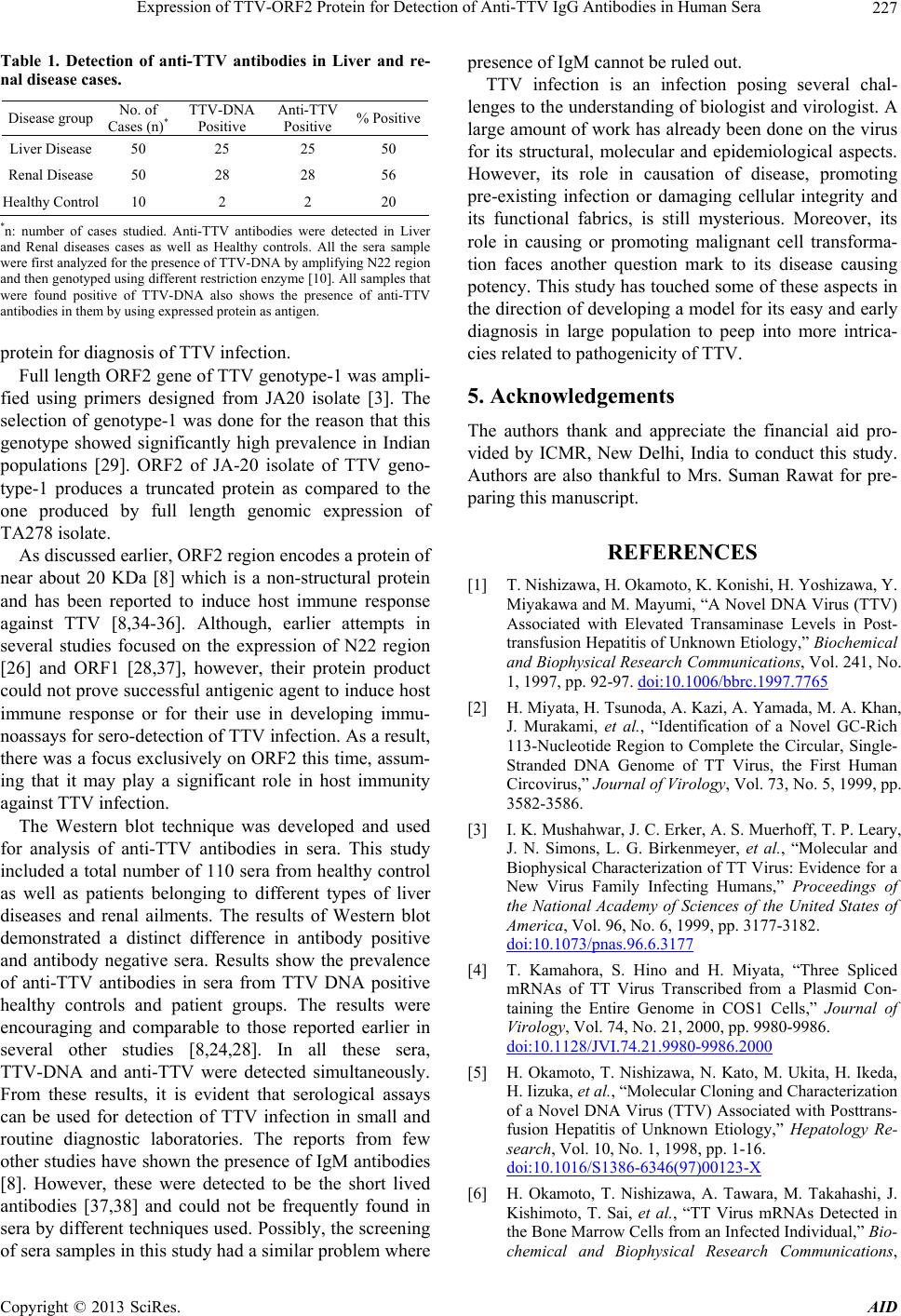 Expression of TTV-ORF2 Protein for Detection of Anti-TTV IgG Antibodies in Human Sera 227 Table 1. Detection of anti-TTV antibodies in Liver and re- nal disease cases. Disease group No. of Cases (n)* TTV-DNA Positive Anti-TTV Positive % Positive Liver Disease 50 25 25 50 Renal Disease 50 28 28 56 Healthy Control 10 2 2 20 *n: number of cases studied. Anti-TTV antibodies were detected in Liver and Renal diseases cases as well as Healthy controls. All the sera sample were first analyzed for the presence of TTV-DNA by amplifying N22 region and then genotyped using different restriction enzyme [10]. All samples that were found positive of TTV-DNA also shows the presence of anti-TTV antibodies in them by using expressed protein as antigen. protein for diagnosis of TTV infection. Full length ORF2 gene of TTV genotype-1 was ampli- fied using primers designed from JA20 isolate [3]. The selection of genotype-1 was done for the reason that this genotype showed significantly high prevalence in Indian populations [29]. ORF2 of JA-20 isolate of TTV geno- type-1 produces a truncated protein as compared to the one produced by full length genomic expression of TA278 isolate. As discussed earlier, ORF2 region encodes a protein of near about 20 KDa [8] which is a non-structural protein and has been reported to induce host immune response against TTV [8,34-36]. Although, earlier attempts in several studies focused on the expression of N22 region [26] and ORF1 [28,37], however, their protein product could not prove successful antigenic agent to induce host immune response or for their use in developing immu- noassays for sero-detection of TTV infection. As a result, there was a focus exclusively on ORF2 this time, assum- ing that it may play a significant role in host immunity against TTV infection. The Western blot technique was developed and used for analysis of anti-TTV antibodies in sera. This study included a total number of 110 sera from healthy control as well as patients belonging to different types of liver diseases and renal ailments. The results of Western blot demonstrated a distinct difference in antibody positive and antibody negative sera. Results show the prevalence of anti-TTV antibodies in sera from TTV DNA positive healthy controls and patient groups. The results were encouraging and comparable to those reported earlier in several other studies [8,24,28]. In all these sera, TTV-DNA and anti-TTV were detected simultaneously. From these results, it is evident that serological assays can be used for detection of TTV infection in small and routine diagnostic laboratories. The reports from few other studies have shown the presence of IgM antibodies [8]. However, these were detected to be the short lived antibodies [37,38] and could not be frequently found in sera by different techniques used. Possibly, the screening of sera samples in this study had a similar problem where presence of IgM cannot be ruled out. TTV infection is an infection posing several chal- lenges to the understanding of biologist and virologist. A large amount of work has already been done on the virus for its structural, molecular and epidemiological aspects. However, its role in causation of disease, promoting pre-existing infection or damaging cellular integrity and its functional fabrics, is still mysterious. Moreover, its role in causing or promoting malignant cell transforma- tion faces another question mark to its disease causing potency. This study has touched some of these aspects in the direction of developing a model for its easy and early diagnosis in large population to peep into more intrica- cies related to pathogenicity of TTV. 5. Acknowledgements The authors thank and appreciate the financial aid pro- vided by ICMR, New Delhi, India to conduct this study. Authors are also thankful to Mrs. Suman Rawat for pre- paring this manuscript. REFERENCES [1] T. Nishizawa, H. Okamoto, K. Konishi, H. Yoshizawa, Y. Miyakawa and M. Mayumi, “A Novel DNA Virus (TTV) Associated with Elevated Transaminase Levels in Post- transfusion Hepatitis of Unknown Etiology,” Biochemical and Biophysical Research Communications, Vol. 241, No. 1, 1997, pp. 92-97. doi:10.1006/bbrc.1997.7765 [2] H. Miyata, H. Tsunoda, A. Kazi, A. Yamada, M. A. Khan, J. Murakami, et al., “Identification of a Novel GC-Rich 113-Nucleotide Region to Complete the Circular, Single- Stranded DNA Genome of TT Virus, the First Human Circovirus,” Journal of Virology, Vol. 73, No. 5, 1999, pp. 3582-3586. [3] I. K. Mushahwar, J. C. Erker, A. S. Muerhoff, T. P. Leary, J. N. Simons, L. G. Birkenmeyer, et al., “Molecular and Biophysical Characterization of TT Virus: Evidence for a New Virus Family Infecting Humans,” Proceedings of the National Academy of Sciences of the United States of America, Vol. 96, No. 6, 1999, pp. 3177-3182. doi:10.1073/pnas.96.6.3177 [4] T. Kamahora, S. Hino and H. Miyata, “Three Spliced mRNAs of TT Virus Transcribed from a Plasmid Con- taining the Entire Genome in COS1 Cells,” Journal of Virology, Vol. 74, No. 21, 2000, pp. 9980-9986. doi:10.1128/JVI.74.21.9980-9986.2000 [5] H. Okamoto, T. Nishizawa, N. Kato, M. Ukita, H. Ikeda, H. Iizuka, et al., “Molecular Cloning and Characterization of a Novel DNA Virus (TTV) Associated with Posttrans- fusion Hepatitis of Unknown Etiology,” Hepatology Re- search, Vol. 10, No. 1, 1998, pp. 1-16. doi:10.1016/S1386-6346(97)00123-X [6] H. Okamoto, T. Nishizawa, A. Tawara, M. Takahashi, J. Kishimoto, T. Sai, et al., “TT Virus mRNAs Detected in the Bone Marrow Cells from an Infected Individual,” Bio- chemical and Biophysical Research Communications, Copyright © 2013 SciRes. AID 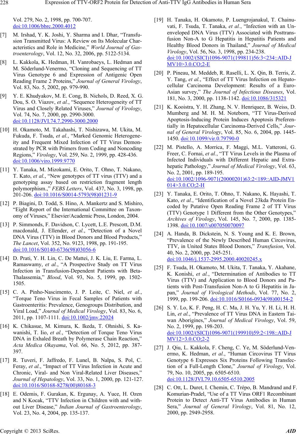 Expression of TTV-ORF2 Protein for Detection of Anti-TTV IgG Antibodies in Human Sera 228 Vol. 279, No. 2, 1998, pp. 700-707. doi:10.1006/bbrc.2000.4012 [7] M. Irshad, Y. K. Joshi, Y. Sharma and I. Dhar, “Transfu- sion Transmitted Virus: A Review on Its Molecular Char- acteristics and Role in Medicine,” World Journal of Gas- troenterology, Vol. 12, No. 32, 2006, pp. 5122-5134. [8] L. Kakkola, K. Hedman, H. Vanrobaeys, L. Hedman and M. Söderlund-Venermo, “Cloning and Sequencing of TT Virus Genotype 6 and Expression of Antigenic Open Reading Frame 2 Proteins,” Journal of General Virology, Vol. 83, No. 5, 2002, pp. 979-990. [9] Y. E. Khudyakov, M. E. Cong, B. Nichols, D. Reed, X. G. Dou, S. O. Viazov, et al., “Sequence Heterogeneity of TT Virus and Closely Related Viruses,” Journal of Virology, Vol. 74, No. 7, 2000, pp. 2990-3000. doi:10.1128/JVI.74.7.2990-3000.2000 [10] H. Okamoto, M. Takahashi, T. Nishizawa, M. Ukita, M. Fukuda, F. Tsuda, et al., “Marked Genomic Heterogene- ity and Frequent Mixed Infection of TT Virus Demon- strated by PCR with Primers from Coding and Noncoding Regions,” Virology, Vol. 259, No. 2, 1999, pp. 428-436. doi:10.1006/viro.1999.9770 [11] Y. Tanaka, M. Mizokami, E. Orito, T. Ohno, T. Nakano, T. Kato, et al., “New genotypes of TT virus (TTV) and a genotyping assay based on restriction fragment length polymorphism.,” FEBS Letters, Vol. 437, No. 3, 1998, pp. 201-206. doi:10.1016/S0014-5793(98)01231-9 [12] P. Biagini, D. Todd, S. Hino, A. Mankertz and S. Mishiro, “Eight Report of the International Committee on Taxon- omy of Viruses,” Elsevier/Academic Press, London, 2004. [13] P. Simmonds, F. Davidson, C. Lycett, L.E. Prescott, D.M. macdonald, J. Ellender, et al., “Detection of a Novel DNA Virus (TTV) in Blood Donors and Blood Products,” The Lancet, Vol. 352, No. 9123, 1998, pp. 191-195. doi:10.1016/S0140-6736(98)03056-6 [14] D. Prati, Y. H. Lin, C. De Mattei, J. K. Liu, E. Farma, L. Ramaswamy, et al., “A Prospective Study on TT Virus Infection in Transfusion-Dependent Patients with Beta- Thalassemia,” Blood, Vol. 93, No. 5, 1999, pp. 1502- 1505. [15] C. A. Pinho-Nascimento, J. P. Leite, C. Niel, et al., “Torque Teno Virus in Fecal Samples of Patients with Gastroenteritis: Prevalence, Genogroups Distribution, and Viral Load,” Journal of Medical Virology, Vol. 83, No. 6, 2011, pp. 1107-1111. doi:10.1002/jmv.22024 [16] K. Chikasue, M. Kimura, K. Ikeda, T. Ohnishi, S. Ka- wanishi, T. Iio, et al., “Detection of Torque Teno Virus DNA in Exhaled Breath by Polymerase Chain Reaction,” Acta Medica Okayama, Vol. 66, No. 5, 2012, pp. 387- 397. [17] R. Tuveri, F. Jaffredo, F. Lunel, B. Nalpa, S. Pol, C. Feray, et al., “Impact of TT Virus Infection in Acute and Chronic, Viral- and Non Viral-Related Liver Diseases,” Journal of Hepatology, Vol. 33, No. 1, 2000, pp. 121-127. doi:10.1016/S0168-8278(00)80168-3 [18] E. Odemis, F. Gurakan, K. Ergunay, A. Yuce, H. Ozen and N Kocak, “TTV Infection in Children with and with- out Liver Disease,” Indian Journal of Gastroenterology, Vol. 23, No. 4, 2004, pp. 135-137. [19] H. Tanaka, H. Okamoto, P. Luengrojanakul, T. Chainu- vati, F. Tsuda, T. Tanaka, et al., “Infection with an Un- enveloped DNA Virus (TTV) Associated with Posttrans- fusion Non-A to G Hepatitis in Hepatitis Patients and Healthy Blood Donors in Thailand,” Journal of Medical Virology, Vol. 56, No. 3, 1998, pp. 234-238. doi:10.1002/(SICI)1096-9071(199811)56:3<234::AID-J MV10>3.0.CO;2-E [20] P. Pineau, M. Meddeb, R. Raselli, L. X. Qin, B. Terris, Z. Y. Tang, et al., “Effect of TT Virus Infection on Hepato- cellular Carcinoma Development: Results of a Euro- Asian survey,” The Journal of Infectious Diseases, Vol. 181, No. 3, 2000, pp. 1138-1142. doi:10.1086/315321 [21] K. Kooistra, Y. H. Zhang, N. V. Henriquez, B. Weiss, D. Mumberg and M. H. M. Noteborn, “TT Virus-Derived Apoptosis-Inducing Protein Induces Apoptosis Preferen- tially in Hepatocellular Carcinoma-Derived Cells,” Jour- nal of General Virology, Vol. 85, No. 6, 2004, pp. 1445- 1450. doi:10.1099/vir.0.79790-0 [22] M. Pistello, A. Morrica, F. Maggi, M.L. Vatteroni, G. Freer, C. Fornai, et al., “TT Virus Levels in the Plasma of Infected Individuals with Different Hepatic and Extra- hepatic Pathology,” Journal of Medical Virology, Vol. 63, No. 2, 2001, pp. 189-195. doi:10.1002/1096-9071(20000201)63:2<189::AID-JMV1 014>3.0.CO;2-H [23] Y. Tanaka, E. Orito, T. Ohno, T. Nakano, K. Hayashi, T. Kato, et al., “Identification of a Novel 23kda Protein En- coded by Putative Open Reading Frame 2 of TT Virus (TTV) Genotype 1 Different from the Other Genotypes,” Archives of Virology, Vol. 145, No. 7, 2000, pp. 1385- 1398. doi:10.1007/s007050070097 [24] A. Handa, B. Dickstein, N. S. Young and K. E. Brown, “Prevalence of the Newly Described Human Circovirus, TTV, in United States Blood Donors,” Transfusion, Vol. 40, No. 2, 2000, pp. 245-251. doi:10.1046/j.1537-2995.2000.40020245.x [25] F. Tsuda, H. Okamoto, M. Ukita, T. Tanaka, Y. Akahane, K. Konishi, et al., “Determination of Antibodies to TT Virus (TTV) and Application to Blood Donors and Pa- tients with Post-Transfusion Non-A to G Hepatitis in Ja- pan,” Journal of Virological Methods, Vol. 77, No. 2, 1999, pp. 199-206. doi:10.1016/S0166-0934(98)00154-2 [26] S. Y. Lo, K. F. Peng, H. C. Ma, J. H. Yu, Y. H. Li, H. H. Lin, et al., “Prevalence of TT Virus DNA in Eastern Tai- wan Aborigines,” Journal of Medical Virology, Vol. 59, No. 2, 1999, pp. 198-203. doi:10.1002/(SICI)1096-9071(199910)59:2<198::AID-J MV12>3.0.CO;2-2 [27] J. Qiu, L. Kakkola, F. Cheng, C. Ye, M. Söderlund-Ven- ermo, K. Hedman, et al., “Human Circovirus TT Virus Genotype 6 Expresses Six Proteins Following Transfec- tion of a Full-Length Clone,” Journal of Virology, Vol. 79, No. 10, 2005, pp. 6505-6510. doi:10.1128/JVI.79.10.6505-6510.2005 [28] C. Ott, L. Duret, I. Chemin, C. Trépo, B. Mandrand and F. Komurian-Pradel, “Use of a TT Virus ORF1 Recombinant Protein to Detect Anti-TT Virus Antibodies in Human Sera,” Journal of General Virology, Vol. 81, No. 12, 2000, pp. 2949-2958. Copyright © 2013 SciRes. AID 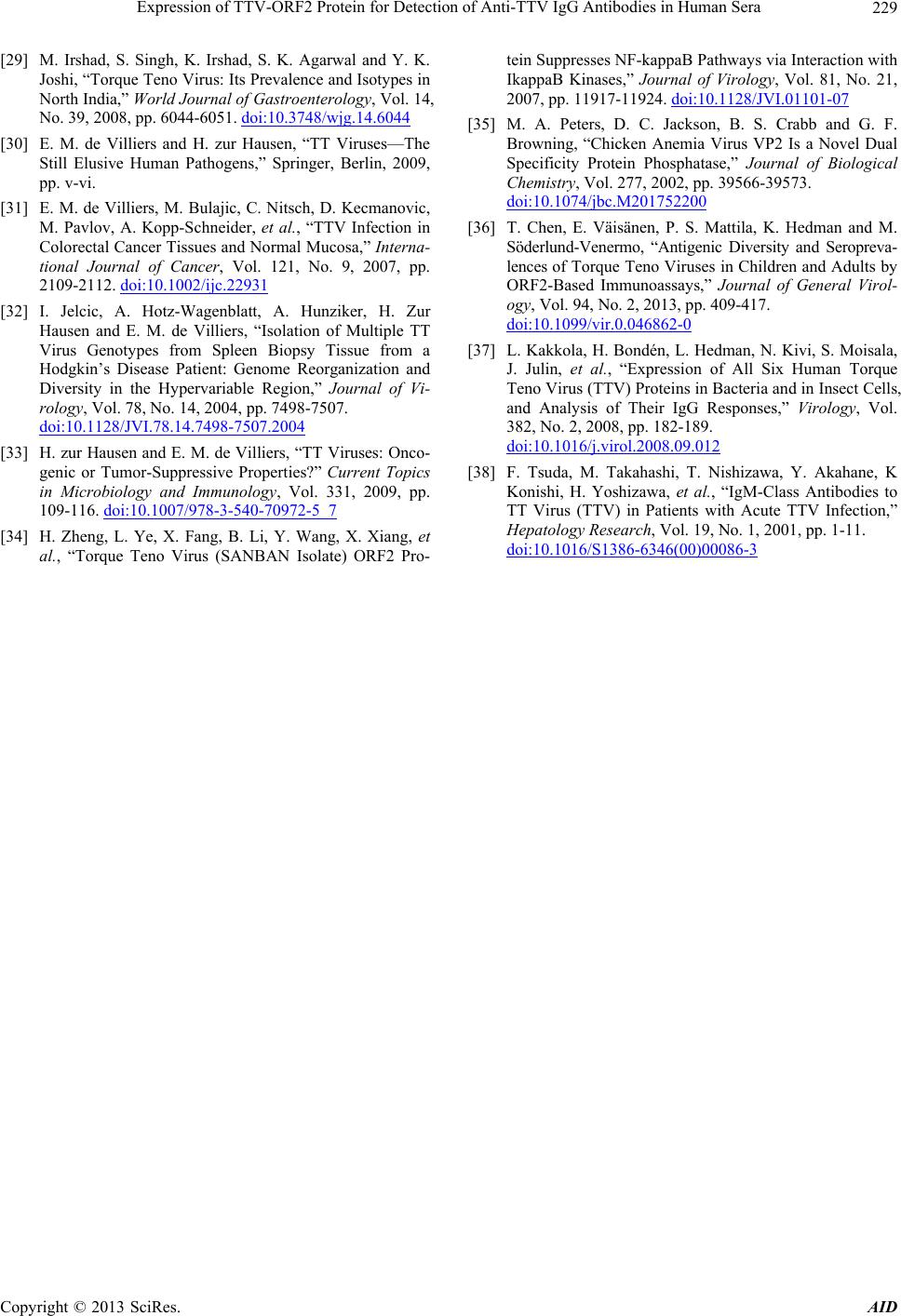 Expression of TTV-ORF2 Protein for Detection of Anti-TTV IgG Antibodies in Human Sera Copyright © 2013 SciRes. AID 229 [29] M. Irshad, S. Singh, K. Irshad, S. K. Agarwal and Y. K. Joshi, “Torque Teno Virus: Its Prevalence and Isotypes in North India,” World Journal of Gastroenterology, Vol. 14, No. 39, 2008, pp. 6044-6051. doi:10.3748/wjg.14.6044 [30] E. M. de Villiers and H. zur Hausen, “TT Viruses—The Still Elusive Human Pathogens,” Springer, Berlin, 2009, pp. v-vi. [31] E. M. de Villiers, M. Bulajic, C. Nitsch, D. Kecmanovic, M. Pavlov, A. Kopp-Schneider, et al., “TTV Infection in Colorectal Cancer Tissues and Normal Mucosa,” Interna- tional Journal of Cancer, Vol. 121, No. 9, 2007, pp. 2109-2112. doi:10.1002/ijc.22931 [32] I. Jelcic, A. Hotz-Wagenblatt, A. Hunziker, H. Zur Hausen and E. M. de Villiers, “Isolation of Multiple TT Virus Genotypes from Spleen Biopsy Tissue from a Hodgkin’s Disease Patient: Genome Reorganization and Diversity in the Hypervariable Region,” Journal of Vi- rology, Vol. 78, No. 14, 2004, pp. 7498-7507. doi:10.1128/JVI.78.14.7498-7507.2004 [33] H. zur Hausen and E. M. de Villiers, “TT Viruses: Onco- genic or Tumor-Suppressive Properties?” Current Topics in Microbiology and Immunology, Vol. 331, 2009, pp. 109-116. doi:10.1007/978-3-540-70972-5_7 [34] H. Zheng, L. Ye, X. Fang, B. Li, Y. Wang, X. Xiang, et al., “Torque Teno Virus (SANBAN Isolate) ORF2 Pro- tein Suppresses NF-kappaB Pathways via Interaction with IkappaB Kinases,” Journal of Virology, Vol. 81, No. 21, 2007, pp. 11917-11924. doi:10.1128/JVI.01101-07 [35] M. A. Peters, D. C. Jackson, B. S. Crabb and G. F. Browning, “Chicken Anemia Virus VP2 Is a Novel Dual Specificity Protein Phosphatase,” Journal of Biological Chemistry, Vol. 277, 2002, pp. 39566-39573. doi:10.1074/jbc.M201752200 [36] T. Chen, E. Väisänen, P. S. Mattila, K. Hedman and M. Söderlund-Venermo, “Antigenic Diversity and Seropreva- lences of Torque Teno Viruses in Children and Adults by ORF2-Based Immunoassays,” Journal of General Virol- ogy, Vol. 94, No. 2, 2013, pp. 409-417. doi:10.1099/vir.0.046862-0 [37] L. Kakkola, H. Bondén, L. Hedman, N. Kivi, S. Moisala, J. Julin, et al., “Expression of All Six Human Torque Teno Virus (TTV) Proteins in Bacteria and in Insect Cells, and Analysis of Their IgG Responses,” Virology, Vol. 382, No. 2, 2008, pp. 182-189. doi:10.1016/j.virol.2008.09.012 [38] F. Tsuda, M. Takahashi, T. Nishizawa, Y. Akahane, K Konishi, H. Yoshizawa, et al., “IgM-Class Antibodies to TT Virus (TTV) in Patients with Acute TTV Infection,” Hepatology Research, Vol. 19, No. 1, 2001, pp. 1-11. doi:10.1016/S1386-6346(00)00086-3 |