

Paper Menu >>
Journal Menu >>
 Advances in Infectious Diseases, 2013, 3, 219-222 http://dx.doi.org/10.4236/aid.2013.33032 Published Online September 2013 (http://www.scirp.org/journal/aid) 219 A Case of Carbapenem Resistant Non-K1/K2 Serotype Klebsiella pneumoniae Liver Abscess Lucy Cheng1, Leung Kristopher Siu2, Tom Chiang1,3 1Department of Medicine, Rutgers New Jersey Medical School, Newark, USA; 2Institute of Infectious Diseases and Vaccinology, National Health Research Institutes, Miaoli, Taiwan; 3Division of Infectious Disease, Veterans Affairs New Jersey Health Care Sys- tem, East Orange, USA. Email: [email protected] Received July 30th, 2013; revised August 15th, 2013; accepted August 20th, 2013 Copyright © 2013 Lucy Cheng et al. This is an open access article distributed under the Creative Commons Attribution License, which permits unrestricted use, distribution, and reproduction in any medium, provided the original work is properly cited. ABSTRACT Klebsiella pneumoniae liver abscess (KPLA) has been described as an invasive syndrome with extrahepatic complica- tions. The majority of KPLA is caused by capsular serotype K1 and K2 isolates. We report a case of carbapenem resis- tant Klebsiella pneumoniae liver abscess. The patient initially presented with infected right above-the-knee amputation and was later found with a large liver abscess. Initial antimicrobial susceptibility showed carbapenem resistant K. pneu- moniae (CRKP). Further molecular workup revealed that the isolate was a less virulent non-K1/K2 serotype, and both rmpA and kfu genes were negative. The lack of outer membrane porins likely contributed to the carbapenem resistance. To our knowledge, this is a first reported case of carbapenem resistant, non-K1/K2 serotype K. pneumoniae liver ab- scess in the United States. Keywords: Liver Abscess; Klebsiella pneumoniae; Capsular Serotype; Carbapenem Resistance 1. Introduction Klebsiella pneumoniae liver abscess (KPLA) has been described as an invasive syndrome with extrahepatic complications via hematologic spread. The hypermu- coviscosity phenotype and capsular serotype (K1 and K2) are the major virulence factors, along with capsular polysaccharide synthesis regulator gene rmpA, iron che- lator aerobactin, and iron uptake gene kfu, which have been described to contribute to the virulence and invasive features of K. pneumoniae [1-3]. To our knowledge, we describe the first case of non-K1/K2 KPLA in the United States. 2. Case Report A 64-year-old African American man presented to the Veterans Affairs New Jersey Healthcare System with an infected right above-the-knee amputation (AKA). The patient’s hospital course was complicated by poor wound healing, wound debridement, revision of the AKA, and a urinary tract infection with Pseudomonas aeruginosa and Klebsiella pneumoniae, for which he was treated with doripenem. After one and a half month of hospitalization, the right AKA began to heal appropriately. On hospital day 49, the patient developed transient right upper quad- rant abdominal pain. He then developed intermittent fe- vers ranging from 100.7˚F to 101˚F, however fever workup including blood and urine cultures, as well as chest X-ray were negative. The patient did not have any further physical complaints and remained hemodynami- cally stable. On hospital day 57, the patient was febrile (tempera- ture, 102˚F), with a heart rate of 121 beats per minute and blood pressure of 155/77 mm·Hg. The physical ex- amination was unremarkable including normal thoracic and abdominal findings. The white cell count was 19,600 cell/μL with 90 percent neutrophils; the hemoglobin level was 8.5 g/dL, and the platelet count was 377,000 cell/μL. The aspartate aminotransferase and alanine aminotrans- ferase were mildly elevated, 38 IU/L and 51 IU/L, re- spectively. The total bilirubin level was 0.6 mg/dL. A computerized axial tomography scan of the thorax and abdomen showed a large loculated abscess in the right lobe of the liver and intrahepatic biliary ductal dilatation (Figure 1). Piperacillin-tazobactam was started empiri- cally, and the patient underwent fine-needle aspiration of the liver abscess and pigtail placement by interventional radiology. Culture of the liver abscess grew carbapenem Copyright © 2013 SciRes. AID 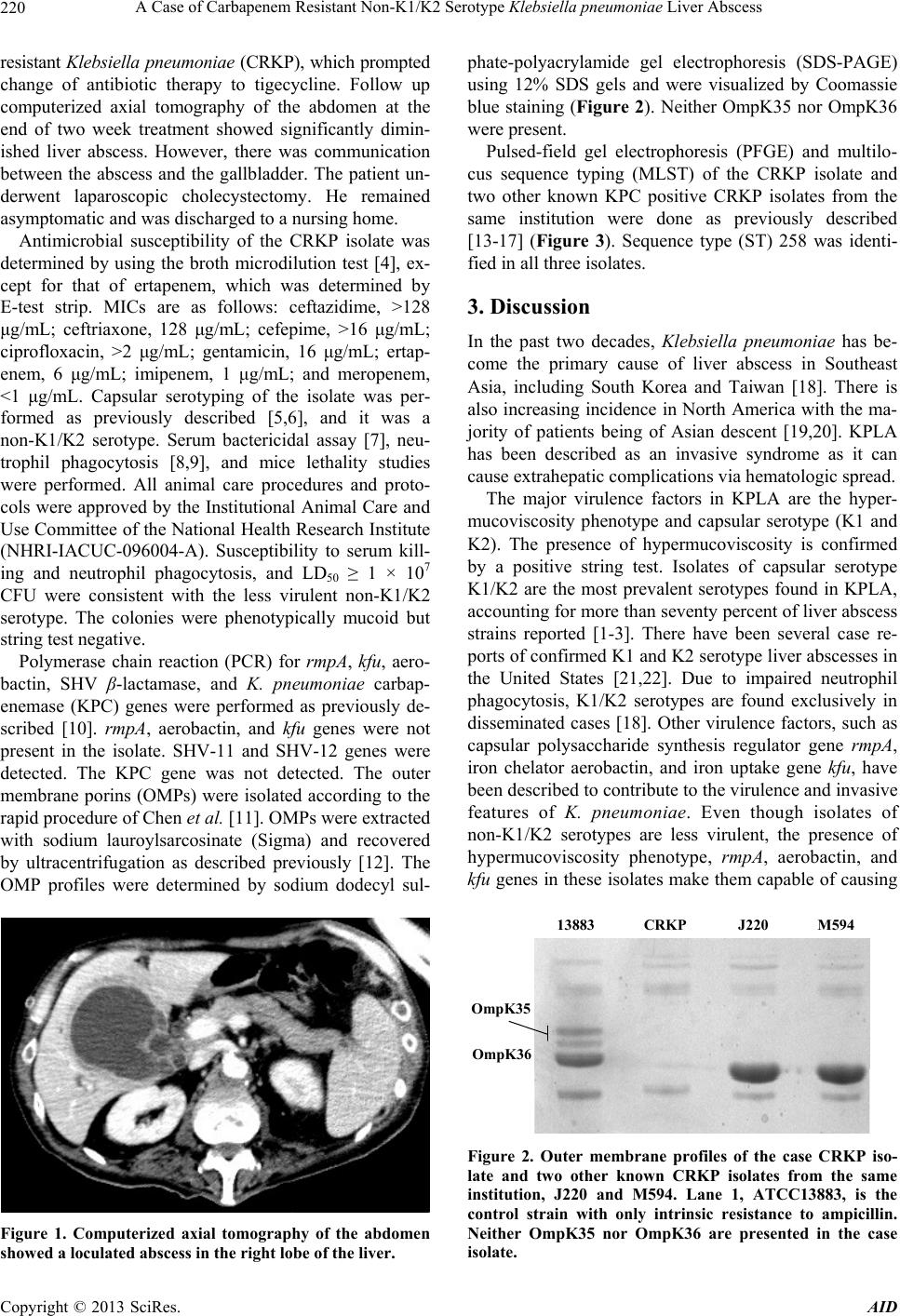 A Case of Carbapenem Resistant Non-K1/K2 Serotype Klebsiella pneumoniae Liver Abscess 220 resistant Klebsiella pneumoniae (CRKP), which prompted change of antibiotic therapy to tigecycline. Follow up computerized axial tomography of the abdomen at the end of two week treatment showed significantly dimin- ished liver abscess. However, there was communication between the abscess and the gallbladder. The patient un- derwent laparoscopic cholecystectomy. He remained asymptomatic and was discharged to a nursing home. Antimicrobial susceptibility of the CRKP isolate was determined by using the broth microdilution test [4], ex- cept for that of ertapenem, which was determined by E-test strip. MICs are as follows: ceftazidime, >128 μg/mL; ceftriaxone, 128 μg/mL; cefepime, >16 μg/mL; ciprofloxacin, >2 μg/mL; gentamicin, 16 μg/mL; ertap- enem, 6 μg/mL; imipenem, 1 μg/mL; and meropenem, <1 μg/mL. Capsular serotyping of the isolate was per- formed as previously described [5,6], and it was a non-K1/K2 serotype. Serum bactericidal assay [7], neu- trophil phagocytosis [8,9], and mice lethality studies were performed. All animal care procedures and proto- cols were approved by the Institutional Animal Care and Use Committee of the National Health Research Institute (NHRI-IACUC-096004-A). Susceptibility to serum kill- ing and neutrophil phagocytosis, and LD50 ≥ 1 × 10⁷ CFU were consistent with the less virulent non-K1/K2 serotype. The colonies were phenotypically mucoid but string test negative. Polymerase chain reaction (PCR) for rmpA, kfu, aero- bactin, SHV β-lactamase, and K. pneumoniae carbap- enemase (KPC) genes were performed as previously de- scribed [10]. rmpA, aerobactin, and kfu genes were not present in the isolate. SHV-11 and SHV-12 genes were detected. The KPC gene was not detected. The outer membrane porins (OMPs) were isolated according to the rapid procedure of Chen et al. [11]. OMPs were extracted with sodium lauroylsarcosinate (Sigma) and recovered by ultracentrifugation as described previously [12]. The OMP profiles were determined by sodium dodecyl sul- Figure 1. Computerized axial tomography of the abdomen showed a loculated abscess in the right lobe of the liver. phate-polyacrylamide gel electrophoresis (SDS-PAGE) using 12% SDS gels and were visualized by Coomassie blue staining (Figure 2). Neither OmpK35 nor OmpK36 were present. Pulsed-field gel electrophoresis (PFGE) and multilo- cus sequence typing (MLST) of the CRKP isolate and two other known KPC positive CRKP isolates from the same institution were done as previously described [13-17] (Figure 3). Sequence type (ST) 258 was identi- fied in all three isolates. 3. Discussion In the past two decades, Klebsiella pneumoniae has be- come the primary cause of liver abscess in Southeast Asia, including South Korea and Taiwan [18]. There is also increasing incidence in North America with the ma- jority of patients being of Asian descent [19,20]. KPLA has been described as an invasive syndrome as it can cause extrahepatic complications via hematologic spread. The major virulence factors in KPLA are the hyper- mucoviscosity phenotype and capsular serotype (K1 and K2). The presence of hypermucoviscosity is confirmed by a positive string test. Isolates of capsular serotype K1/K2 are the most prevalent serotypes found in KPLA, accounting for more than seventy percent of liver abscess strains reported [1-3]. There have been several case re- ports of confirmed K1 and K2 serotype liver abscesses in the United States [21,22]. Due to impaired neutrophil phagocytosis, K1/K2 serotypes are found exclusively in disseminated cases [18]. Other virulence factors, such as capsular polysaccharide synthesis regulator gene rmpA, iron chelator aerobactin, and iron uptake gene kfu, have been described to contribute to the virulence and invasive features of K. pn eumoniae . Even though isolates of non-K1/K2 serotypes are less virulent, the presence of hypermucoviscosity phenotype, rmpA, aerobactin, and kfu genes in these isolates make them capable of causing 13883 CRKP J220 M594 OmpK35 OmpK36 Figure 2. Outer membrane profiles of the case CRKP iso- late and two other known CRKP isolates from the same institution, J220 and M594. Lane 1, ATCC13883, is the control strain with only intrinsic resistance to ampicillin. Neither OmpK35 nor OmpK36 are presented in the case isolate. Copyright © 2013 SciRes. AID 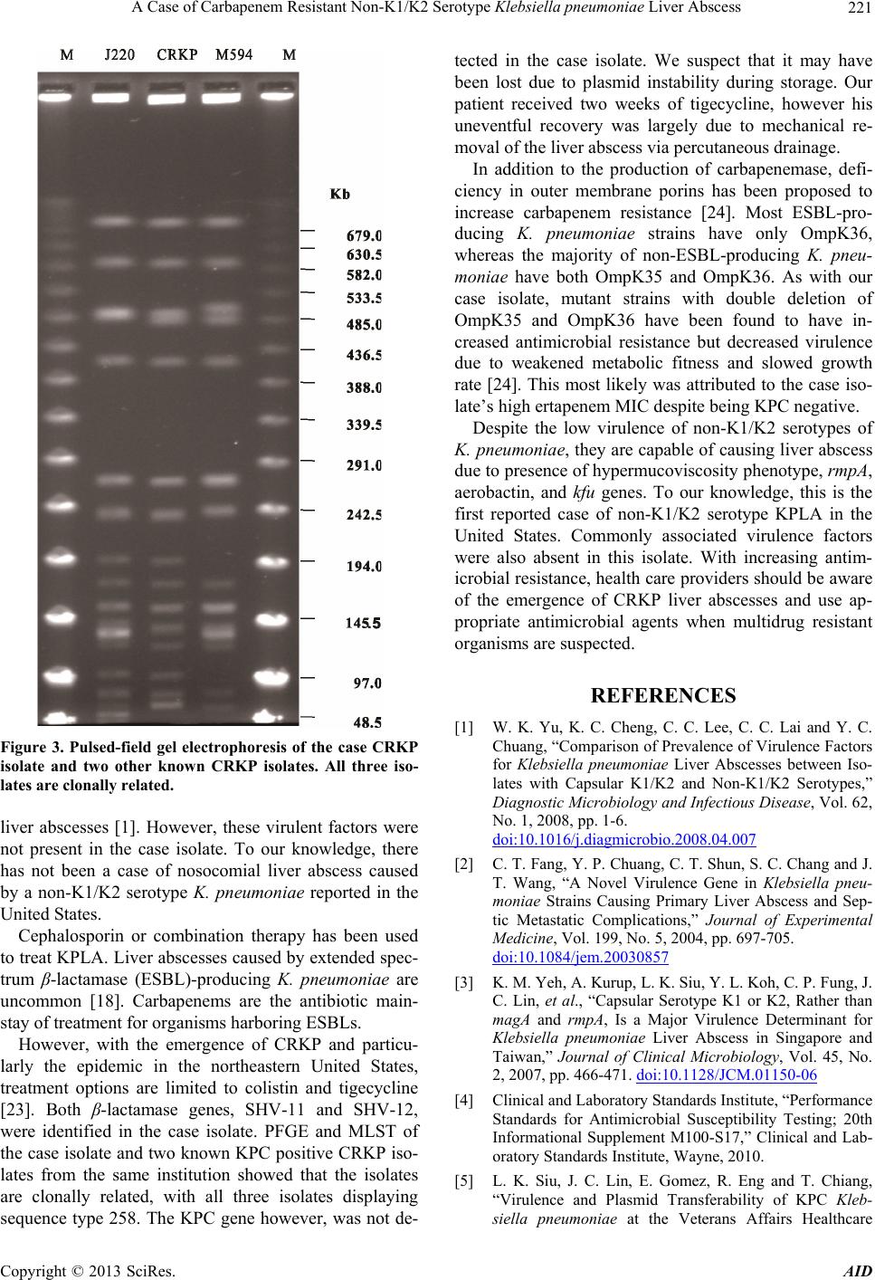 A Case of Carbapenem Resistant Non-K1/K2 Serotype Klebsiella pneumoniae Liver Abscess 221 Figure 3. Pulsed-field gel electrophoresis of the case CRKP isolate and two other known CRKP isolates. All three iso- lates are clonally related. liver abscesses [1]. However, these virulent factors were not present in the case isolate. To our knowledge, there has not been a case of nosocomial liver abscess caused by a non-K1/K2 serotype K. pneumoniae reported in the United States. Cephalosporin or combination therapy has been used to treat KPLA. Liver abscesses caused by extended spec- trum β-lactamase (ESBL)-producing K. pneumoniae are uncommon [18]. Carbapenems are the antibiotic main- stay of treatment for organisms harboring ESBLs. However, with the emergence of CRKP and particu- larly the epidemic in the northeastern United States, treatment options are limited to colistin and tigecycline [23]. Both β-lactamase genes, SHV-11 and SHV-12, were identified in the case isolate. PFGE and MLST of the case isolate and two known KPC positive CRKP iso- lates from the same institution showed that the isolates are clonally related, with all three isolates displaying sequence type 258. The KPC gene however, was not de- tected in the case isolate. We suspect that it may have been lost due to plasmid instability during storage. Our patient received two weeks of tigecycline, however his uneventful recovery was largely due to mechanical re- moval of the liver abscess via percutaneous drainage. In addition to the production of carbapenemase, defi- ciency in outer membrane porins has been proposed to increase carbapenem resistance [24]. Most ESBL-pro- ducing K. pneumoniae strains have only OmpK36, whereas the majority of non-ESBL-producing K. pneu- moniae have both OmpK35 and OmpK36. As with our case isolate, mutant strains with double deletion of OmpK35 and OmpK36 have been found to have in- creased antimicrobial resistance but decreased virulence due to weakened metabolic fitness and slowed growth rate [24]. This most likely was attributed to the case iso- late’s high ertapenem MIC despite being KPC negative. Despite the low virulence of non-K1/K2 serotypes of K. pneumoniae, they are capable of causing liver abscess due to presence of hypermucoviscosity phenotype, rmpA, aerobactin, and kfu genes. To our knowledge, this is the first reported case of non-K1/K2 serotype KPLA in the United States. Commonly associated virulence factors were also absent in this isolate. With increasing antim- icrobial resistance, health care providers should be aware of the emergence of CRKP liver abscesses and use ap- propriate antimicrobial agents when multidrug resistant organisms are suspected. REFERENCES [1] W. K. Yu, K. C. Cheng, C. C. Lee, C. C. Lai and Y. C. Chuang, “Comparison of Prevalence of Virulence Factors for Klebsiella pneumoniae Liver Abscesses between Iso- lates with Capsular K1/K2 and Non-K1/K2 Serotypes,” Diagnostic Microbiology and Infectious Disease, Vol. 62, No. 1, 2008, pp. 1-6. doi:10.1016/j.diagmicrobio.2008.04.007 [2] C. T. Fang, Y. P. Chuang, C. T. Shun, S. C. Chang and J. T. Wang, “A Novel Virulence Gene in Klebsiella pneu- moniae Strains Causing Primary Liver Abscess and Sep- tic Metastatic Complications,” Journal of Experimental Medicine, Vol. 199, No. 5, 2004, pp. 697-705. doi:10.1084/jem.20030857 [3] K. M. Yeh, A. Kurup, L. K. Siu, Y. L. Koh, C. P. Fung, J. C. Lin, et al., “Capsular Serotype K1 or K2, Rather than magA and rmpA, Is a Major Virulence Determinant for Klebsiella pneumoniae Liver Abscess in Singapore and Taiwan,” Journal of Clinical Microbiology, Vol. 45, No. 2, 2007, pp. 466-471. doi:10.1128/JCM.01150-06 [4] Clinical and Laboratory Standards Institute, “Performance Standards for Antimicrobial Susceptibility Testing; 20th Informational Supplement M100-S17,” Clinical and Lab- oratory Standards Institute, Wayne, 2010. [5] L. K. Siu, J. C. Lin, E. Gomez, R. Eng and T. Chiang, “Virulence and Plasmid Transferability of KPC Kleb- siella pneumoniae at the Veterans Affairs Healthcare Copyright © 2013 SciRes. AID 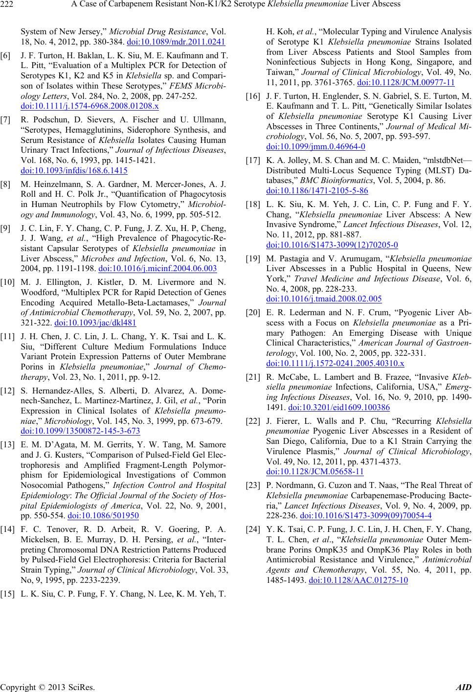 A Case of Carbapenem Resistant Non-K1/K2 Serotype Klebsiella pneumoniae Liver Abscess Copyright © 2013 SciRes. AID 222 System of New Jersey,” Microbial Drug Resistance, Vol. 18, No. 4, 2012, pp. 380-384. doi:10.1089/mdr.2011.0241 [6] J. F. Turton, H. Baklan, L. K. Siu, M. E. Kaufmann and T. L. Pitt, “Evaluation of a Multiplex PCR for Detection of Serotypes K1, K2 and K5 in Klebsiella sp. and Compari- son of Isolates within These Serotypes,” FEMS Microbi- ology Letters, Vol. 284, No. 2, 2008, pp. 247-252. doi:10.1111/j.1574-6968.2008.01208.x [7] R. Podschun, D. Sievers, A. Fischer and U. Ullmann, “Serotypes, Hemagglutinins, Siderophore Synthesis, and Serum Resistance of Klebsiella Isolates Causing Human Urinary Tract Infections,” Journal of Infectious Diseases, Vol. 168, No. 6, 1993, pp. 1415-1421. doi:10.1093/infdis/168.6.1415 [8] M. Heinzelmann, S. A. Gardner, M. Mercer-Jones, A. J. Roll and H. C. Polk Jr., “Quantification of Phagocytosis in Human Neutrophils by Flow Cytometry,” Microbiol- ogy and Immunology, Vol. 43, No. 6, 1999, pp. 505-512. [9] J. C. Lin, F. Y. Chang, C. P. Fung, J. Z. Xu, H. P, Cheng, J. J. Wang, et al., “High Prevalence of Phagocytic-Re- sistant Capsular Serotypes of Klebsiella pneumoniae in Liver Abscess,” Microbes and Infection, Vol. 6, No. 13, 2004, pp. 1191-1198. doi:10.1016/j.micinf.2004.06.003 [10] M. J. Ellington, J. Kistler, D. M. Livermore and N. Woodford, “Multiplex PCR for Rapid Detection of Genes Encoding Acquired Metallo-Beta-Lactamases,” Journal of Antimicrobial Chemotherapy, Vol. 59, No. 2, 2007, pp. 321-322. doi:10.1093/jac/dkl481 [11] J. H. Chen, J. C. Lin, J. L. Chang, Y. K. Tsai and L. K. Siu, “Different Culture Medium Formulations Induce Variant Protein Expression Patterns of Outer Membrane Porins in Klebsiella pneumoniae,” Journal of Chemo- therapy, Vol. 23, No. 1, 2011, pp. 9-12. [12] S. Hernandez-Alles, S. Alberti, D. Alvarez, A. Dome- nech-Sanchez, L. Martinez-Martinez, J. Gil, et al., “Porin Expression in Clinical Isolates of Klebsiella pneumo- niae,” Microbiology, Vol. 145, No. 3, 1999, pp. 673-679. doi:10.1099/13500872-145-3-673 [13] E. M. D’Agata, M. M. Gerrits, Y. W. Tang, M. Samore and J. G. Kusters, “Comparison of Pulsed-Field Gel Elec- trophoresis and Amplified Fragment-Length Polymor- phism for Epidemiological Investigations of Common Nosocomial Pathogens,” Infection Control and Hospital Epidemiology: The Official Journal of the Society of Hos- pital Epidemiologists of America, Vol. 22, No. 9, 2001, pp. 550-554. doi:10.1086/501950 [14] F. C. Tenover, R. D. Arbeit, R. V. Goering, P. A. Mickelsen, B. E. Murray, D. H. Persing, et al., “Inter- preting Chromosomal DNA Restriction Patterns Produced by Pulsed-Field Gel Electrophoresis: Criteria for Bacterial Strain Typing,” Journal of Clinical Microbiology, Vol. 33, No, 9, 1995, pp. 2233-2239. [15] L. K. Siu, C. P. Fung, F. Y. Chang, N. Lee, K. M. Yeh, T. H. Koh, et al., “Molecular Typing and Virulence Analysis of Serotype K1 Klebsiella pneumoniae Strains Isolated from Liver Abscess Patients and Stool Samples from Noninfectious Subjects in Hong Kong, Singapore, and Taiwan,” Journal of Clinical Microbiology, Vol. 49, No. 11, 2011, pp. 3761-3765. doi:10.1128/JCM.00977-11 [16] J. F. Turton, H. Englender, S. N. Gabriel, S. E. Turton, M. E. Kaufmann and T. L. Pitt, “Genetically Similar Isolates of Klebsiella pneumoniae Serotype K1 Causing Liver Abscesses in Three Continents,” Journal of Medical Mi- crobiology, Vol. 56, No. 5, 2007, pp. 593-597. doi:10.1099/jmm.0.46964-0 [17] K. A. Jolley, M. S. Chan and M. C. Maiden, “mlstdbNet— Distributed Multi-Locus Sequence Typing (MLST) Da- tabases,” BMC Bioinformatics, Vol. 5, 2004, p. 86. doi:10.1186/1471-2105-5-86 [18] L. K. Siu, K. M. Yeh, J. C. Lin, C. P. Fung and F. Y. Chang, “Klebsiella pneumoniae Liver Abscess: A New Invasive Syndrome,” Lancet Infectious Diseases, Vol. 12, No. 11, 2012, pp. 881-887. doi:10.1016/S1473-3099(12)70205-0 [19] M. Pastagia and V. Arumugam, “Klebsiella pneumoniae Liver Abscesses in a Public Hospital in Queens, New York,” Travel Medicine and Infectious Disease, Vol. 6, No. 4, 2008, pp. 228-233. doi:10.1016/j.tmaid.2008.02.005 [20] E. R. Lederman and N. F. Crum, “Pyogenic Liver Ab- scess with a Focus on Klebsiella pneumoniae as a Pri- mary Pathogen: An Emerging Disease with Unique Clinical Characteristics,” American Journal of Gastroen- terology, Vol. 100, No. 2, 2005, pp. 322-331. doi:10.1111/j.1572-0241.2005.40310.x [21] R. McCabe, L. Lambert and B. Frazee, “Invasive Kleb- siella pneumoniae Infections, California, USA,” Emerg- ing Infectious Diseases, Vol. 16, No. 9, 2010, pp. 1490- 1491. doi:10.3201/eid1609.100386 [22] J. Fierer, L. Walls and P. Chu, “Recurring Klebsiella pneumoniae Pyogenic Liver Abscesses in a Resident of San Diego, California, Due to a K1 Strain Carrying the Virulence Plasmis,” Journal of Clinical Microbiology, Vol. 49, No. 12, 2011, pp. 4371-4373. doi:10.1128/JCM.05658-11 [23] P. Nordmann, G. Cuzon and T. Naas, “The Real Threat of Klebsiella pneumoniae Carbapenemase-Producing Bacte- ria,” Lancet Infectious Diseases, Vol. 9, No. 4, 2009, pp. 228-236. doi:10.1016/S1473-3099(09)70054-4 [24] Y. K. Tsai, C. P. Fung, J. C. Lin, J. H. Chen, F. Y. Chang, T. L. Chen, et al., “Klebsiella pneumoniae Outer Mem- brane Porins OmpK35 and OmpK36 Play Roles in both Antimicrobial Resistance and Virulence,” Antimicrobial Agents and Chemotherapy, Vol. 55, No. 4, 2011, pp. 1485-1493. doi:10.1128/AAC.01275-10 |