

Paper Menu >>
Journal Menu >>
 Advances in Infectious Diseases, 2013, 3, 205-209 http://dx.doi.org/10.4236/aid.2013.33030 Published Online September 2013 (http://www.scirp.org/journal/aid) 205 Bacterial Contamination of Blood and Blood Products at Mbarara Regional Blood Bank in Rural South Western Uganda G. B. Matte Aloysius1,2*, Bazira Joel1, Richard Apecu2, Boum Yap II3, Frederick Byarugaba1 1Department of Microbiology, Faculty of Medicine, Mbarara University of Science and Technology (MUST), Mbarara, Uganda; 2Department of Medical Laboratory Science, Faculty of Medicine, Mbarara University of Science and Technology, Mbarara, Uganda. 3Epicentre Mbarara Research Base, Mbarara, Uganda; Email: *[email protected] Received July 9th, 2013; revised August 7th, 2013; accepted August 16th, 2013 Copyright © 2013 G. B. Matte Aloysius et al. This is an open access article distributed under the Creative Commons Attribution License, which permits unrestricted use, distribution, and reproduction in any medium, provided the original work is properly cited. ABSTRACT Background: Screening blood donors has practically eliminated viral and bacterial pathogens in blood used for trans- fusion. However, transfusion-associated bacterial sepsis remains an important health-care concern and the commonest cause of transfusion-related fatality in resource limited settings. Data on bacterial contamination of blood are scarce while the demand of blood transfusion is continuously growing. Therefore we conducted a study to determine the prevalence and typ e of bacterial con tamination in donor blood and bloo d produ cts, at the Mbarara Regio nal Blood Ban k. Methodology: A total of 510 units of screened blood and blood products consisting of refrigerated whole blood and packed cells were randomly sampled following aseptic procedures from Mbarara Regional Blood Bank. Two samples from each unit were collected in universal containers containing Brain Heart Infusion Broth and incubated at 37˚C for up to 7 days. Subcultures were carried out on Blood agar, Chocolate agar and MacConkey agar. Isolates were identified by standard microbiologic techniques and drug susceptibility testing was performed by Kirby Bauer disc diffusion method. Results: Of the 510 samples collected between June and October 2012, 18 (3.5%) samples showed growth. The contaminants were Staphylococcus aureus 17/18 (94.4%) and Streptococcus viridans 1/18 (5.6%). Isolates were sensitive to erythomycin, ampicillin, chloramphenicol and ciproflox acin and resistant to penicillin and cloxacillin. Con- clusion: Blood and blood products from Mbarara Regional Blood have unacceptable levels of bacterial contamination that can affect patient safety especially in an area with high malaria endemicity. Therefore it is critical to improve hy- giene precautions in order to minimize bacterial contamination and ensure patient safety. Keywords: Bacterial Contamination; Blood/Blood Products; Staph ylococci 1. Introduction Blood transfusion is a medical intervention intended to provide safe blood or blood components in a cost effec- tive way to patients who require blood and/or blood products. However, blood and blood components for transfusion can be a source of infection to recipients arising from contamination of these products by a variety of transmissible agents. Since the 1980’s when the hu- man immunodeficiency virus (HIV) was recognized, rigorous screening of blood before it is supplied to re- cipients was instituted and accepted worldwide [1]. Uganda adopted this rigorous screening of blood in order to provide safe blood to her population. Blood is screened for viruses including the Human Immunodefi- ciency virus (HIV), Hepatitis B virus (HBV), Hepatitis C (HCV) and for Treponema pallidum, a bacterium. It has been documented that bacteria can cause morbidity and mortality from blood and blood transfusion components [2,3]. In developed countries, transfusion of blood and blood components has a low but known infectious risk for pa- tients and remains a threat [4,5]. In the United States, bacterial contamination is said to account for 15.9% of all transfusion related fatalities, and is considered the second commonest cause of death from blood transfusion after clerical errors [4,6]. Recent data indicate that bacte- rial contamination has declined by about 50% or more *Corresponding a uthor. Copyright © 2013 SciRes. AID 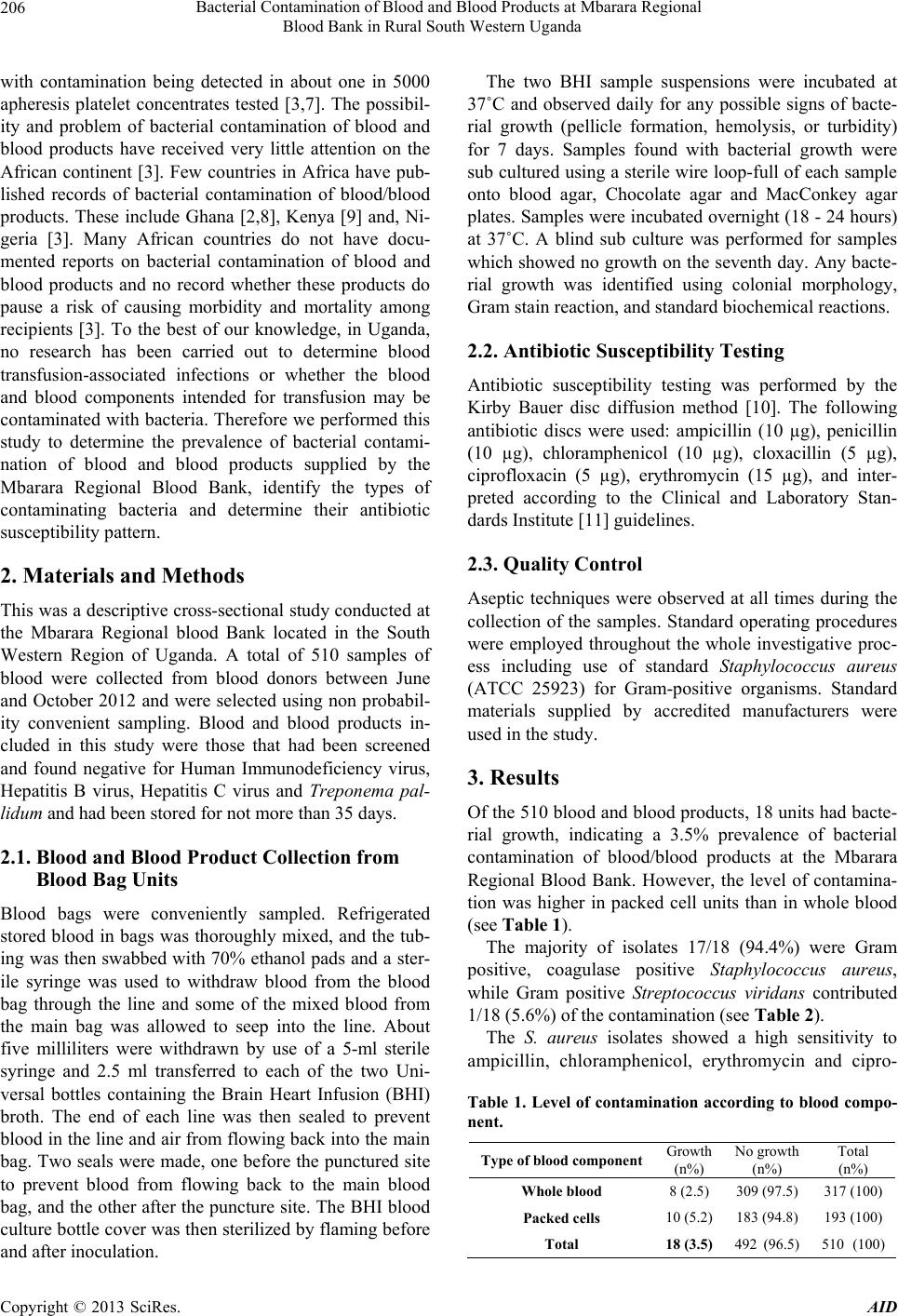 Bacterial Contamination of Blood and Blood Products at Mbarara Regional Blood Bank in Rural South Western Uganda 206 with contamination being detected in about one in 5000 apheresis platelet concentrates tested [3,7]. The possibil- ity and problem of bacterial contamination of blood and blood products have received very little attention on the African continent [3]. Few countries in Africa have pub- lished records of bacterial contamination of blood/blood products. These include Ghana [2,8], Kenya [9] and, Ni- geria [3]. Many African countries do not have docu- mented reports on bacterial contamination of blood and blood products and no record whether these products do pause a risk of causing morbidity and mortality among recipients [3]. To the best of our knowledge, in Uganda, no research has been carried out to determine blood transfusion-associated infections or whether the blood and blood components intended for transfusion may be contaminated with bacteria. Therefore we performed this study to determine the prevalence of bacterial contami- nation of blood and blood products supplied by the Mbarara Regional Blood Bank, identify the types of contaminating bacteria and determine their antibiotic susceptibility pattern. 2. Materials and Methods This was a descriptive cross-sectional study conducted at the Mbarara Regional blood Bank located in the South Western Region of Uganda. A total of 510 samples of blood were collected from blood donors between June and October 2012 and were selected using non probabil- ity convenient sampling. Blood and blood products in- cluded in this study were those that had been screened and found negative for Human Immunodeficiency virus, Hepatitis B virus, Hepatitis C virus and Treponema pal- lidum and had been stored for not more than 35 days. 2.1. Blood and Blood Product Collection from Blood Bag Units Blood bags were conveniently sampled. Refrigerated stored blood in bags was thoroughly mixed, and the tub- ing was then swabbed with 70% ethanol pads and a ster- ile syringe was used to withdraw blood from the blood bag through the line and some of the mixed blood from the main bag was allowed to seep into the line. About five milliliters were withdrawn by use of a 5-ml sterile syringe and 2.5 ml transferred to each of the two Uni- versal bottles containing the Brain Heart Infusion (BHI) broth. The end of each line was then sealed to prevent blood in the line and air from flowing back into the main bag. Two seals were made, one before the punctured site to prevent blood from flowing back to the main blood bag, and the other after the puncture site. The BHI blood culture bottle cover was then sterilized by flaming b efore and after inoculation. The two BHI sample suspensions were incubated at 37˚C and observed daily for any possible signs of bacte- rial growth (pellicle formation, hemolysis, or turbidity) for 7 days. Samples found with bacterial growth were sub cultured using a sterile wire loop-full of each sample onto blood agar, Chocolate agar and MacConkey agar plates. Samples were incubated overnight (18 - 24 hours) at 37˚C. A blind sub culture was performed for samples which showed no growth on the seventh day. Any bacte- rial growth was identified using colonial morphology, Gram stain reaction, and standard biochemical reactions. 2.2. Antibiotic Susceptibility Testing Antibiotic susceptibility testing was performed by the Kirby Bauer disc diffusion method [10]. The following antibiotic discs were used: ampicillin (10 µg), penicillin (10 µg), chloramphenicol (10 µg), cloxacillin (5 µg), ciprofloxacin (5 µg), erythromycin (15 µg), and inter- preted according to the Clinical and Laboratory Stan- dards Institute [11] guidelines. 2.3. Quality Control Aseptic techniques were observed at all times during the collection of the samples. Standard operating procedures were employed throughout the whole investigative proc- ess including use of standard Staphylococcus aureus (ATCC 25923) for Gram-positive organisms. Standard materials supplied by accredited manufacturers were used in the study. 3. Results Of the 510 blood and blood products, 18 units had bacte- rial growth, indicating a 3.5% prevalence of bacterial contamination of blood/blood products at the Mbarara Regional Blood Bank. However, the level of contamina- tion was higher in packed cell units than in whole blood (see Table 1). The majority of isolates 17/18 (94.4%) were Gram positive, coagulase positive Staphylococcus aureus, while Gram positive Streptococcus viridans contributed 1/18 (5.6%) of the contamination (see Table 2). The S. aureus isolates showed a high sensitivity to ampicillin, chloramphenicol, erythromycin and cipro- Table 1. Level of contamination according to blood compo- nent. Type of blood componentGrowth (n%) No growth (n%) Total (n%) Whole blood 8 (2.5) 309 (97.5) 317 (100) Packed cells 10 (5.2) 183 (94.8) 193 (100) Total 18 (3.5) 492 (96.5) 510 (100) Copyright © 2013 SciRes. AID 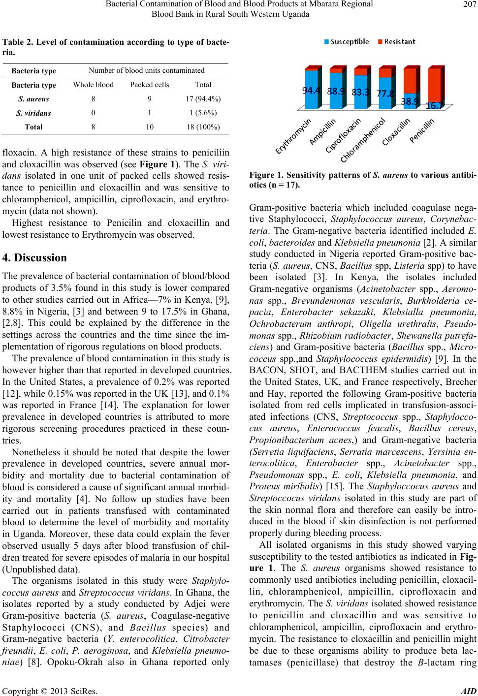 Bacterial Contamination of Blood and Blood Products at Mbarara Regional Blood Bank in Rural South Western Uganda 207 Table 2. Level of contamination according to type of bacte- ria. Bacteria type Number of blood units contamina te d Bacteria type Whole blood Packed cells Total S. aureus 8 9 17 (94.4%) S. viridans 0 1 1 (5.6%) Total 8 10 18 (100%) floxacin. A high resistance of these strains to peniciliin and cloxacillin was observed (see Figure 1). The S. viri- dans isolated in one unit of packed cells showed resis- tance to penicillin and cloxacillin and was sensitive to chloramphenicol, ampicillin, ciprofloxacin, and erythro- mycin (data not shown). Highest resistance to Penicilin and cloxacillin and lowest resistance to Erythromycin was observed. 4. Discussion The prevalence of bacterial contamination of blood/blood products of 3.5% found in this study is lower compared to other studies carried out in Africa—7% in Kenya, [9], 8.8% in Nigeria, [3] and between 9 to 17.5% in Ghana, [2,8]. This could be explained by the difference in the settings across the countries and the time since the im- plementation of rigorous regulations on blood products. The prevalence of blood contamination in this study is however higher than that reported in developed countries. In the United States, a prevalence of 0.2% was reported [12], while 0.15% was reported in the UK [13], and 0.1% was reported in France [14]. The explanation for lower prevalence in developed countries is attributed to more rigorous screening procedures practiced in these coun- tries. Nonetheless it should be noted that despite the lower prevalence in developed countries, severe annual mor- bidity and mortality due to bacterial contamination of blood is considered a cause of significant annual morbid- ity and mortality [4]. No follow up studies have been carried out in patients transfused with contaminated blood to determine the level of morbidity and mortality in Uganda. Moreover, these data could explain the fever observed usually 5 days after blood transfusion of chil- dren treated for severe episodes of malaria in our hospital (Unpubli shed data). The organisms isolated in this study were Staphylo- coccus aureus and Streptococcus viridans. In Ghana, the isolates reported by a study conducted by Adjei were Gram-positive bacteria (S. aureus, Coagulase-negative Staphylococci (CNS), and Bacillus species) and Gram-negative bacteria (Y. enterocolitica, Citrobacter freundii, E. coli, P. aeroginosa, and Klebsiella pneumo- niae) [8]. Opoku-Okrah also in Ghana reported only Figure 1. Sensitivity patterns of S. aureus to various antibi- otics (n = 17). Gram-positive bacteria which included coagulase nega- tive Staphylococci, Staphylococcus aureus, Corynebac- teria. The Gram-negative bacteria identified included E. coli, bactero ides an d Klebsiella pneumonia [2]. A similar study conducted in Nigeria reported Gram-positive bac- teria (S. aureus, CNS, Bacillus spp, Listeria spp) to have been isolated [3]. In Kenya, the isolates included Gram-negative organisms (Acinetobacter spp., Aeromo- nas spp., Brevundemonas vescularis, Burkholderia ce- pacia, Enterobacter sekazaki, Klebsialla pneumonia, Ochrobacterum anthropi, Oligella urethralis, Pseudo- monas spp., Rhizob ium radiobacter, Shewanella putrefa- ciens) and Gram-positive bacteria (Ba cillus spp., Micro- coccus spp.,and Staphylococcus epidermidis) [9]. In the BACON, SHOT, and BACTHEM studies carried out in the United States, UK, and France respectively, Brecher and Hay, reported the following Gram-positive bacteria isolated from red cells implicated in transfusion-associ- ated infections (CNS, Streptococcus spp., Staphylocco- cus aureus, Enterococcus feacalis, Bacillus cereus, Propionibacterium acnes,) and Gram-negative bacteria (Serretia liquifaciens, Serratia marcescens, Yersinia en- terocolitica, Enterobacter spp., Acinetobacter spp., Pseudomonas spp., E. coli, Klebsiella pneumonia, and Proteus miribalis) [15]. The Staphyloccocus aureus and Streptoccocus viridans isolated in this study are part of the skin normal flora and therefore can easily be intro- duced in the blood if skin disinfection is not performed properly duri n g bl eeding process. All isolated organisms in this study showed varying susceptibility to the tested antibio tics as indicated in Fig- ure 1. The S. aureus organisms showed resistance to commonly used antibiotics including penicillin, cloxacil- lin, chloramphenicol, ampicillin, ciprofloxacin and erythromycin. The S. viridans isolated showed resistance to penicillin and cloxacillin and was sensitive to chloramphenicol, ampicillin, ciprofloxacin and erythro- mycin. The resistance to cloxacillin and penicillin might be due to these organisms ability to produce beta lac- tamases (penicillase) that destroy the B-lactam ring Copyright © 2013 SciRes. AID  Bacterial Contamination of Blood and Blood Products at Mbarara Regional Blood Bank in Rural South Western Uganda 208 found in these antibiotics, rendering the antibiotics inef- fective. In Ghana, it was reported that all Gram-positive isolates were resistant to cefuroxime, penicillin, ampicil- lin, and cotrimoxazole but sensitive to cefotoxime, tetra- cycline, erythromycin, and gentamycin [2,8]. Similarly, all the Gram-negative organisms isolated were resistant to cefotoxime (except Y. enterocolitica), tetracycline, ampicillin, cefuroxime, cotrimoxazole, and chloram- phenicol but sensitive to amikacin and gentamycin. A study in Nigeria reported resistance to antibiotics tested (ampicillin, cotrimoxazile, erythromycin, penicillin, tet- racycline, rifampicin) except gentamycin, and ceftri- oxone that ranged from 50% to 100% [3]. One possible explanation for the high resistance of donor blood isolates may be associated with the ease of procuring antibiotics over the counter in Uganda, self medication, and shortfalls in infection control [16]. An- other possible cause of high resistance could be due to misuse of antibiotics by cattle farmers, Amanya, (unpub- lished) reported bacterial resistance to antibiotics on bacteria isolated from milk which included S. aureus, Klebsiella pneumonia, S. agalactiae, and E. coli which showed resistance to the commonly used antibiotics in- cluding tetracycline, gentamycin and penicillin. This can imply that people consuming milk and milk products may acquire bacterial strains from milk and milk prod- ucts that are already resistant to the commonly used anti- biotics. 5. Conclusion Bacterial contamination of blood was found to be 3.5%. Staphylococcus aureus was found to be the major con- taminant with 17 out of 18 organisms isolated (94.4%) while Streptococcus viridans contributed 1/18 (5.6%). The isolates are known to be part of the skin normal flora. The organisms showed increased resistance to penicillin and cloxacillin but susceptible to ampicillin, chloram- phenicol, erythromycin and ciprofloxacin. 6. Recommendation While the Mbarara Regional Blood Bank is doing a commendable job in provision of safe blood and blood products emphasis should be put on ensuring proper dis- infectio n o f phlebotomy site s . Policy makers should ensure emphasis of blood safety during phlebotomy by providing quality disinfectants. Routine antibiotic susceptibility testing is recommended in all cases of post-transfusion sepsis. 7. Acknowledgements We appreciate the financial, material and moral support offered to us by Mbarara University of Science and Technology, Department of Microbiology and; Epicen- tre Mbarara Research Base. Special thanks to Dr. At- wine Daniel of Epicentre for the support towards the data analysis of our study . REFERENCES [1] American Association of Blood Banks, “Comprehensive Report on Blood Collection and Transfusion in the United States in 2001,” National Blood Data Resource Center, Bethesda, 2003. [2] C. Opoku-Okrah, P. Feglo, N. Amidu and M. P. Dakorah, “Bacterial Contamination of Donor Blood at Tamale Teaching Hospital , Gha n a, ” African Health Sciences, Vol. 9, No. 1, 2009, pp. 13-18. [3] A. R. Bolarinwa, A. A. Oladipo, W. O. Baba tunde and B . A. Aramide, “Bacterial Contamination of Blood and Blood Components in a Tertiary Hospital Setting in Ni- geria,” International Journal of Infection Control, Vol. 7, No. 1, 2011, pp. 1-6. [4] C. D. Hillyer, C. D. Josephson, M. A. Blajchman, J. G. Vostal, J. S. Epstein and J. L. Goodman, “Bacterial Con- tamination of Blood Components: Risks, Strategies, and Regulation: Joint ASH and AABB Educational Session in Transfusion Medicine,” Hematology (American Society of Hematology Education Program), Vol. 2003, No. 1, 2003, pp. 575-589. doi:10.1182/asheducation-2003.1.575 [5] C. P. Engelbreit, H. W. Reesink, M. A. Blajchman, et al., “Bacterial Contamination of Blood Components,” Vox Sanguinis, Vol. 78, No. 1, 2008, pp. 59-67. [6] P. A. Hoppe, “Interim Measure for Detection of Bacteri- ally Contaminated Red Cell Components,” Transfusion, Vol. 32, No. 3, 1992, pp. 199-201. [7] C. T. Fang, L. A. Chambers, J. Kennedy, A. Strupp, M. C. Fucci, J. A. Janas, Y. Tang, C. A. Hapip, T. B. Lawrence and R. Y. Dodd, “Detection of Bacterial Contamination in Apheresis Platelet Products,” Transfusion, Vol. 45, No. 12, 2004, pp. 1845-1852. [8] A. A. Adjei, K. G. Kuma, Y. Tettey, F. P. Ayeh-Kumi, J. Opintan, F. Apeagyei, O. J. Ankrah, K. T. Adiku and E. G. Narter-Olaga, “Bacterial Contamination of Blood and Blood Components in Three Major Blood Transfusion Centers, Accra, Ghana,” Japanese Journal of Infectious Diseases, Vol. 62, No. 4, 2009, pp. 265-269. [9] O. Hassall, K. Maitland, L. Pole, et al., “Bacterial Con- tamination of Paediatric Whole Blood Transfusions in a Kenyan Hospital,” Transfusion, Vol. 49, No. 12, 2009, pp. 2594-2598. [10] A. W. Bauer, W. M. M. Kirby, J. C. Sherris, et al., “Anti- biotic Susceptibility Testing by a Standard Single Disc Diffusion Method,” American Journal of Clinical Pa- thology, Vol. 46, 1966, pp. 493-496. http://202.114.65.51/fzjx/wsw/newindex/wswfzjs/pdf/105 bauer.pdf [11] CLSI—Performance Standards for Antimicrobial Suscep- tibility Testing, “Twenty-First Information Supplement,” 2011. Copyright © 2013 SciRes. AID  Bacterial Contamination of Blood and Blood Products at Mbarara Regional Blood Bank in Rural South Western Uganda Copyright © 2013 SciRes. AID 209 http://antimicrobianos.com.ar/ATB/wp-content/uploads/2 012/.../M100S22E.pdf [12] M. Kuehnert, V. R. Roth, N. R. Haley, et al., “Transfu- sion Transmitted Bacterial Infection in the United States, 1998 through 2000,” Transfusion, Vol. 41, No. 12, 2001, pp. 1493-1499. doi:10.1046/j.1537-2995.2001.41121493.x [13] E. M. Love, D. Asher, C. L. J. Atterbury, C. Chapman, H. Cohen, H. Jones, D. R. Norfolk, J. Revill, K. Soldan, A. Todd and L. M. Williamson, “Serious Hazard of Transfu- sion. SHOT Report for 2000-2001,” 2002. http//www.shot.demon.co.uk [14] P. L. Perez, R. Salmi, G. Follea, et al., “Determinants of Transfusion-Associated Bacterial Contamination: Results of the French BACTHEM Case-Control Study,” Transfu- sion, Vol. 41, No. 7, 2001, pp. 862-871. doi:10.1046/j.1537-2995.2001.41070862.x [15] M. E. Brecher and S. N. Hay, “Bacterial Contamination of Blood Components,” Clinical Microbiology Reviews, Vol. 18, No. 1, 2005, pp. 195-204. doi:10.1128/CMR.18.1.195-204.2005 [16] I. N. Okeke, R. La xminarayan, Z. A. Bhutta, A. G. Duse, P. Jenkins, F. O’Brien, A. Pablos-Mendez and K. Klug- man, “Antimicrobial Resistance in Developing Countries. Part I: Recent Trends and Current Status,” The Lancet In- fectious Diseases, Vol. 5, No. 8, 2005, pp. 481-493. |