

Paper Menu >>
Journal Menu >>
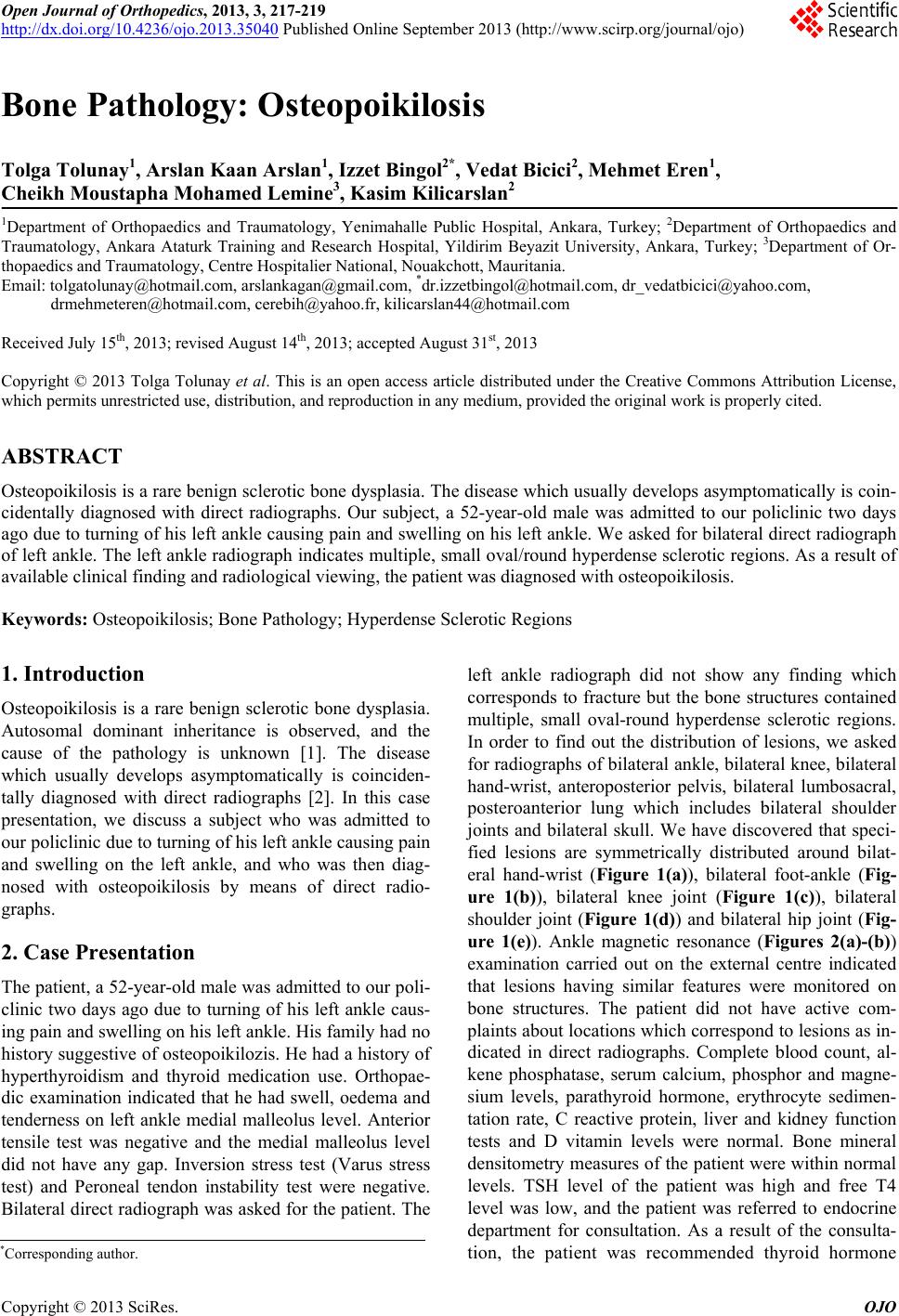 Open Journal of Orthopedics, 2013, 3, 217-219 http://dx.doi.org/10.4236/ojo.2013.35040 Published Online September 2013 (http://www.scirp.org/journal/ojo) 217 Bone Pathology: Osteopoikilosis Tolga Tolunay1, Arslan Kaan Arslan1, Izzet Bingol2*, Vedat Bicici2, Mehmet Eren1, Cheikh Moustapha Mohamed Lemine3, Kasim Kilicarslan2 1Department of Orthopaedics and Traumatology, Yenimahalle Public Hospital, Ankara, Turkey; 2Department of Orthopaedics and Traumatology, Ankara Ataturk Training and Research Hospital, Yildirim Beyazit University, Ankara, Turkey; 3Department of Or- thopaedics and Traumatology, Centre Hospitalier National, Nouakchott, Mauritania. [email protected], cerebih@yahoo.fr, [email protected] Received July 15th, 2013; revised August 14th, 2013; accepted August 31st, 2013 Copyright © 2013 Tolga Tolunay et al. This is an open access article distributed under the Creative Commons Attribution License, which permits unrestricted use, distribution, and reproduction in any medium, provided the original work is properly cited. ABSTRACT Osteopoikilosis is a rare benign sclerotic bone dysplasia. The disease which usually develops asymptomatically is coin- cidentally diagnosed with direct radiographs. Our subject, a 52-year-old male was admitted to our policlinic two days ago due to turning of his left ankle causing pain and swelling on his left ankle. We asked for bilateral direct radiograph of left ankle. The left ankle radiograph indicates multiple, small oval/round hyperdense sclerotic regions. As a result of available clinical finding and radiological viewing, the patient was diagnosed with osteopoikilosis. Keywords: Osteopoikilosis; Bone Pathology; Hyperdense Sclerotic Regions 1. Introduction Osteopoikilosis is a rare benign sclerotic bone dysplasia. Autosomal dominant inheritance is observed, and the cause of the pathology is unknown [1]. The disease which usually develops asymptomatically is coinciden- tally diagnosed with direct radiographs [2]. In this case presentation, we discuss a subject who was admitted to our policlinic due to turning of his left ankle causing pain and swelling on the left ankle, and who was then diag- nosed with osteopoikilosis by means of direct radio- graphs. 2. Case Presentation The patient, a 52-year-old male was admitted to our poli- clinic two days ago due to turning of his left ankle caus- ing pain and swelling on his left ankle. His family had no history suggestive of osteopoikilozis. He had a history of hyperthyroidism and thyroid medication use. Orthopae- dic examination indicated that he had swell, oedema and tenderness on left ankle medial malleolus level. Anterior tensile test was negative and the medial malleolus level did not have any gap. Inversion stress test (Varus stress test) and Peroneal tendon instability test were negative. Bilateral direct radiograph was asked for the patient. The left ankle radiograph did not show any finding which corresponds to fracture but the bone structures contained multiple, small oval-round hyperdense sclerotic regions. In order to find out the distribution of lesions, we asked for radiographs of bilateral ankle, bilateral knee, bilateral hand-wrist, anteroposterior pelvis, bilateral lumbosacral, posteroanterior lung which includes bilateral shoulder joints and bilateral skull. We have discovered that speci- fied lesions are symmetrically distributed around bilat- eral hand-wrist (Figure 1(a)), bilateral foot-ankle (Fig- ure 1(b)), bilateral knee joint (Figure 1(c)), bilateral shoulder joint (Figure 1(d)) and bilateral hip joint (Fig- ure 1(e)). Ankle magnetic resonance (Figures 2(a)-(b)) examination carried out on the external centre indicated that lesions having similar features were monitored on bone structures. The patient did not have active com- plaints about locations which correspond to lesions as in- dicated in direct radiographs. Complete blood count, al- kene phosphatase, serum calcium, phosphor and magne- sium levels, parathyroid hormone, erythrocyte sedimen- tation rate, C reactive protein, liver and kidney function tests and D vitamin levels were normal. Bone mineral densitometry measures of the patient were within normal levels. TSH level of the patient was high and free T4 level was low, and the patient was referred to endocrine department for consultation. As a result of the consulta- tion, the patient was recommended thyroid hormone *Corresponding author. Copyright © 2013 SciRes. OJO 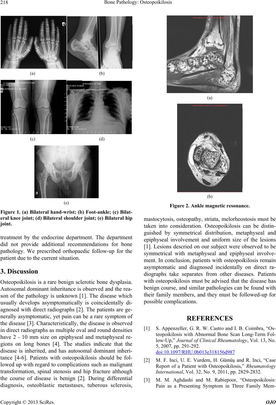 Bone Pathology: Osteopoikilosis 218 (a) (b) (c) (d) (e) Figure 1. (a) Bilateral hand-wrist; (b) Foot-ankle; (c) Bilat- eral knee joint; (d) Bilateral shoulder joint; (e) Bilateral hip joint. treatment by the endocrine department. The department did not provide additional recommendations for bone pathology. We prescribed orthopaedic follow-up for the patient due to the current situation. 3. Discussion Osteopoikilosis is a rare benign sclerotic bone dysplasia. Autosomal dominant inheritance is observed and the rea- son of the pathology is unknown [1]. The disease which usually develops asymptomatically is coincidentally di- agnosed with direct radiographs [2]. The patients are ge- nerally asymptomatic, yet pain can be a rare symptom of the disease [3]. Characteristically, the disease is observed in direct radiographs as multiple oval and round densities have 2 - 10 mm size on epiphyseal and metaphyseal re- gions on long bones [4]. The studies indicate that the disease is inherited, and has autosomal dominant inheri- tance [4-6]. Patients with osteopoikilosis should be fol- lowed up with regard to complications such as malignant transformation, spinal stenosis and hip fracture although the course of disease is benign [2]. During differential diagnosis, osteoblastic metastases, tuberous sclerosis, (a) (b) Figure 2. Ankle magnetic resonance. mastocytosis, osteopathy, striata, melorheostosis must be taken into consideration. Osteopoikilosis can be distin- guished by symmetrical distribution, metaphyseal and epiphyseal involvement and uniform size of the lesions [1]. Lesions descried on our subject were observed to be symmetrical with metaphyseal and epiphyseal involve- ment. In conclusion, patients with osteopoikilosis remain asymptomatic and diagnosed incidentally on direct ra- diographs take separates from other diseases. Patients with osteopoikilosis must be advised that the disease has benign course, and similar pathologies can be found with their family members, and they must be followed-up for possible complications. REFERENCES [1] S. Appenzeller, G. R. W. Castro and I. B. Coimbra, “Os- teopoikilosis with Abnormal Bone Scan Long-Term Fol- low-Up,” Journal of Clinical Rheumatology, Vol. 13, No. 5, 2007, pp. 291-292. doi:10.1097/RHU.0b013e318156d987 [2] M. F. Inci, U. E. Vurdem, H. Gümüş and R. Inci, “Case Report of a Patient with Osteopoikilosis,” Rheumatology International, Vol. 32, No. 9, 2011, pp. 2829-2832. [3] M. M. Aghdashi and M. Rabiepoor, “Osteopoikilosis: Pain as a Presenting Symptom in Three Family Mem- Copyright © 2013 SciRes. OJO  Bone Pathology: Osteopoikilosis Copyright © 2013 SciRes. OJO 219 bers,” Clinical Medicine Insights: Arthritis and Muscu- loskeletal Disorders, Vol. 4, 2011, pp. 29-32. doi:10.4137/CMAMD.S7035 [4] I. T. Benli, S. Akalin, E. Boysan, E. F. Mumcu, M. Ki and D. Türkolu, “Epidemiological, Clinical and Radio- logical Aspects of Osteopoikilosis,” Journal of Bone & Joint Surgery, Vol. 74, No. 4, 1992, pp. 504-506. [5] S. Baasanjav, A. Jamsheer, M. Kolanczyk, D. Horn, K. Hoffmann, A. Latos-Bielenska, S. Mundlos and T. Latos, “Osteopoikilosis and Multiple Exostoses Caused by No- vel Mutations in LEMD3 and EXT1 Genes Respectively- Coincidence within One Family, Baasanjav et al.,” BMC Medical Genetics, Vol. 11, 2010, p. 110. doi:10.1186/1471-2350-11-110 [6] E. Ben-Asher, E. Zelzer and D. Lancet, “LEMD3: The Gene Responsible for Bone Density Disorders (Osteo- poikilosis),” Israel Medical Association Journal, Vol. 7, No. 4, 2005, pp. 273-274. |