

Paper Menu >>
Journal Menu >>
 International Journal of Clinical Medicine, 2013, 4, 375-377 http://dx.doi.org/10.4236/ijcm.2013.49067 Published Online September 2013 (http://www.scirp.org/journal/ijcm) 375 Impact of Ocular Compression on Ocular Surface Bacterial Contamination Amir Reza Daneshmand Eslami1*, Nikolai M. Sergienko1, Ivan P. Bilko2 1Department of Ophthalmology, National Medical Academy of Postgraduate Education, Kiev, Ukraine; 2Department of Microbiol- ogy and Epidemiology, National Medical Academy of Postgraduate Education, Kiev, Ukraine. Email: *[email protected] Received June 12th, 2013; revised July 16th, 2013; accepted August 5th, 2013 Copyright © 2013 Amir Reza Daneshmand Eslami et al. This is an open access article distributed under the Creative Commons At- tribution License, which permits unrestricted use, distribution, and reproduction in any medium, provided the original work is prop- erly cited. ABSTRACT Background: To study the impact of preoperativ e ocular compression on bacterial contamination of the lid margin and conjunctival sac. Methods: This study evaluated 106 eyes from 106 patients undergoing cataract surgery. Preopera- tively, all of the patients received one drop of 0.5% levofloxacin eye drops 5 times per day for 4 days. The patients were divided into 2 groups after receiving retrobulbar anaesthesia. The study group (75 eyes) received 15 ± 2 minutes of ocular compression, and the control group (31 eyes) remained with their eyes closed for 15 ± 2 minutes without the ap- plication of ocular compression or digital massage. Following the device removal (study group only) and the opening of the eyes, bacterial cultures were taken from the lid margin and conjunctival sac. Results: The lid margin was positive for bacteria in 23 eyes (30.7%) in the study group vs. 3 eyes (9.7%) in the control group (P = 0.0223), and the con junc- tival sac was positive for bacteria in 16 eyes (21.3%) in the study group vs. 5 eyes (16.1%) in the control group (P = 0.5409). Conclusion: Preoperative ocular compression is associated with the release of meibomian gland secretions and bacterial contamination of the eyelid edges. Keywords: Bacteria; Conjunctival Sac; Endophthalmitis; Lid Margin; Ocular Compression 1. Introduction Postoperative endophthalmitis (POE) remains a rare but severe complication in modern cataract surgery [1]. The most common sites of ocular bacterial contamination leading to POE include the eyelids, skin and conjunctiva [2,3]. The lid margins, which carry bacterial flora to the skin surface, and meibomian gland secretions also play an important role in POE development. However, it remains unclear whether preoperative ocular compres- sion (OC), which is performed to reduce intraocular pres- sure (IOP), can promote bacterial contamination of the eyelids and conjunctiva. Although in recent years OC has become less popular, this technique is still commonly performed. Pressure delivered via the modified Honan’s balloon is realised not on ly on the eyeball but also on the lid margin and may provoke the release of meibomian gland secretions that contain bacteria. The aim of this study was to determine the impact of preoperative ocular compression on bacterial contamina- tion in the lid margin and the conjunctival sac. 2. Materials and Methods 2.1. Study Sample This study evaluated 106 eyes from 106 patients under- going cataract surgery. The exclusion criteria included systemic or local infection, con junctivitis, b lepharitis, da- cryocystitis, meibomian gland dysfunction and diabetes mellitus. Preoperatively, all of the patients received one drop of 0.5% levofloxacin ophthalmic solution (Ofta- quix®, Santen Oy, Niittyhaankatu 20, 33720 Tampere, Finland) 5 times per day for 4 days. The patients were placed in the supine position prior to treatment and remained undisturbed throughout the test period. Retrobulbar anaesthesia was administered with a single injection of 0.5% bupivacaine hydrochloride, 2% lidocaine hydrochloride, and 0.5% hyaluronidase (3.75 IU/ml). The volume of the block ranged from 3.5 to 5.5 ml, depending on the patient’s weight. The patients were divided into two groups. The study group (75 eyes of 75 patients) received retrobulbar anaesthesia in addition to the preoperative application of *Corresponding a uthor. Copyright © 2013 SciRes. IJCM 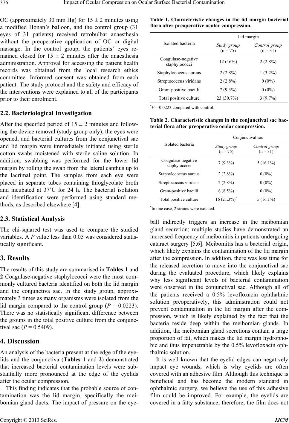 Impact of Ocular Compression on Ocular Surface Bacterial Contaminat i on 376 OC (approximately 30 mm Hg) for 15 ± 2 minutes using a modified Honan’s balloon, and the control group (31 eyes of 31 patients) received retrobulbar anaesthesia without the preoperative application of OC or digital massage. In the control group, the patients’ eyes re- mained closed for 15 ± 2 minutes after the anaesthesia administration. Approval for accessing the patient health records was obtained from the local research ethics committee. Informed consent was obtained from each patient. The study protocol and the safety and efficacy of the intervention s were explained to all of the participants prior to their enrolment. 2.2. Bacteriological Investigation After the specified period of 15 ± 2 minutes and follow- ing the device removal (study group only), the eyes were opened, and bacterial cultures from the conjunctival sac and lid margin were immediately initiated using sterile cotton swabs moistened with sterile saline solution. In addition, swabbing was performed for the lower lid margin by rolling the swab from the lateral canthus up to the lacrimal point. The samples from each eye were placed in separate tubes containing thioglycolate broth and incubated at 37˚C for 24 h. The bacterial isolation and identification were performed using standard me- thods, as described elsewhere [4]. 2.3. Statistical Analysis The chi-squared test was used to compare the studied variables. A P value less than 0.05 was considered statis- tically significant. 3. Results The results of this study are summarised in Tables 1 and 2 Coagulase-negative staphylococci were the most com- monly cultured bacteria identified on both the lid margin and the conjunctiva sac. In the study group, approxi- mately 3 times as many organisms were isolated from the lid margin compared to the control group (P = 0.0223). There was no statistically significant difference between the groups in the total positive culture from the conjunc- tival sac (P = 0.5409). 4. Discussion An analysis of the bacteria present at the edge of the eye- lids and the conjunctiva (Tables 1 and 2) demonstrated that increased bacterial contamination levels were sub- stantially more pronounced at the edge of the eyelids after the ocular compression. This finding indicates that the probable source of con- tamination was the lid margin, specifically the mei- bomian gland ducts. The impact of pressure on the eye- Table 1. Characteristic changes in the lid margin bacterial flora after preoperative ocular compression. Lid margin Isolated bacteria Study group (n = 75) Control group (n = 31) Coagulase-negative staphylococci 12 (16%) 2 (2.8%) Staphylococcus aureus 2 (2.8%) 1 (3.2%) Streptococcus viridans 2 (2.8%) 0 (0%) Gram-positive bacilli 7 (9.3%) 0 (0%) Total positive culture 23 (30.7%)* 3 (9.7%) *P = 0.0223 compared with control. Table 2. Characteristic changes in the conjunctival sac bac- terial flora after preoperative ocular compression. Conjunctival sac Isolated bacteria Stu dy group (n = 75) Control group (n = 31) Coagulase-negative staphylococci 7 (9.3%) 5 (16.1%) Staphylococcus aureus 2 (2.8%) 0 (0%) Streptococcus viridans 2 (2.8%) 0 (0%) Gram-posi t iv e bacilli 6 (8.5%) 0 (0%) Total positive culture 16 (21.3%)* 5 (16.1%) *In one cas e, 2 strains were isolated. ball indirectly triggers an increase in the meibomian gland secretion; multiple studies have demonstrated an increased frequency of meibomitis in patients undergoing cataract surgery [5,6]. Meibomitis has a bacterial origin, which likely exp lains the contamination o f the lid margin after the compression. In addition, there was less time for the released secretion to move into the conjunctival sac during the evaluated procedure, which likely explains why less significant levels of bacterial contamination were observed in the conjunctival sac. Although all of the patients received a 0.5% levofloxacin ophthalmic solution preoperatively, this administration could not prevent contamination in the lid margin after the com- pression, which is likely explained by the fact that the bacteria reside deep within the meibomian glands. In addition, the meibomian gland secretions contain a large proportion of fat, which makes the lid margin hydropho- bic and thus impenetrable by the 0.5% levofloxacin oph- thalmic solution. It is well known that the eyelid edges can negatively impact eye wounds, which is why eyelids are often covered with an adhesive film. Although this technique is beneficial and has become the modern standard in ophthalmic surgery, we believe the use of this adhesive film could be improved. For example, the eyelids are covered in a fatty substance; therefore, the film does not Copyright © 2013 SciRes. IJCM  Impact of Ocular Compression on Ocular Surface Bacterial Contaminat i o n Copyright © 2013 SciRes. IJCM 377 reliably adhere to the eyelid s. Moreover, the liquid filling the conjunctive sac washes the free edge of the eyelids and transfers bacteria from the lids to the surgical wound. We studied the effect of ocular compression using the modified Honan’s balloon, although the use of an eyelid speculum may have a similar but less dramatic effect. Presumably, the factors that contribute to the release of meibomian gland secretions may be related to the ma- nipulations performed during retrobulbar or peribulbar anaesthesia. Theoretically, even eye movements during surgery and immediately after surgery (performed while the patient is under topical anaesthesia) could place pressure on the meibomian glands. Improperly applying antibiotics on the first postoperative day might also result in negative consequences. Garcia-Arumi further reported that the POE rate was higher following the administra- tion of local anaesthesia compared to retrobulbar anaes- thesia [7]. The most effective method of suppressing bacterial surface contamination may be via the isolation of the lid margin during surgery. We initiated this type of study and evaluated the effects of covering the lid edges with textile napkins impreg nated with 0.02% aqueous so lution of chlorhex idine. The results indicated that the continued presence of a strong antiseptic intraoperatively reduced the POE rate [8]. 5. Conclusion Preoperative ocular compression performed with the goal of reducing IOP was associated with the release of mei- bomian gland secretions and bacterial contamination of the eyelids. Thus, reliable intraoperative isolation of the eyelids and the postoperative application of antibiotics are required for effective POE prophylactic measures. 6. Acknowledgements The authors thank the individuals who participate in this study. None of the authors had financial or personal con- flict of interest with regard to this study. This paper was derived from a PhD thesis. REFERENCES [1] J. C. Lloyd and R. Braga-Mele, “Incidence of Postopera- tive Endophthalmitis in a High-Volume Cataract Surgi- centre in Canada,” Canadian Journal Ophthalmology, Vol. 44, No. 3, 2009, pp. 288-292. [2] R. G. Ariyasu, T. Nakamura, N. D. Trousdale and R. E. Smith, “Intraoperative Bacterial Contamination of the Aqueous Humor,” Ophthalmic Surgery, Vol. 24, No. 6, 1993, pp. 367-374. [3] M. G. Speaker, F. A Milch, M. K. Shah, W. Eisner and B. N. Kreiswirth, “Role of External Bacterial Flora in the Pathogenesis of Acute Postoperative Endophthalmitis,” Ophthalmology, Vol. 98, No. 5, 1991, pp. 639-649. [4] G. L. Wood, “Microbiology,” In: K. D. McClatchey, Ed., Clinical Laboratory Medicine, Lippincott Williams & Wilkins, Philadelphia, 2002, pp. 1024-1175. [5] F. A. Bucci Jr., “Prevention of Endophthalmitis and Ocu- la r Absorp tion of Fluor oqui nolo nes,” Ocula r Surge ry News, November 2003, pp. 5-8. [6] E. Knop, N. Knop, T. Millar, H. Obata and D. A. Sullivan, “The International Workshop on Meibomian Gland Dys- function: Report of the Subcommittee on Anatomy, Phy- siology, and Pathophysiology of the Meibomian Gland,” Investigative Ophthalmology & Visual Science, Vol. 52, No. 4, 2011, pp. 1938-1978. doi:10.1167/iovs.10-6997c [7] J. Garcia-Arumi, A. Fonollosa, L. Sararols, F. Fina, V. Martinez-Castillo, A. Boixadera, M. A. Zapata and M. Campins, “Topical Anesthesia: Possible Risk Factor for Endophthalmitis after Cataract Extraction,” Journal Cata- ract Refract Surgery, Vol. 33, No. 6, 2007, pp. 989-992. doi:10.1016/j.jcrs.2007.02.030 [8] N. M. Sergienko, Y. N. Kondratenko, N. V. Chumak and A. Daneshmand, “Results of Prophylaxis of Bacterial Endophthalmitis in Cataract Surgery,” Proceedings of the Joint Congress of SOE/AAO, Geneva, June 2011, p. 51. |