

Paper Menu >>
Journal Menu >>
 International Journal of Otolaryngology and Head & Neck Surgery, 2013, 2, 160-164 http://dx.doi.org/10.4236/ijohns.2013.25035 Published Online September 2013 (http://www.scirp.org/journal/ijohns) Comparison of Transtympanic and Extratympanic Electrocochleography Alan K. Dobbs1, Wesley W. O. Krueger2, Sheryl Bishop1 1University of Texas Medical Branch, Galveston, USA, 2Ear Institute of Texas, San Antonio, USA Email: [email protected] Received April 2, 2013; revised May 10, 2013; accepted June 12, 2013 Copyright © 2013 Alan K. Dobbs et al. This is an open access article distributed under the Creative Commons Attribution License, which permits unrestricted use, distribution, and reproduction in any medium, provided the original work is properly cited. ABSTRACT Electrocochleography (ECoG) has been an important tool in the diagnosis of Meniere’s disease or endolymphatic hy- drops. There are two methods employed, transtympanic and extratympanic. Many have regarded the results of these methods as being equally reliable. The purpose of this study is to determine any differences in sensitivity between the two methods. In this study patients with known endolymphatic hydrops or Meniere’s disease underwent ECoG testing with both the extratympanic method and the transtympanic method on the same day in the same ear. The results show a significant difference between the two methods, with the transtympanic wave values being smaller and therefore more sensitive than the extratympanic method. In addition, transtympanic ECoG resulted in better waveform morphology and better correlation with the audiometric findings in endolymphatic hydrops and Meniere’s disease. The results emphasize the superior role of transtympanic ECoG over extratympanic ECoG as a valuable component in the confirmation of Meniere’s disease or endolymphatic hydrops along with history and audiometric findings characteristic of the disease. Implications of the study promote the use of transtympanic ECoG rather than extratympanic ECoG in patients with symptoms suggestive of Meniere’s disease or endolymphatic hydrops. Keywords: Meniere’s Disease; Endolymphatic Hydrops; Transtympanic Electrocochleography; Extratympanic Electrocochleog raphy; ECoG 1. Introduction Electrocochleography (ECoG) has been an important tool in the diagnosis and monitoring of Meniere’s disease/ endolymphatic hydrops and includes the components of the summating potential (SP) and action potential (AP) generated by the cochlea and auditory nerve respectively. The generated waveform potentials of each can then be evaluated and compared. The SP is elevated relative to the AP and therefore produces the more pronounced ele- vated SP/AP ratio found in endolymphatic hydrops or Meniere’s disease [1]. Electrocochleography is a near-field cochlear evoked- potential measurement. The recording needle electrode may be placed on the cochlear promontory transtympani- cally (TT) or placed within the medial external auditory canal/TM extratympanically (ET). The nearer in prox- imity to the recording electrode is placed in the cochlea, the more robust the evoked potentials will be. There are two techniques for obtaining the waveforms: transtym- panic (TT) and extratympanic (ET). The proper place- ment has been considered vital in obtaining reproducible and well-formed waveforms with the best location being the round window niche [2]. However, Krueger and Wagner demonstrated that the same waveforms may be obtained by placing the needle at the promontory, lateral, or medial niches [3]. The trans-tympanic method is a much more precise method and involves placing an electrode through the tympanic membrane (TM) to the promontory [4]. The ET method involves placing an electrode in the ear canal or on the surface of the TM. The ET method is less invasive, however the TT method has typically been quite safe and well received by patients [1]. Each method involves the delivery of a stimulus as either a tone burst or a click. The electrode array consists of three electrodes. The primary/non-inverting recording electrode (+) is in the canal, the inverting electrode is on the forehead or tragus, and a ground electrode is utilized [4]. Once the stimulus is delivered, a waveform is created. The SP is produced from the cochlear hair cells and the stria vascularis. The AP is created from the auditory C opyright © 2013 SciRes. IJOHNS 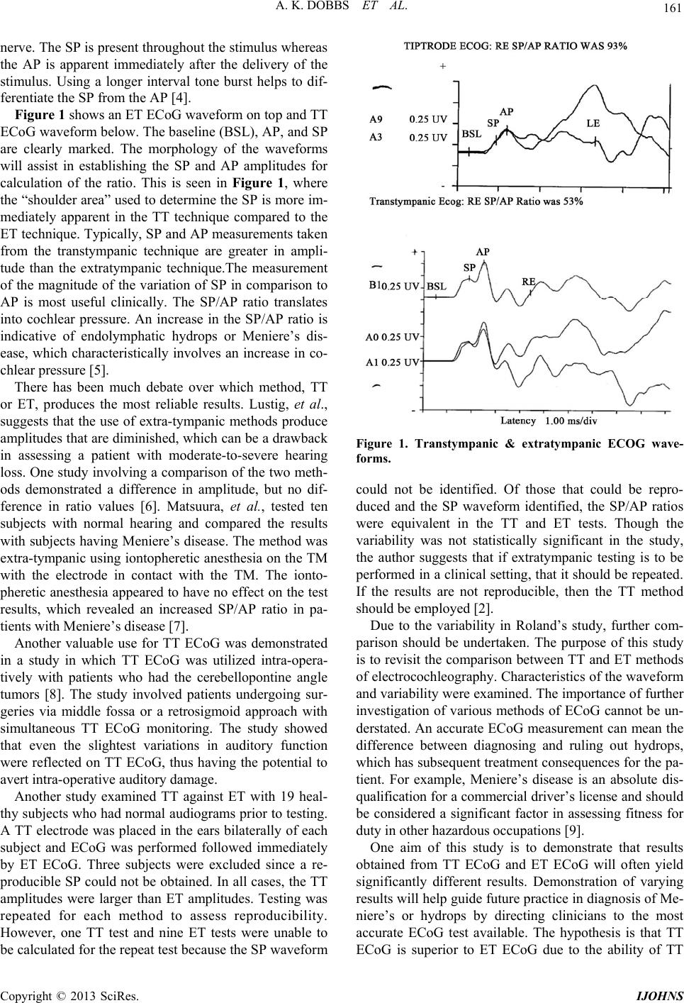 A. K. DOBBS ET AL. 161 nerve. The SP is present throu ghout the stimulus wh ereas the AP is apparent immediately after the delivery of the stimulus. Using a longer interval tone burst helps to dif- ferentiate the SP from the AP [4]. Figure 1 shows an ET ECoG wavefor m on top and TT ECoG waveform below. The baseline (BSL), AP, and SP are clearly marked. The morphology of the waveforms will assist in establishing the SP and AP amplitudes for calculation of the ratio. This is seen in Figure 1, where the “shoulder area” used to determine the SP is more im- mediately apparent in the TT technique compared to the ET technique. Typically, SP and AP measurements taken from the transtympanic technique are greater in ampli- tude than the extratympanic technique.The measurement of the magnitude of the variation of SP in comparison to AP is most useful clinically. The SP/AP ratio translates into cochlear pressure. An increase in the SP/AP ratio is indicative of endolymphatic hydrops or Meniere’s dis- ease, which characteristically involves an increase in co- chlear pressure [5 ] . There has been much debate over which method, TT or ET, produces the most reliable results. Lustig, et al., suggests that the use of extra-tympanic methods produce amplitudes that are diminished, which can be a drawback in assessing a patient with moderate-to-severe hearing loss. One study involving a comparison of the two meth- ods demonstrated a difference in amplitude, but no dif- ference in ratio values [6]. Matsuura, et al., tested ten subjects with normal hearing and compared the results with subjects having Meniere’s disease. The method was extra-tympanic using iontop heretic anesthesia on the TM with the electrode in contact with the TM. The ionto- pheretic anesthesia appeared to have no effect on the test results, which revealed an increased SP/AP ratio in pa- tients with Meniere’s disease [7]. Another valuable use for TT ECoG was demonstrated in a study in which TT ECoG was utilized intra-opera- tively with patients who had the cerebellopontine angle tumors [8]. The study involved patients undergoing sur- geries via middle fossa or a retrosigmoid approach with simultaneous TT ECoG monitoring. The study showed that even the slightest variations in auditory function were reflected on TT ECoG, thus having the potential to avert intra-operative auditory damage. Another study examined TT against ET with 19 heal- thy subjects who had normal audiograms prior to testing. A TT electrode was placed in the ears bilaterally of each subject and ECoG was performed followed immediately by ET ECoG. Three subjects were excluded since a re- producible SP could not be obtained. In all cases, the TT amplitudes were larger than ET amplitudes. Testing was repeated for each method to assess reproducibility. However, one TT test and nine ET tests were unable to be calculated for the repeat test because the SP waveform Figure 1. Transtympanic & extratympanic ECOG wave- forms. could not be identified. Of those that could be repro- duced and the SP waveform identified, the SP/AP ratios were equivalent in the TT and ET tests. Though the variability was not statistically significant in the study, the author suggests that if extratympanic testing is to be performed in a clinical setting, that it should be repeated. If the results are not reproducible, then the TT method should be employed [2]. Due to the variability in Roland’s study, further com- parison should be undertaken. The purpose of this study is to revisit the comparison between TT and ET methods of electrocochleography. Characteristics of the waveform and variability were ex amined. The importance o f further investigation of various methods of ECoG cannot be un- derstated. An accurate ECoG measurement can mean the difference between diagnosing and ruling out hydrops, which has subsequent treatment consequences for the pa- tient. For example, Meniere’s disease is an absolute dis- qualification for a commercial driver’s license and should be considered a significant factor in assessing fitness for duty in other hazardous occupations [9]. One aim of this study is to demonstrate that results obtained from TT ECoG and ET ECoG will often yield significantly different results. Demonstration of varying results will help guide future practice in diagnosis of Me- niere’s or hydrops by directing clinicians to the most accurate ECoG test available. The hypothesis is that TT ECoG is superior to ET ECoG due to the ability of TT Copyright © 2013 SciRes. IJOHNS  A. K. DOBBS ET AL. 162 ECoG to produce waveforms of lesser amplitude and a smaller amount of variability, and could therefore aid in the diagnosis of endolymphatic hydropsor Meniere’s dis- ease to a more accurate level. The American Academy of Head and Neck Surgeons (AAO-HNS) created guidelines for the diagnosis of Me- niere’s disease. The guidelines state that, for a definitive diagnosis of Meniere’s disease, there should be at least two episodes of vertigo lasting 20 minutes of more, au- diometrically documented hearing loss on at least one occasion, tinnitus or aural fullness in the affected ear, and that other causes be excluded [10]. Despite the guidelines, some practices use only ET ECoG and make a diagnosis of hydrops or Meniere’s disease based only on these re- sults. If the ET ECoG lacks the sensitivity to aid in the diagnosis a better method should be considered. The aim of this study is to determine if there is a significant dif- ference in ET and TT ECoG methods. The hypothesis is that TT ECoG will demonstrate a higher sensitivity than ET ECoG. 2. Methods The current study is a secondary data analysis of medical records from 51 private practice neurotology clinic pa- tients in San Antonio, Texas. Patients were selected based on a history compatible with endolymphatic hydrops or Meniere’s disease and either a unilateral or bilateral hear- ing loss. After having an audiogram, all subjects con- sented for evaluation by both TT ECoG and ET ECG as part of their clinical assessment. The study was approved by the IRB at the University of Texas Medical Branch. Hearing data was collected and recorded in the patient’s medical record between May of 2004 and February of 2008. A Tiptrode was used for the extratympanic method of testing and the Viking Nicolet needle electrode was used for the transtympanic method. Patients who had conductive hearing loss or prior otologic surgery were excluded from this evaluation. Each patient met the crite- ria for Meniere’s diseaseas set forth by the American Academy of Otolaryngology—Head Neck Surgeons (AAO-HNS), which includes vertigo, hearing loss, and tinnitus or aural fullness. Protocol Patients had an audiogram, followed by ET ECoG and subsequent TT ECoG. All testing occurred on the same day for each patient to avoid variability in day-to-day hearing performance. The audiogram was performed in the sound booth with headphones and a bone conducting transducer. First, the air and bone conduction audiogram was performed by delivering tones through ear inserts followed by conducting transducer. ET ECoG evaluations were performed first using the ET Tiptrode placed on the tympanic membrane of the af- fected ear. This was followed immediately by TT ECoG testing. The recording needle was placed using otomi- croscopy and secured in place with a foam insert to pre- vent migration. Topical phenol was placed on the TM being tested and the Nicolet TT needle electrode pierced the TM and was placed on the promontory, near the round window niche. ECoG responses were evoked by click stimulus. ET and TT ECoGs were individually recorded for each ear. As a reference, patients were rated according to the American Speech Hearing Associations’ classifications based on the audiogram and the results were associated with the ECoG results from the respective patients. The audiograms for each patient were suggestive of Me- niere’s disease having a low tone hearing loss. Medical records for all 51 patients were initially iden- tified by chart number alone, and then recoded into a randomly generated four-digit identification number and password protected. ET waveforms were obtainable for 47 (n = 47) of the 51 tests. TT waveforms were obtain- able for 46 of the 51 tests. Comparisons were made only on those waveforms that were reproducible. 3. Plan of Analyses Specific Aim: To explore the differences between TT ECoG and ET ECoG hearing assessment and correlate these waveforms to the hearing loss (e.g. to determine which technique would be better correlated with Me- niere’s disease or endolymphatic hydrops and the associ- ated hearing loss). Hypothesis: TT ECoG offers greater sensitivity to ET ECoG as demonstrated by TT ECoG waveforms of sig- nificantly lesser amplitude. Analyses included paired t-tests to compare between methods on measures of amplitude and variability. 4. Results SP/AP ratios were obtained from ECoG testing. Paired t-tests were conducted comparing transtympanicand ex- tratympanic responses in each ear. SP/AP ratios for the transtympanic method revealed mean values significantly (p < 0.000) smaller compared to SP/AP ratios obtained with the extratympanic method in both ears (right: t = 4.744, df = 21, m = 0.3509, sd = 0.0375 versus m = 0.05368, sd = 0.1255, respectively; left: t = 5.767, df = 21, m = 0.3145, sd = 0.1278 versus 0.04764, sd = 0.0906, respectively) indicating greater sensitivity for transtym- panic assessment, see Figure 2. 5. Discussion Diagnostic sensitivity is enhanced with the use of the Copyright © 2013 SciRes. IJOHNS 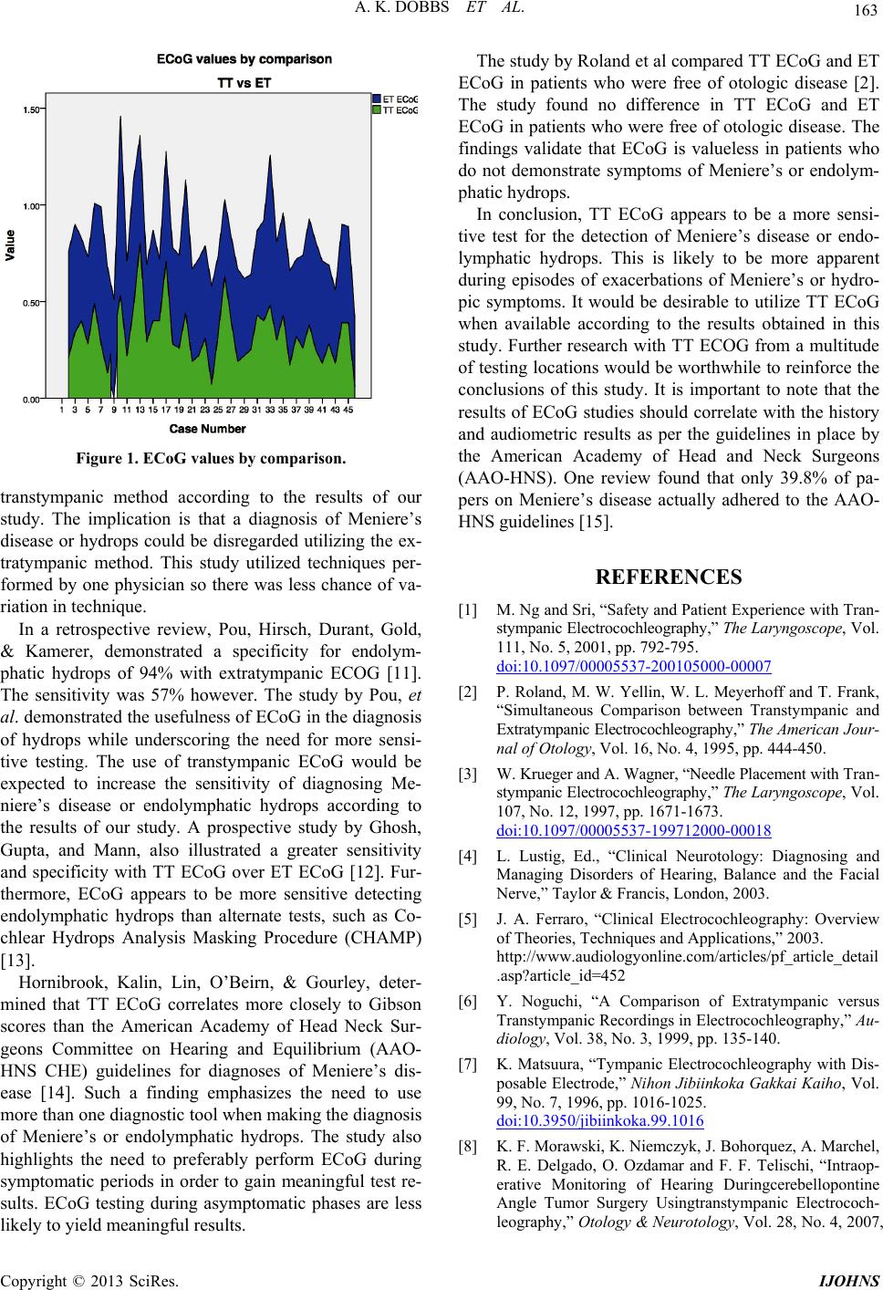 A. K. DOBBS ET AL. 163 Figure 1. ECoG values by comparison. transtympanic method according to the results of our study. The implication is that a diagnosis of Meniere’s disease or hydrops could be disregarded utilizing the ex- tratympanic method. This study utilized techniques per- formed by one physician so there was less chance of va- riation in technique. In a retrospective review, Pou, Hirsch, Durant, Gold, & Kamerer, demonstrated a specificity for endolym- phatic hydrops of 94% with extratympanic ECOG [11]. The sensitivity was 57% however. The study by Pou, et al. demonstrated the usefulness of ECoG in the diagnosis of hydrops while underscoring the need for more sensi- tive testing. The use of transtympanic ECoG would be expected to increase the sensitivity of diagnosing Me- niere’s disease or endolymphatic hydrops according to the results of our study. A prospective study by Ghosh, Gupta, and Mann, also illustrated a greater sensitivity and specificity with TT ECoG over ET ECoG [12]. Fur- thermore, ECoG appears to be more sensitive detecting endolymphatic hydrops than alternate tests, such as Co- chlear Hydrops Analysis Masking Procedure (CHAMP) [13]. Hornibrook, Kalin, Lin, O’Beirn, & Gourley, deter- mined that TT ECoG correlates more closely to Gibson scores than the American Academy of Head Neck Sur- geons Committee on Hearing and Equilibrium (AAO- HNS CHE) guidelines for diagnoses of Meniere’s dis- ease [14]. Such a finding emphasizes the need to use more than one diagnostic tool when making the diagnosis of Meniere’s or endolymphatic hydrops. The study also highlights the need to preferably perform ECoG during symptomatic periods in order to gain meaningful test re- sults. ECoG testing during asymptomatic phases are less likely to yield meaningful results. The study by Roland et al compared TT ECoG and ET ECoG in patients who were free of otologic disease [2]. The study found no difference in TT ECoG and ET ECoG in patients who were free of otologic disease. The findings validate that ECoG is valueless in patients who do not demonstrate symptoms of Meniere’s or endolym- phatic hydrops. In conclusion, TT ECoG appears to be a more sensi- tive test for the detection of Meniere’s disease or endo- lymphatic hydrops. This is likely to be more apparent during episodes of exacerbations of Meniere’s or hydro- pic symptoms. It would be desirable to utilize TT ECoG when available according to the results obtained in this study. Further research with TT ECOG from a multitude of testing locations would be worthwhile to reinforce the conclusions of this study. It is important to note that the results of ECoG studies should correlate with the history and audiometric results as per the guidelines in place by the American Academy of Head and Neck Surgeons (AAO-HNS). One review found that only 39.8% of pa- pers on Meniere’s disease actually adhered to the AAO- HNS guidel ines [15]. REFERENCES [1] M. Ng and Sri, “Safety and Patient Experience with Tran- stympanic Electrocochleography,” The Laryngosc ope, Vol . 111, No. 5, 2001, pp. 792-795. doi:10.1097/00005537-200105000-00007 [2] P. Roland, M. W. Yellin, W. L. Meyerhoff and T. Frank, “Simultaneous Comparison between Transtympanic and Extratympanic Electroc ochleography,” The American Jo ur - nal of Otology, Vol. 16, No. 4, 1995, pp. 444-450. [3] W. Krueger and A. Wagner, “Needle Placement with Tran- stympanic Electrocochleography,” The Laryngosc ope, Vol. 107, No. 12, 1997, pp. 1671-1673. doi:10.1097/00005537-199712000-00018 [4] L. Lustig, Ed., “Clinical Neurotology: Diagnosing and Managing Disorders of Hearing, Balance and the Facial Nerve,” Taylor & Francis, London, 2003. [5] J. A. Ferraro, “Clinical Electrocochleography: Overview of Theories, Techniques and Applications,” 2003. http://www.audiologyonline.com/articles/pf_article_detail .asp?article_id=452 [6] Y. Noguchi, “A Comparison of Extratympanic versus Transtympanic Recordings in Electrocochleography,” Au- diology, Vol. 38, No. 3, 1999, pp. 135-140. [7] K. Matsuura, “Tympanic Electrocochleography with Dis- posable Electrode,” Nihon Jibiinkoka Gakkai Kaiho, Vol. 99, No. 7, 1996, pp. 1016-1025. doi:10.3950/jibiinkoka.99.1016 [8] K. F. Morawski, K. Niemczyk, J. Bohorquez, A. Marchel, R. E. Delgado, O. Ozdamar and F. F. Telischi, “Intraop- erative Monitoring of Hearing Duringcerebellopontine Angle Tumor Surgery Usingtranstympanic Electrococh- leography,” Otology & Neurotology, Vol. 28, No. 4, 2007, Copyright © 2013 SciRes. IJOHNS  A. K. DOBBS ET AL. Copyright © 2013 SciRes. IJOHNS 164 pp. 541-545. [9] H. Booker, “U.S. Department of Transportation—Federal Highway Administration,” In: Conference on Neurologic Disorders and Commerical Drivers, US Department of Transportation, Washington DC, 1988, pp. 1-47. [10] Committee on Hearing and Equilibrium, “Committee on Hearing and Equilibrium Guidelines for the Diagnosis and Evaluation of Therapy in Meniere’s Disease,” Oto- laryngology—Head and Neck Surgery, Vol. 113, No. 3, 1995, pp. 181-185. doi:10.1016/S0194-5998(95)70102-8 [11] A. M. Pou, B. E. Hirsch, J. D. Durant, S. R. Gold and D. B. Kamerer, “The Efficacy of Tympanic Electrocochleo- graphy in the Diagnosis of Endolymphatic Hydrops,” American Journal of Otology, Vol. 17, No. 4, 1996, pp. 607-611. [12] S. Ghosh, A. K. Gupta and S. S. Mann, “Can Electro- cochleography in Meniere’s Disease Be Noninvasive?” Journal of Otolaryngology, Vol. 31, No. 6, 2002, pp. 371- 375. doi:10.2310/7070.2002.34383 [13] D. Zack-Williams, R. M. Angelo and Y. Qing, “A Com- parison of Electrocochleography and High-Pass Noise Masking of Auditory Brainstem Response for Diagnosis of Meniere’s Disease,” International Journal of Audiol- ogy,Vol. 51, No. 10, 2012, pp. 783-787. doi:10.3109/14992027.2012.699199 [14] J. Hornibrook, C. Kalin, E. Lin, G. A. O’Beirn and J. Gourley, “Transtympanic Electrocochleography for the Diagnosis of Ménière’s Disease,” International Journal of Otolaryngology, 2012, pp. 1-11. doi:10.1155/2012/852714 [15] M. A. Thorp, Z. P. Shehab, M. L. Bance and J. A. Rutka, “Committee on Hearing and Equilibrium Guidelines for the Diagnosis and Evaluation of Therapy in Meniere’s Disease,” Clinical Otolaryngology, Vol. 28, No. 3, 2003, pp. 173-176. doi:10.1046/j.1365-2273.2003.00687.x |