

Paper Menu >>
Journal Menu >>
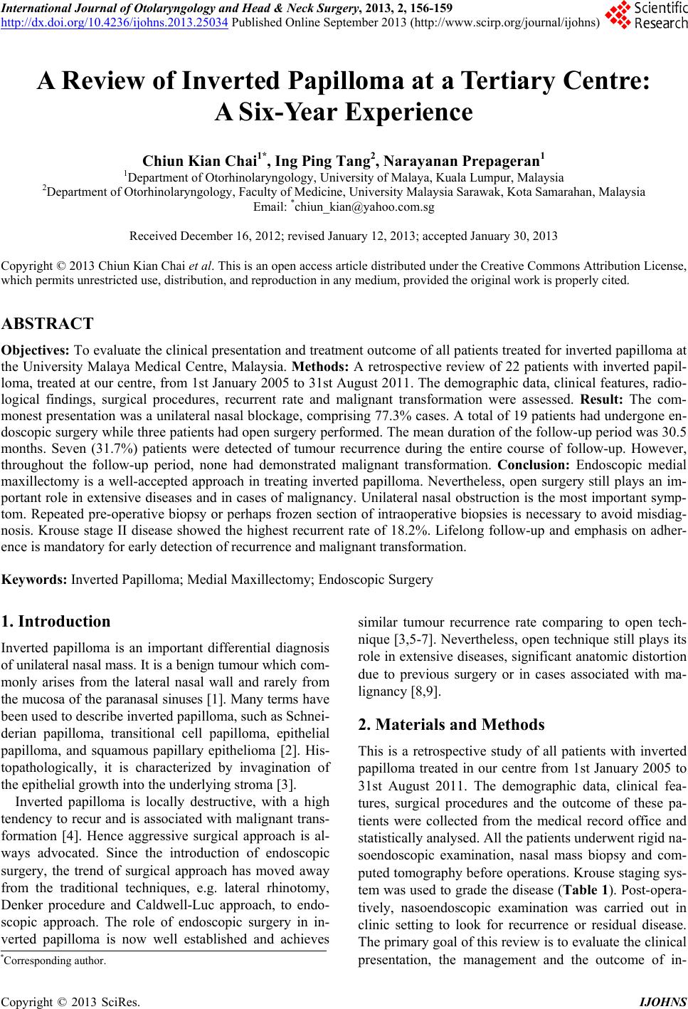 International Journal of Otolaryngology and Head & Neck Surgery, 2013, 2, 156-159 http://dx.doi.org/10.4236/ijohns.2013.25034 Published Online September 2013 (http://www.scirp.org/journal/ijohns) A Review of Inverted Papilloma at a Tertiary Centre: A Six-Year Experience Chiun Kian Chai1*, Ing Ping Tang2, Narayanan Prepageran1 1Department of Otorhinolaryngology, University of Malaya, Kuala Lumpur, Malaysia 2Department of Otorhinolaryngology, Faculty of Medicine, University Malaysia Sarawak, Kota Samarahan, Malaysia Email: *[email protected] Received December 16, 2012; revised January 12, 2013; accepted January 30, 2013 Copyright © 2013 Chiun Kian Chai et al. This is an open access article distributed under the Creative Commons Attribution License, which permits unrestricted use, distribution, and reproduction in any medium, provided the original work is properly cited. ABSTRACT Objectives: To ev aluate the clinical presentation and treatment outcome of all patients treated for inverted papilloma at the University Malaya Medical Centre, Malaysia. Methods: A retrospective review of 22 patients with inverted papil- loma, treated at our centre, from 1st Januar y 2005 to 31st August 2011. The demographic data, clinical features, r adio- logical findings, surgical procedures, recurrent rate and malignant transformation were assessed. Result: The com- monest presentation was a unilateral nasal blockage, comprising 77.3% cases. A total of 19 patients had undergone en- doscopic surgery while three patients had open surgery performed. The mean duration of the follow-up period was 30.5 months. Seven (31.7%) patients were detected of tumour recurrence during the entire course of follow-up. However, throughout the follow-up period, none had demonstrated malignant transformation. Conclusion: Endoscopic medial maxillectomy is a well-accepted approach in treating inverted papilloma. Nevertheless, open surgery still plays an im- portant role in extensive diseases and in cases of malignancy. Unilateral nasal obstruction is the most important symp- tom. Repeated pre-operative biopsy or perhaps frozen section of intraoperative biopsies is necessary to avoid misdiag- nosis. Krouse stage II disease showed the highest recurrent rate of 18.2%. Lifelong follow-up and emphasis on adher- ence is mandatory for early detection of recurrence and malignant transformation. Keywords: Inverted Papilloma; Medial Maxillectomy; Endoscopic Surgery 1. Introduction Inverted papilloma is an important differential diagnosis of unilateral nasal mass. It is a benign tumour which co m- monly arises from the lateral nasal wall and rarely from the mucosa of the paranasal sinuses [1]. Many terms ha ve been used to describe inverted papilloma, such as Schnei- derian papilloma, transitional cell papilloma, epithelial papilloma, and squamous papillary epithelioma [2]. His- topathologically, it is characterized by invagination of the epithelial growth into the underlying stroma [3]. Inverted papilloma is locally destructive, with a high tendency to recur and is associated with malignant trans- formation [4]. Hence aggressive surgical approach is al- ways advocated. Since the introduction of endoscopic surgery, the trend of surgical approach has moved away from the traditional techniques, e.g. lateral rhinotomy, Denker procedure and Caldwell-Luc approach, to endo- scopic approach. The role of endoscopic surgery in in- verted papilloma is now well established and achieves similar tumour recurrence rate comparing to open tech- nique [3,5-7]. Nevertheless, open techniqu e still plays its role in extensive diseases, significant anatomic distortion due to previous surgery or in cases associated with ma- lignancy [8,9] . 2. Materials and Methods This is a retrospective study of all patients with inverted papilloma treated in our centre from 1st January 2005 to 31st August 2011. The demographic data, clinical fea- tures, surgical procedures and the outcome of these pa- tients were collected from the medical record office and statistically analysed. All the patients underwent rigid na- soendoscopic examination, nasal mass biopsy and com- puted tomography before operations. Krouse staging sys- tem was used to grade the disease (Table 1). Post-opera- tively, nasoendoscopic examination was carried out in clinic setting to look for recurrence or residual disease. The primary goal of this review is to evaluate the clinical presentation, the management and the outcome of in- *Corresponding author. C opyright © 2013 SciRes. IJOHNS 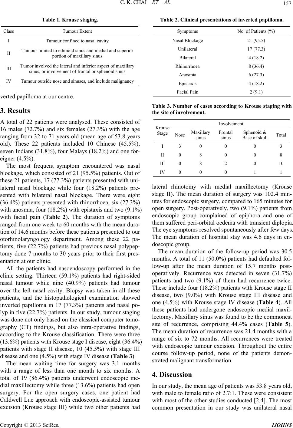 C. K. CHAI ET AL. 157 Table 1. Krouse staging. Class Tumour Extent I Tumour confined to nasal cavity II Tumour limited to ethmoid sinus and medial and superior portion of maxillary sinus III Tumor involved t he lateral and inferior aspect of maxillary sinus, or involveme nt of frontal or sphenoid si nus IV Tumour outside nose and sinuses, and inclu de malignancy verted papilloma at our cen tre. 3. Results A total of 22 patients were analysed. These consisted of 16 males (72.7%) and six females (27.3%) with the age ranging from 32 to 71 years old (mean age of 53.8 years old). These 22 patients included 10 Chinese (45.5%), seven Indians (31.8%), four Malays (18.2%) and one for- eigner (4.5%). The most frequent symptom encountered was nasal blockage, which consisted of 21 (95.5%) patients. Out of these 21 patients, 17 (77 .3%) patients p resented with un i- lateral nasal blockage while four (18.2%) patients pre- sented with bilateral nasal blockage. There were eight (36.4%) patients presented with rhinorrhoea, six (27.3%) with anosmia, four (18.2%) with ep istaxis and two (9.1%) with facial pain (Table 2). The duration of symptoms ranged from one week to 60 months with the mean dura- tion of 14.6 months before these patients presented to our otorhinolaryngology department. Among these 22 pa- tients, five (22.7%) patients had previous nasal polypep- tomy done 7 months to 30 years prior to their first pres- entation at our clinic. All the patients had nasoendoscopy performed in the clinic setting. Thirteen (59.1%) patients had right-sided nasal tumour while nine (40.9%) patients had tumour over the left nasal cavity. Biopsy was taken in all these patients, and the histopathological examination showed inverted papilloma in 17 (77.3%) patients and nasal po- lyp in five (22.7%) patients. In our study, tumour staging was done not only b ased on the classical computer tomo- graphy (CT) findings, but also intra-operative findings, according to the Krouse classification. There were three (13.6%) patients with Krouse stage I disease, eight (36 . 4 %) patients with stage II disease, 10 (45.5%) with stage III disease and one (4.5%) with stage IV disease (Table 3). The mean waiting time for surgery was 3.1 months with a range of less than one month to six months. A total of 19 (86.4%) patients underwent endoscopic me- dial maxillectomy while three (13.6%) patients had open surgery. For the open surgery cases, one patient had Caldwell Luc approach with endoscopic-assisted tumour excision (Krouse stage III) while two other patients had Table 2. Clinical presentations of inverted papilloma. Symptoms No. of Patients (%) Nasal Blockage 21 (95.5) Unilateral 17 (77.3) Bilateral 4 (18.2) Rhinorrhoea 8 (36.4) Anosmia 6 (27.3) Epistaxis 4 (18.2) Facial Pain 2 (9.1) Table 3. Number of cases according to Krouse staging with the site of involvement. Involvement Krouse Stage Nose Maxillary sinus Frontal sinus Sphenoid & Base of skullTotal I 3 0 0 0 3 II 0 8 0 0 8 III 0 8 2 0 10 IV 0 0 0 1 1 lateral rhinotomy with medial maxillectomy (Krouse stage II). The mean duration of surgery was 102.4 min- utes for endoscopic surgery, compared to 165 minutes for open surgery. Post-operatively, two (9.1%) patients from endoscopic group complained of epiphora and one of them suffered peri-orbital oedema with transient diplopia. The eye symptoms resolved spontaneously after few days. The mean duration of hospital stay was 4.6 days in en- doscopic group. The mean duration of the follow-up period was 30.5 months. A total of 11 (50.0%) patients had defaulted fol- low-up after the mean duration of 15.7 months post- operatively. Recurrence was detected in seven (31.7%) patients and two (9.1%) of them had recurrence twice. These include four (18.2%) patients with Krouse stage II disease, two (9.0%) with Krouse stage III disease and one (4.5%) with Krouse stage IV disease (Table 4). All these patients had undergone endoscopic medial maxil- lectomy. Maxillary sinus was found to be the commonest site of recurrence, comprising 44.4% cases (Table 5). The mean duration of recurrence was 21.4 months with a range of six to 72 months. All recurrences were treated with endoscopic tumour excision. Throughout the entire course follow-up period, none of the patients demon- strated malignant transformation. 4. Discussion In our study, the mean age of patients was 53.8 years old, with male to female ratio of 2.7:1. These were consistent with most of the other studies conducted [2,4]. The most common presentation in our study was unilateral nasal Copyright © 2013 SciRes. IJOHNS 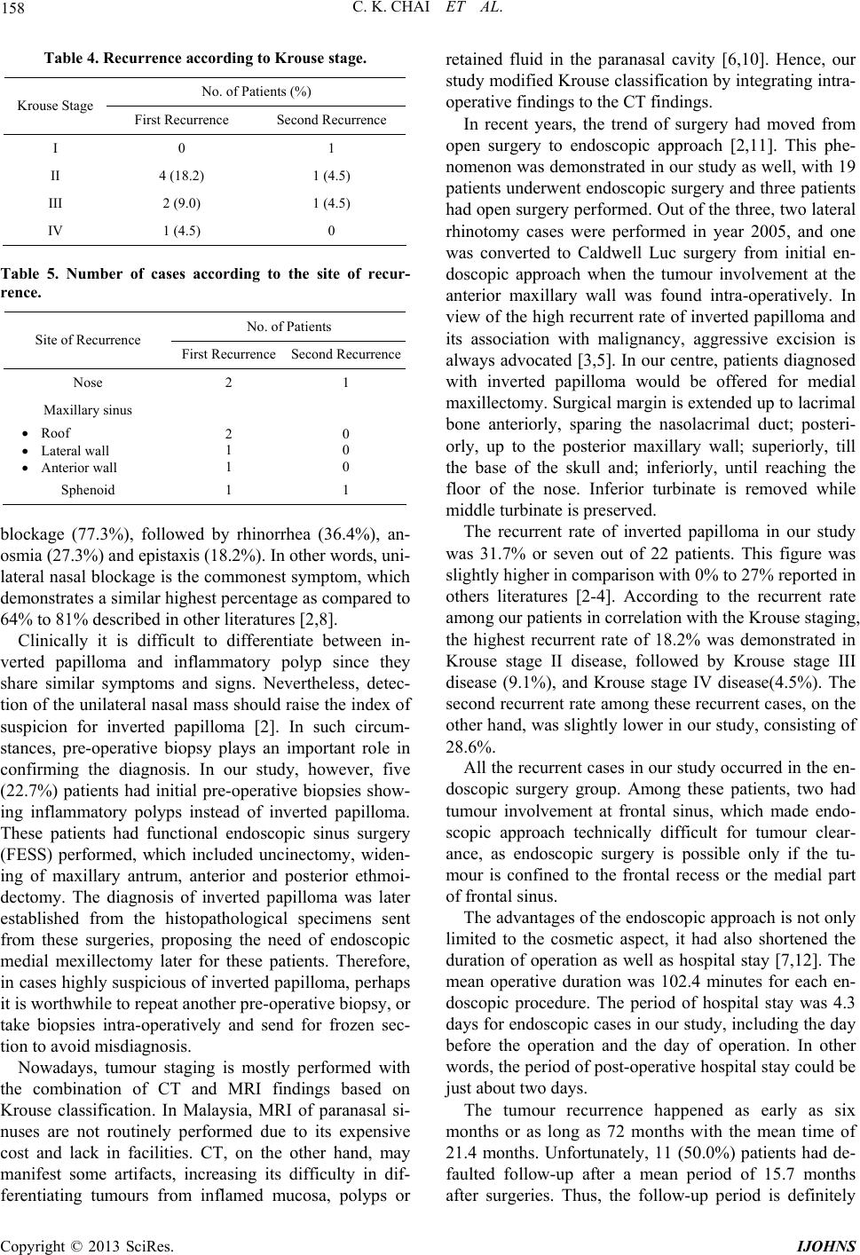 C. K. CHAI ET AL. 158 Table 4. Recurrence according to Krouse stage. No. of Patients (%) Krouse Stage First Recurrence Second Recurrence I 0 1 II 4 (18.2) 1 (4.5) III 2 (9.0) 1 (4.5) IV 1 (4.5) 0 Table 5. Number of cases according to the site of recur- rence. No. of Patients Site of Recurrence First Recurrence Second Recurrence Nose 2 1 Maxillary sinus Roof Lateral wall Anterior wall 2 1 1 0 0 0 Sphenoid 1 1 blockage (77.3%), followed by rhinorrhea (36.4%), an- osmia (27.3%) and epistaxis (18.2%). In other words, uni- lateral nasal blockage is the co mmonest symptom, which demonstrates a similar highest percentage as compared to 64% to 81% described in other literatures [2,8]. Clinically it is difficult to differentiate between in- verted papilloma and inflammatory polyp since they share similar symptoms and signs. Nevertheless, detec- tion of the unilateral nasal mass should raise the index of suspicion for inverted papilloma [2]. In such circum- stances, pre-operative biopsy plays an important role in confirming the diagnosis. In our study, however, five (22.7%) patients had initial pre-operative biopsies show- ing inflammatory polyps instead of inverted papilloma. These patients had functional endoscopic sinus surgery (FESS) performed, which included uncinectomy, widen- ing of maxillary antrum, anterior and posterior ethmoi- dectomy. The diagnosis of inverted papilloma was later established from the histopathological specimens sent from these surgeries, proposing the need of endoscopic medial mexillectomy later for these patients. Therefore, in cases highly suspicious of inverted pap illoma, perhaps it is worthwhile to repeat another pre-operative biop sy, or take biopsies intra-operatively and send for frozen sec- tion to avoid misdiagnosis. Nowadays, tumour staging is mostly performed with the combination of CT and MRI findings based on Krouse classification. In Malaysia, MRI of paranasal si- nuses are not routinely performed due to its expensive cost and lack in facilities. CT, on the other hand, may manifest some artifacts, increasing its difficulty in dif- ferentiating tumours from inflamed mucosa, polyps or retained fluid in the paranasal cavity [6,10]. Hence, our study modified Krouse classification by integrating intra- operative findings to the CT findings. In recent years, the trend of surgery had moved from open surgery to endoscopic approach [2,11]. This phe- nomenon was demonstrated in our study as well, with 19 patients underwent endoscopic surgery and three patients had open surgery performed. Out of the three, two lateral rhinotomy cases were performed in year 2005, and one was converted to Caldwell Luc surgery from initial en- doscopic approach when the tumour involvement at the anterior maxillary wall was found intra-operatively. In view of the high recurrent rate of inverted papilloma and its association with malignancy, aggressive excision is always advocated [3 ,5]. In our centre, patients diagnosed with inverted papilloma would be offered for medial maxillectomy. Surgical margin is ex tended up to lacrimal bone anteriorly, sparing the nasolacrimal duct; posteri- orly, up to the posterior maxillary wall; superiorly, till the base of the skull and; inferiorly, until reaching the floor of the nose. Inferior turbinate is removed while middle turbinate is preserved. The recurrent rate of inverted papilloma in our study was 31.7% or seven out of 22 patients. This figure was slightly higher in comparison with 0% to 27% reported in others literatures [2-4]. According to the recurrent rate among our patients in correlation with the Krouse staging, the highest recurrent rate of 18.2% was demonstrated in Krouse stage II disease, followed by Krouse stage III disease (9.1%), and Krouse stage IV disease(4.5%). The second recurrent rate among these recurrent cases, on the other hand, was slightly lower in our stud y, consisting of 28.6%. All the recurrent cases in our study occurred in the en- doscopic surgery group. Among these patients, two had tumour involvement at frontal sinus, which made endo- scopic approach technically difficult for tumour clear- ance, as endoscopic surgery is possible only if the tu- mour is confined to the frontal recess or the medial part of frontal sinus. The advantages of the endoscopic approach is not only limited to the cosmetic aspect, it had also shortened the duration of operation as well as hospital stay [7,12]. The mean operative duration was 102.4 minutes for each en- doscopic procedure. The period of hospital stay was 4.3 days for endoscopic cases in our study, including the day before the operation and the day of operation. In other words, the period of post-operative hospital stay could be just about two days. The tumour recurrence happened as early as six months or as long as 72 months with the mean time of 21.4 months. Unfortunately, 11 (50.0%) patients had de- faulted follow-up after a mean period of 15.7 months after surgeries. Thus, the follow-up period is definitely Copyright © 2013 SciRes. IJOHNS  C. K. CHAI ET AL. Copyright © 2013 SciRes. IJOHNS 159 inadequate to detect tumour recurrence in this context. Malignancy is another main concern, which makes long- term follow-up necessary, with the risk of malignant transformation ranging from 5% to 21% as well as the mean duration of 52 months to develop carcinoma, re- ported in many other literatures [3,7,13]. Currently there are no universal guidelines for follow-up regime after surgery. Nevertheless, some authors had suggested for quarterly follow-up in the first two years, six monthly in the subsequent two years and yearly thereafter [3]. In other words, long-term follow up is mandatory and ad- herence to follow-up should always be emphasized to all patients [10]. 5. Conclusion Endoscopic medial maxillectomy is a well-accepted ap- proach in treating inverted papilloma. Nevertheless, open surgery still plays an important role in extensiv e diseases and in cases of malignancy. Unilateral nasal obstruction is the most important symptom. Repeated pre-operative biopsy or perhaps a frozen section of intraoperative biop- sies is necessary to avoid misdiagnosis. Krouse stage II disease showed the highest recurrent rate of 18.2%. Life- long follow-up and emphasis on adherence is mandatory for early detection of recurrence and malignant transfor- mation. REFERENCES [1] E. Pasquini, V. Sciarretta, G. Farneti, G. C. Modugno and A. R. Ceroni, “Inverted Papilloma: Report of 89 Cases,” American Journal of Otolaryngology, Vol. 25, No. 3, 2004, pp. 178-185. doi:10.1016/j.amjoto.2004.01.004 [2] M. A. Thorp, M. F. Oyarzabal-Amiqo, J. H. du Plessis and S. L. Sellars, “Inverted Papilloma: A Review of 53 Cases,” Laryngoscope, Vol. 111, No. 8, 2001, pp. 1401- 1405. doi:10.1097/00005537-200108000-00016 [3] S. G. Wolfe, R. J. Schlosser, W. E. Bolger, D. C. Lanza and D. W. Kennedy, “Endoscopic and Endoscope-As- sisted Resections of Inverted Sinonasal Papillomas,” Otolaryngology—Head and Neck Surgery, Vol. 131, No. 3, 2004, pp. 174-179. doi:10.1016/j.otohns.2004.05.011 [4] H. Katori and M. Tsukuda, “Staging of Surgical Ap- proach of Sinonasal Inverted Papilloma,” Auris Nasus Larynx, Vol. 32, No. 3, 2005, pp. 257-263. doi:10.1016/j.anl.2005.03.015 [5] R. J. Schlosser, J. C. Mason and C. W. Gross, “Aggres- sive Endoscopic Resection of Inverted Papilloma: An Up- date,” Otolaryngology—Head and Neck Surgery, Vol. 125, No. 1, 2001, pp. 49-53. doi:10.1067/mhn.2001.116789 [6] T. J. Lee, S. F. Huang, L. A. Lee and C. C. Huang, “En- doscopic Surgery for Recurrent Inverted Papilloma,” La- ryngoscope, Vol. 114, No. 1, 2004, pp. 106-112. doi:10.1097/00005537-200401000-00019 [7] J. M. Busquets and P. H. Hwang, “Endoscopic Resection of Sinonasal Inveted Papilloma: A Meta-Analysis,” Oto- laryngology—Head and Neck Surgery, Vol. 134, No. 3, 2006, pp. 476-482. doi:10.1016/j.otohns.2005.11.038 [8] W. Lawson and Z. M. Patel, “The Evolution of Manage- ment for Inverted Papilloma: An Analysis of 200 Cases,” Otolaryngology—Head and Neck Surgery, Vol. 140, No. 3, 2009, pp. 330-335. doi:10.1016/j.otohns.2008.11.010 [9] W. M. Mendenhall, R. W. Hinerman, R. S. Malyapa, J. W. Werning, R. J. Amdur, D. B. Villaret and N. P. Men- denhall, “Inverted Papilloma of Nasal Cavity and Para- nasal Sinuses,” American Journal of Clinical Oncology, Vol. 30, No. 5, 2007, pp. 560-563. doi:10.1097/COC.0b013e318064c711 [10] G. Eggers, J. Muhling and S. Hassfeld, “Inverted Papil- loma of Paranasal Sinuses,” Journal of Cranio-Maxillo- facial Surgery, Vol. 35, No. 1, 2007, pp. 21-29. doi:10.1016/j.jcms.2006.10.003 [11] F. Carta, B. Verillaud and P. Herman, “Role of Endo- scopic Approach in the Management of Inverted Papil- loma,” Current Opinion in Otolaryngology & Head and Neck Surgery, Vol. 19, No. 1, 2011, pp. 21-24. doi:10.1097/MOO.0b013e3283425213 [12] P. Peng and G. Har-El, “Management of Inverted Papil- lomas of the Nose and Paranasal Sinuses,” American Journal of Otolaryngology, Vol. 27, No. 4, 2006, pp. 233- 237. doi:10.1016/j.amjoto.2005.11.005 [13] C. von Buchwald and P. J. Bradley, “Risk of Malignancy in Inverted Papilloma of the Nose and Paranasal Sin u s es,” Current Opinion in Otolaryngology & Head and Neck Surgery, Vol. 15, No. 2, 2007, pp. 95-98. doi:10.1097/MOO.0b013e3280803d9b |