

Paper Menu >>
Journal Menu >>
 American Journal of Plant Sciences, 2013, 4, 1725-1730 http://dx.doi.org/10.4236/ajps.2013.49211 Published Online September 2013 (http://www.scirp.org/journal/ajps) Influence of Desiccation Time on Survival and Regeneration of Embryonic Axes of Groundnut (Arachis hypogaea L.) Immersed in Liquid Nitrogen M. M. Abdulmalik, I. S. Usman, J. D. Olarewaju, D. A. Aba Department of Plant Science, Ahmadu Bello University, Zaria, Nigeria. Email: [email protected] Received June 17th, 2013; revised July 17th, 2013; accepted August 5th, 2013 Copyright © 2013 M. M. Abdulmalik et al. This is an open access article distributed under the Creative Commons Attribution Li- cense, which permits unrestricted use, distribution, and reproduction in any medium, provided the original work is properly cited. ABSTRACT Cryopreservation, the storage of biological materials in liquid nitrogen (LN), is a useful method for long term conserva- tion of plant germplasm. This study was carried out with the objective of establishing an efficient desiccation technique for successful cryopreservation and recovery of embryonic axes of groundnut. Embryonic axes of four groundnut (Ara- chis hypogaea L.) genotypes were evaluated. The excised embryonic axes were dehydrated by air current of a laminar air flow cabinet for different duration (0, 1, 2, 3, 4 & 5 hrs) before being plunged in LN (−196˚C) and held for 1 hr. Samples were thawed in water bath at 40˚C for 2 min, thereafter cultured on MS medium supplemented with 15 mg/L BAP for recovery. Highest survival (96.67% - 100%) and shoot formation (91.67% - 96.67%) were obtained at an av- erage moisture content of 17% after 4 - 5 hr desiccation. Among the genotypes evaluated, Samnut 22 and Samnut 23 recorded the highest survival and shoot formation. This technique therefore appears promising for cryopreservation of groundnut germplasm. Keywords: Cryopreservation; Groundnut; Embryonic Axes; Desiccation 1. Introduction Groundnut (Arachis hypogaea L.) is an important source of protein and edible oil in the world. Nigeria ranks third after India and China in terms of production [1]. Conser- vation of groundnut ensures availability of germplasm for future breeding needs, and seeds are the most pre- ferred propagule used by seed bank curators for its stor- age. However, even under seed bank condition, long term storage of groundnut is not feasible as viability losses frequently occur [2]. This is because of its high oil con- tent (45% - 50%), which makes it more perishable and prone to rapid loss of both quality and viability in storage [3]. For this reason, groundnut germplasm is maintained by planting every season in the Institute for Agricultural Research (IAR). This is not only laborious, time con- suming and expensive, but also plants are exposed to the possible risk of pest, disease and environmental stresses. Newly improved genotypes of crops are fast replacing traditional genotypes or landraces which are often the source of diversity that breeders use for crop improve- ment. The need to conserve groundnut and other crop biodiversity therefore becomes imperative. Thus, cryo- preservation should be considered as important compli- mentary strategy for ex situ conservation. Cryopreserva- tion is used for long-term storage at ultralow temperature of −196˚C [4]. Cell division and metabolic activities are stopped when plants are exposed to ultra-low tempera- tures, allowing storage without alteration for an indefi- nite period of time [5]. Desiccation of excised embryos and embryonic axes is one of the most practicable tech- niques for cryopreservation [6]. Desiccation technique has been applied to a wide range of plant taxa which in- clude embryonic axes of citrus [7,8] almond [9] and em- bryos of maize [10]. The present work is aimed at estab- lishing an efficient desiccation technique for cryopreser- vation of embryonic axes of groundnut. 2. Materials and Methods Seeds of four groundnut (Arachis hypogaea L.) geno- types were obtained from the groundnut breeding unit of IAR and used for this experiment. The seeds were sur- face sterilized by sequential treatment for 5 min in 70% Copyright © 2013 SciRes. AJPS 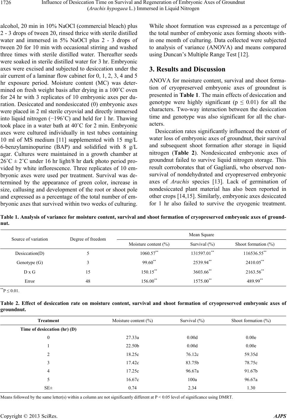 Influence of Desiccation Time on Survival and Regeneration of Embryonic Axes of Groundnut (Arachis hypogaea L.) Immersed in Liquid Nitrogen 1726 alcohol, 20 min in 10% NaOCl (commercial bleach) plus 2 - 3 drops of tween 20, rinsed thrice with sterile distilled water and immersed in 5% NaOCl plus 2 - 3 drops of tween 20 for 10 min with occasional stirring and washed three times with sterile distilled water. Thereafter seeds were soaked in sterile distilled water for 3 hr. Embryonic axes were excised and subjected to desiccation under the air current of a laminar flow cabinet for 0, 1, 2, 3, 4 and 5 hr exposure period. Moisture content (MC) was deter- mined on fresh weight basis after drying in a 100˚C oven for 24 hr with 3 replicates of 10 embryonic axes per du- ration. Desiccated and nondesiccated (0) embryonic axes were placed in 2 ml sterile cryovial and directly immersed into liquid nitrogen (−196˚C) and held for 1 hr. Thawing took place in a water bath at 40˚C for 2 min. Embryonic axes were cultured individually in test tubes containing 10 ml of MS medium [11] supplemented with 15 mg/L 6-benzylaminopurine (BAP) and solidified with 8 g/L agar. Cultures were maintained in a growth chamber at 26˚C ± 2˚C under 16 hr light/8 hr dark photo period pro- vided by white inflorescence. Three replicates of 10 em- bryonic axes were used per treatment. Survival was de- termined by the appearance of green color, increase in size, callusing and development of the root or shoot pole and expressed as a percentage of the total number of em- bryonic axes that survived within two weeks of culturing. While shoot formation was expressed as a percentage of the total number of embryonic axes forming shoots with- in one month of culturing. Data collected were subjected to analysis of variance (ANOVA) and means compared using Duncan’s Multiple Range Test [12]. 3. Results and Discussion ANOVA for moisture content, survival and shoot forma- tion of cryopreserved embryonic axes of groundnut is presented in Table 1. The main effects of desiccation and genotype were highly significant (p ≤ 0.01) for all the characters. Two-way interaction between the desiccation time and genotype was also significant for all the char- acters. Desiccation rates significantly influenced the extent of water loss of embryonic axes of groundnut, their survival and subsequent shoot formation after storage in liquid nitrogen (Table 2). Nondesiccated embryonic axes of groundnut failed to survive liquid nitrogen storage. This result corroborates that of Gagliardi, who observed non- survival of nondehydrated and cryopreserved embryonic axes of Arachis species [13]. Lack of germination of nondesiccated plant material has also been reported in other crops [14,15]. Similarly, embryonic axes desiccated for 1 hr also failed to survive the cryogenic treatment. Table 1. Analysis of variance for moisture content, survival and shoot formation of cryopreserve d embryonic axes of ground- nut. Mean Square Source of variation Degree of freedom Moisture content (%) Survival (%) Shoot formation (%) Desiccation(D) 5 1060.57** 131597.01** 116536.55** Genotype (G) 3 99.60** 2539.94** 2410.05** D x G 15 150.15** 3603.66** 2163.56** Error 48 156.00** 1575.00** 489.99** **P ≤ 0.01. Table 2. Effect of desiccation rate on moisture content, survival and shoot formation of cryopreserved embryonic axes of groundnut. Treatment Moisture content (%) Survival (%) Shoot formation (%) Time of desiccation (hr) (D) 0 27.33a 0.00d 0.00e 1 22.50b 0.00d 0.00e 2 18.25c 76.12c 59.35d 3 17.42c 83.75b 78.75c 4 17.25c 96.67a 91.67b 5 16.67c 100a 96.67a SE± 0.74 2.34 1.30 Mens followed by the same letter(s) within a column are not significantly different at P < 0.05 level of significance using DMRT. a Copyright © 2013 SciRes. AJPS 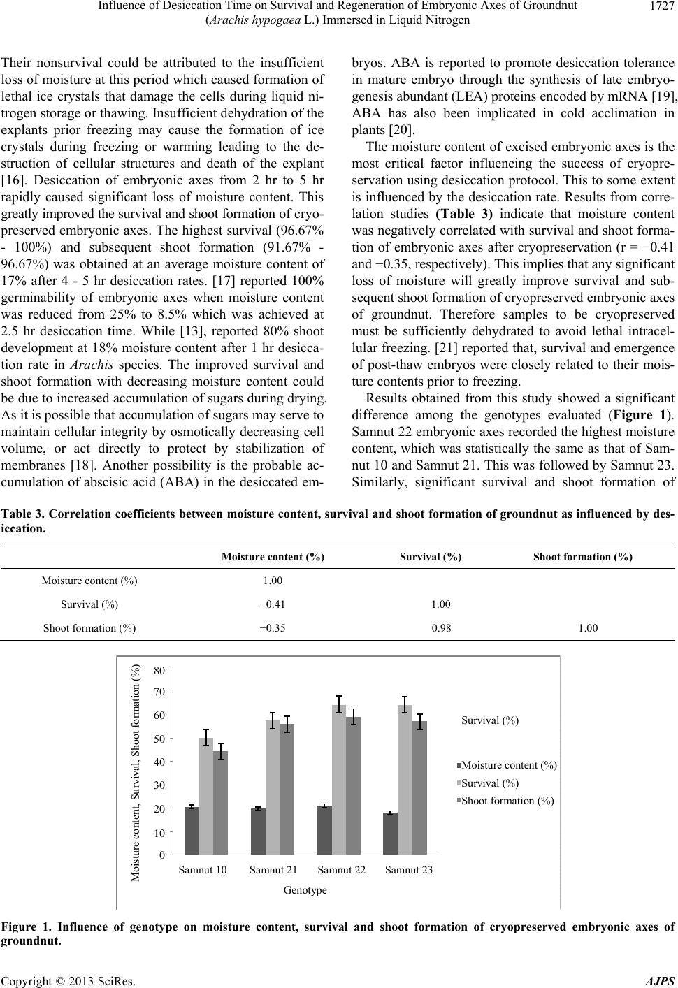 Influence of Desiccation Time on Survival and Regeneration of Embryonic Axes of Groundnut (Arachis hypogaea L.) Immersed in Liquid Nitrogen 1727 Their nonsurvival could be attributed to the insufficient loss of moisture at this period which caused formation of lethal ice crystals that damage the cells during liquid ni- trogen storage or thawing. Insufficient dehydration of the explants prior freezing may cause the formation of ice crystals during freezing or warming leading to the de- struction of cellular structures and death of the explant [16]. Desiccation of embryonic axes from 2 hr to 5 hr rapidly caused significant loss of moisture content. This greatly improved the survival and shoot formation of cryo- preserved embryonic axes. The highest survival (96.67% - 100%) and subsequent shoot formation (91.67% - 96.67%) was obtained at an average moisture content of 17% after 4 - 5 hr desiccation rates. [17] reported 100% germinability of embryonic axes when moisture content was reduced from 25% to 8.5% which was achieved at 2.5 hr desiccation time. While [13], reported 80% shoot development at 18% moisture content after 1 hr desicca- tion rate in Arachis species. The improved survival and shoot formation with decreasing moisture content could be due to increased accumulation of sugars during drying. As it is possible that accumulation of sugars may serve to maintain cellular integrity by osmotically decreasing cell volume, or act directly to protect by stabilization of membranes [18]. Another possibility is the probable ac- cumulation of abscisic acid (ABA) in the desiccated em- bryos. ABA is reported to promote desiccation tolerance in mature embryo through the synthesis of late embryo- genesis abundant (LEA) proteins encoded by mRNA [19], ABA has also been implicated in cold acclimation in plants [20]. The moisture content of excised embryonic axes is the most critical factor influencing the success of cryopre- servation using desiccation protocol. This to some extent is influenced by the desiccation rate. Results from corre- lation studies (Table 3) indicate that moisture content was negatively correlated with survival and shoot forma- tion of embryonic axes after cryopreservation (r = −0.41 and −0.35, respectively). This implies that any significant loss of moisture will greatly improve survival and sub- sequent shoot formation of cryopreserved embryonic axes of groundnut. Therefore samples to be cryopreserved must be sufficiently dehydrated to avoid lethal intracel- lular freezing. [21] reported that, survival and emergence of post-thaw embryos were closely related to their mois- ture contents prior to freezing. Results obtained from this study showed a significant difference among the genotypes evaluated (Figure 1). Samnut 22 embryonic axes recorded the highest moisture content, which was statistically the same as that of Sam- nut 10 and Samnut 21. This was followed by Samnut 23. Similarly, significant survival and shoot formation of Table 3. Correlation coefficie nts between moisture content, surv ival and shoot formation of groundnut as influenced by des- iccation. Moisture content (%) Survival (%) Shoot formation (%) Moisture content (%) 1.00 Survival (%) −0.41 1.00 Shoot formation (%) −0.35 0.98 1.00 0 Samnut 10 10 20 30 40 50 60 70 80 Samnut 21Samnut 22Samnut 23 Genotype Shoot formation (%) Moisture content (%) Survival (%) Survival (%) Moisture content, Survival, Shoot formation (%) Figure 1. Influence of genotype on moisture content, survival and shoot formation of cryopreserved embryonic axes of groundnut. Copyright © 2013 SciRes. AJPS 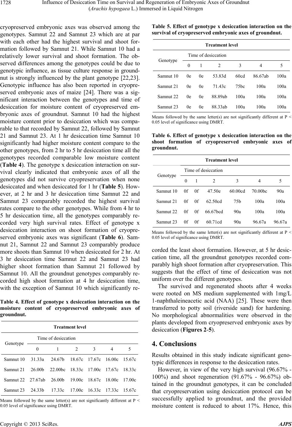 Influence of Desiccation Time on Survival and Regeneration of Embryonic Axes of Groundnut (Arachis hypogaea L.) Immersed in Liquid Nitrogen 1728 cryopreserved embryonic axes was observed among the genotypes. Samnut 22 and Samnut 23 which are at par with each other had the highest survival and shoot for- mation followed by Samnut 21. While Samnut 10 had a relatively lower survival and shoot formation. The ob- served differences among the genotypes could be due to genotypic influence, as tissue culture response in ground- nut is strongly influenced by the plant genotype [22,23]. Genotypic influence has also been reported in cryopre- served embryonic axes of maize [24]. There was a sig- nificant interaction between the genotypes and time of desiccation for moisture content of cryopreserved em- bryonic axes of groundnut. Samnut 10 had the highest moisture content prior to desiccation which was compa- rable to that recorded by Samnut 22, followed by Samnut 21 and Samnut 23. At 1 hr desiccation time Samnut 10 significantly had higher moisture content compare to the other genotypes, from 2 hr to 5 hr desiccation time all the genotypes recorded comparable low moisture content (Table 4). The genotype x desiccation interaction on sur- vival clearly indicated that embryonic axes of all the genotypes did not survive cryopreservation when none desiccated and when desiccated for 1 hr (Table 5 ). How- ever, at 2 hr and 3 hr desiccation time Samnut 22 and Samnut 23 comparably recorded the highest survival rates compare to the other genotypes. While from 4 hr to 5 hr desiccation time, all the genotypes comparably re- corded very high survival rates. Effect of genotype x desiccation interaction on shoot formation of cryopre- served embryonic axes was significant (Table 6). Sam- nut 21, Samnut 22 and Samnut 23 comparably produce more shoots than Samnut 10 when desiccated for 2 hr. At 3 hr desiccation time Samnut 22 and Samnut 23 had higher shoot formation than Samnut 21 followed by Samnut 10. All the groundnut genotypes comparably re- corded high shoot formation at 4 hr desiccation time, with the exception of Samnut 10 which significantly re- Table 4. Effect of genotype x desiccation interaction on the moisture content of cryopreserved embryonic axes of groundnut. Treatment level Time of desiccation Genotype 0 1 2 3 4 5 Samnut 10 31.33a 24.67b 18.67c 17.67c 16.00c 15.67c Samnut 21 26.00b 22.00bc 18.33c 17.00c 17.67c 18.33c Samnut 22 27.67ab 26.00b 19.00c 18.67c 18.00c 17.00c Samnut 23 24.33b 17.33c 17.00c 16.33c 17.33c 15.67c Means followed by the same letter(s) are not significantly different at P < 0.05 level of significance using DMRT. Table 5. Effect of genotype x desiccation interaction on the survival of cryopreserved embryonic axes of groundnut. Treatment level Time of desiccation Genotype 0 12 3 4 5 Samnut 100e0e53.83d 60cd 86.67ab 100a Samnut 210e 0e71.43c 75bc 100a 100a Samnut 220e 0e88.89ab 100a 100a 100a Samnut 230e 0e88.33ab 100a 100a 100a Means followed by the same letter(s) are not significantly different at P < 0.05 level of significance using DMRT. Table 6. Effect of genotype x desiccation interaction on the shoot formation of cryopreserved embryonic axes of groundnut. Treatment level Time of desiccation Genotype 0 12 3 4 5 Samnut 100f 0f47.50e 60.00cd 70.00bc 90a Samnut 210f 0f62.50cd 75b 100a 100a Samnut 220f0f66.67bcd 90a 100a 100a Samnut 230f 0f60.71cd 90a 96.67a 96.67a Means followed by the same letter(s) are not significantly different at P < 0.05 level of significance using DMRT. corded the least shoot formation. However, at 5 hr desic- cation time, all the groundnut genotypes recorded com- parably high shoot formation after cryopreservation. This suggests that the effect of time of desiccation was not uniform over the different genotypes. The survived and regenerated shoots after 4 weeks were rooted on MS medium supplemented with 1mg/L 1-naphthaleineacetic acid (NAA) [25]. These were then transferred to potty soil (riverside sand) for hardening. No morphological abnormalities were observed in the plants developed from cryopreserved embryonic axes by desiccation (Figures 2-5). 4. Conclusions Results obtained in this study indicate significant geno- typic differences in response to the desiccation rates. However, in view of the very high survival (96.67% - 100%) and shoot regeneration (91.67% - 96.67%) ob- tained in the groundnut genotypes, it can be concluded that cryopreservation using desiccation protocol can be successfully applied to groundnut, and the provided moisture content is reduced to about 17%. Hence, this Copyright © 2013 SciRes. AJPS 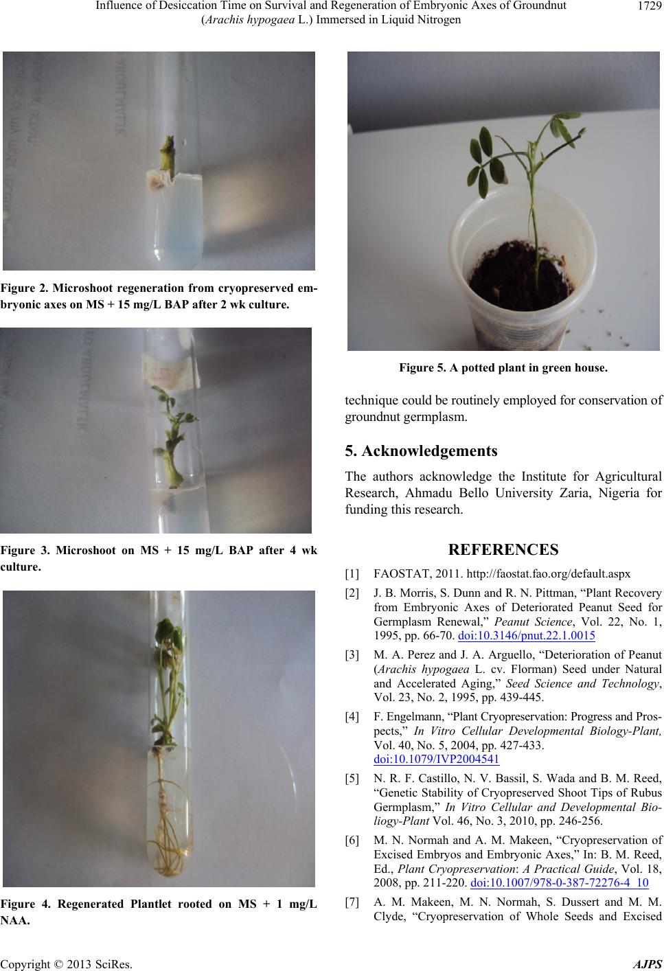 Influence of Desiccation Time on Survival and Regeneration of Embryonic Axes of Groundnut (Arachis hypogaea L.) Immersed in Liquid Nitrogen 1729 Figure 2. Microshoot regeneration from cryopreserved em- bryonic axes on MS + 15 mg/L BAP after 2 wk culture. Figure 3. Microshoot on MS + 15 mg/L BAP after 4 wk culture. Figure 4. Regenerated Plantlet rooted on MS + 1 mg/L NAA. Figure 5. A potted plant in green house. technique could be routinely employed for conservation of groundnut germplasm. 5. Acknowledgements The authors acknowledge the Institute for Agricultural Research, Ahmadu Bello University Zaria, Nigeria for funding this research. REFERENCES [1] FAOSTAT, 2011. http://faostat.fao.org/default.aspx [2] J. B. Morris, S. Dunn and R. N. Pittman, “Plant Recovery from Embryonic Axes of Deteriorated Peanut Seed for Germplasm Renewal,” Peanut Science, Vol. 22, No. 1, 1995, pp. 66-70. doi:10.3146/pnut.22.1.0015 [3] M. A. Perez and J. A. Arguello, “Deterioration of Peanut (Arachis hypogaea L. cv. Florman) Seed under Natural and Accelerated Aging,” Seed Science and Technology, Vol. 23, No. 2, 1995, pp. 439-445. [4] F. Engelmann, “Plant Cryopreservation: Progress and Pros- pects,” In Vitro Cellular Developmental Biology-Plant, Vol. 40, No. 5, 2004, pp. 427-433. doi:10.1079/IVP2004541 [5] N. R. F. Castillo, N. V. Bassil, S. Wada and B. M. Reed, “Genetic Stability of Cryopreserved Shoot Tips of Rubus Germplasm,” In Vitro Cellular and Developmental Bio- liogy-Plant Vol. 46, No. 3, 2010, pp. 246-256. [6] M. N. Normah and A. M. Makeen, “Cryopreservation of Excised Embryos and Embryonic Axes,” In: B. M. Reed, Ed., Plant Cryopreservation: A Practical Guide, Vol. 18, 2008, pp. 211-220. doi:10.1007/978-0-387-72276-4_10 [7] A. M. Makeen, M. N. Normah, S. Dussert and M. M. Clyde, “Cryopreservation of Whole Seeds and Excised Copyright © 2013 SciRes. AJPS 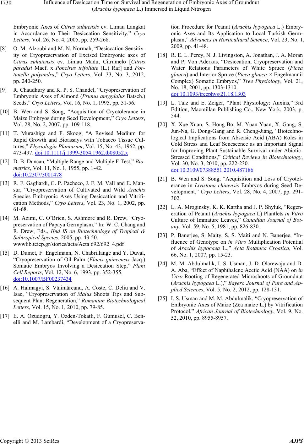 Influence of Desiccation Time on Survival and Regeneration of Embryonic Axes of Groundnut (Arachis hypogaea L.) Immersed in Liquid Nitrogen Copyright © 2013 SciRes. AJPS 1730 Embryonic Axes of Citrus suhuensis cv. Limau Langkat in Accordance to Their Desiccation Sensitivity,” Cryo Letters, Vol. 26, No. 4, 2005, pp. 259-268. [8] O. M. Alzoubi and M. N. Normah, “Desiccation Sensitiv- ity of Cryopreservation of Excised Embryonic axes of Citrus suhuiensis cv. Limau Madu, Citrumelo [Citrus paradisi Macf. x Poncirus trifoliate (L.) Raf] and For- tunella polyandra,” Cryo Letters, Vol. 33, No. 3, 2012, pp. 240-250. [9] R. Chaudhury and K. P. S. Chandel, “Cryopreservation of Embryonic Axes of Almond (Prunus amygdalus Batsch.) Seeds,” Cryo Letters, Vol. 16, No. 1, 1995, pp. 51-56. [10] B. Wen and S. Song, “Acquisition of Cryotolerance in Maize Embryos during Seed Development,” Cryo Letters, Vol. 28, No. 2, 2007, pp. 109-118. [11] T. Murashige and F. Skoog, “A Revised Medium for Rapid Growth and Bioassays with Tobacco Tissue Cul- tures,” Physiologia Plantarum, Vol. 15, No. 43, 1962, pp. 473-497. doi:10.1111/j.1399-3054.1962.tb08052.x [12] D. B. Duncan, “Multiple Range and Multiple F-Test,” Bio- metrics, Vol. 11, No. 1, 1955, pp. 1-42. doi:10.2307/3001478 [13] R. F. Gagliardi, G. P. Pacheco, J. F. M. Vall and E. Man- sur, “Cryopreservation of Cultivated and Wild Arachis Species Embryonic Axes Using Desiccation and Vitrifi- cation Methods,” Cryo Letters, Vol. 23, No. 1, 2002, pp. 61-68. [14] M. Azimi, C. O’Brien, S. Ashmore and R. Drew, “Cryo- preservation of Papaya Germplasm,” In: W. C. Chang and R. Drew, Eds., IInd IS on Biotechnology of Tropical & Subtropical Species, 2005, pp. 43-50. wwwlib.teiep.gr/stories/acta/Acta 692/692_4.pdf [15] D. Dumet, F. Engelmann, N. Chabrillange and Y. Duval, “Cryopreservation of Oil Palm (Elaeis guineensis Jacq.) Somatic Embryos Involving a Desiccation Step,” Plant Cell Reports, Vol. 12, No. 6, 1993, pp. 352-355. doi:10.1007/BF00237434 [16] A. Halmagyi, S. Vălimăreanu, A. Coste, C. Deliu and V. Isac, “Cryopreservation of Malus Shoots Tips and Sub- sequent Plant Regeneration,” Romanian Biotechnological Letters, Vol. 15, No. 1, 2010, pp. 79-85. [17] E. A. Ozudogru, Y. Ozden-Tokatli, F. Gumusel, C. Ben- elli and M. Lambardi, “Development of a Cryopreserva- tion Procedure for Peanut (Arachis hypogaea L.) Embry- onic Axes and Its Application to Local Turkish Germ- plasm,” Advances in Horticultural Science, Vol. 23, No. 1, 2009, pp. 41-48. [18] R. E. L. Percy, N. J. Livingston, A. Jonathan, J. A. Moran and P. Von Aderkas, “Desiccation, Cryopreservation and Water Relations Parameters of White Spruce (Picea glauca) and Interior Spruce (Picea glauca × Engelmannii Complex) Somatic Embryos,” Tree Physiology, Vol. 21, No. 18, 2001, pp. 1303-1310. doi:10.1093/treephys/21.18.1303 [19] L. Taiz and E. Zeiger, “Plant Physiology: Auxins,” 3rd Edition, Macmillan Publishing Co., New York, 2003, p. 544. [20] X. Xue-Xuan, S. Hong-Bo, M. Yuan-Yuan, X. Gang, S. Jun-Na, G. Dong-Gang and R. Cheng-Jiang, “Biotechno- logical Implications from Abscisic Acid (ABA) Roles in Cold Stress and Leaf Senescence as an Important Signal for Improving Plant Sustainable Survival under Abiotic- Stressed Conditions,” Critical Reviews in Biotechnology, Vol. 30, No. 3, 2010, pp. 222-230. doi:10.3109/07388551.2010.487186 [21] B. Wen and S. Song, “Acquisition and Loss of Cryotol- erance in Livistona chinensis Embryos during Seed De- velopment,” Cryo Letters, Vol. 28, No. 4, 2007, pp. 291- 302. [22] L. A. Mroginsky, K. K. Kartha and J. P. Shyluk, “Regen- eration of Peanut (Arachis hypogaea L) Plantlets in Vitro Culture of Immature Leaves,” Canadian Journal of Bot- any, Vol. 59, No. 5, 1981, pp. 826-830. [23] P. Banerjee, S. Maity, S. S. Maiti and N. Banerjee, “In- fluence of Genotype on in Vitro Multiplication Potential of Arachis hypogaea L.,” Acta Botanica Croatica, Vol. 66, No. 1, 2007, pp. 15-23. [24] M. M. Abdulmalik, I. S. Usman, J. D. Olarewaju and D. A. Aba, “Effect of Naphthalene Acetic Acid (NAA) on in Vitro Rooting of Regenerated Microshoots of Groundnut (Arachis hypogaea L.),” Bayero Journal of Pure and Ap- plied Sciences, Vol. 5, No. 2, 2012, pp. 128-131. [25] I. S. Usman and M. M. Abdulmalik, “Cryopreservation of Embryonic Axes of Maize (Zea maize L.) by Vitrification Protocol,” African Journal of Biotechnology, Vol. 9, No. 52, 2010, pp. 8955-8957. |