

Paper Menu >>
Journal Menu >>
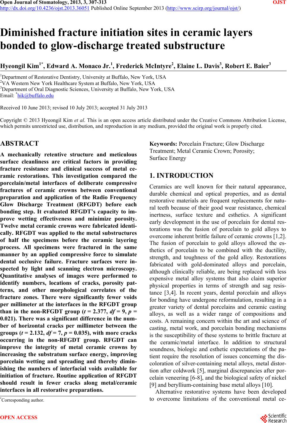 Open Journal of Stomatology, 2013, 3, 307-313 OJST http://dx.doi.org/10.4236/ojst.2013.36051 Published Online September 2013 (http://www.scirp.org/journal/ojst/) Diminished fracture initiation sites in ceramic layers bonded to glow-discharge treated substructure Hyeongil Kim1*, Edward A. Monaco Jr.1, Frederick McIntyre2, Elaine L. Davis3, Robert E. Baier3 1Department of Restorative Dentistry, University at Buffalo, New York, USA 2VA Western New York Healthcare System at Buffalo, New York, USA 3Department of Oral Diagnostic Sciences, University at Buffalo, New York, USA Email: *[email protected] Received 10 June 2013; revised 10 July 2013; accepted 31 July 2013 Copyright © 2013 Hyeongil Kim et al. This is an open access article distributed under the Creative Commons Attribution License, which permits unrestricted use, distribution, and reproduction in any medium, provided the original work is properly cited. ABSTRACT A mechanically retentive structure and meticulous surface cleanliness are critical factors in providing fracture resistance and clinical success of metal ce- ramic restorations. This investigation compared the porcelain/metal interfaces of deliberate compressive fractures of ceramic crowns between conventional preparation and application of the Radio Frequency Glow Discharge Treatment (RFGDT) before each bonding step. It evaluated RFGDT’s capacity to im- prove wetting effectiveness and minimize porosity. Twelve metal ceramic crowns were fabricated identi- cally. RFGDT was applied to the metal substructures of half the specimens before the ceramic layering process. All specimens were fractured in the same manner by an applied compressive force to simulate dental occlusive failure. Fracture surfaces were in- spected by light and scanning electron microscopy. Quantitative analyses of images were performed to identify numbers, locations of cracks, porosity pat- terns, and other morphological correlates of the fracture zones. There were significantly fewer voids per millimeter at the interfaces in the RFGDT group than in the non-RFGDT group (t = 2.377, df = 9, p = 0.021). There was a significant difference in the num- ber of horizontal cracks per millimeter between the groups (t = 2.132, df = 7, p = 0.035), with more cracks occurring in the non-RFGDT group. RFGDT can improve the integrity of metal ceramic crowns by increasing the substratum surface energy, improving porcelain wetting and spreading and thereby dimin- ishing the numbers of interfacial voids available for initiation of fracture. Routine application of RFGDT should result in fewer cracks along metal/ceramic interfaces in all restorative preparations. Keywords: Porcelain Fracture; Glow Discharge Treatment; Metal Ceramic Crown; Porosity; Surface Energy 1. INTRODUCTION Ceramics are well known for their natural appearance, durable chemical and optical properties, and as dental restorative materials are frequent replacements for natu- ral teeth because of their good wear resistance, chemical inertness, surface texture and esthetics. A significant early development in the use of porcelain for dental res- torations was the fusion of porcelain to gold alloys to overcome inherent brittle failure of ceramic crowns [1,2]. The fusion of porcelain to gold alloys allowed the es- thetics of porcelain to be combined with the ductility, strength, and toughness of the gold alloy. Restorations fabricated with gold-dominated alloys and porcelain, although clinically reliable, are being replaced with less expensive metal alloy systems that also claim superior physical properties in terms of strength and sag resis- tance [3,4]. In recent years, dental porcelain and alloys for bonding have undergone reformulation, resulting in a greater variety of dental porcelains and ceramic casting alloys, as well as a wider range of compositions and costs. A remaining concern within the art and science of casting, metal work, and porcelain bonding mechanisms is the susceptibility of these systems to brittle fracture at the ceramic/metal interface. In addition to structural soundness, biologic and esthetic expectations of the pa- tient require the resolution of issues concerning the dis- coloration of silver-containing metal alloys, metal distor- tion after coldwork [5], marginal discrepancies after por- celain veneering [6-8], and the biological safety of nickel [9] and beryllium-containing base metal alloys [10]. Alternative restorative systems have been developed to overcome limitations of the conventional metal ce- *Corresponding author. OPEN ACCESS 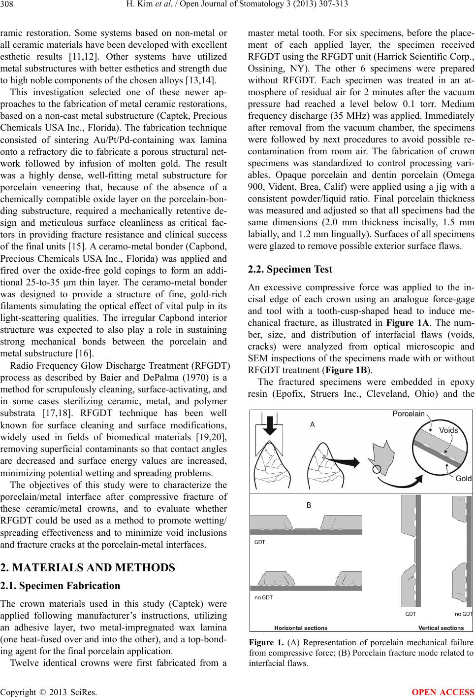 H. Kim et al. / Open Journal of Stomatology 3 (2013) 307-313 308 ramic restoration. Some systems based on non-metal or all ceramic materials have been developed with excellent esthetic results [11,12]. Other systems have utilized metal substructures with better esthetics and strength due to high noble components of the chosen alloys [13,14]. This investigation selected one of these newer ap- proaches to the fabrication of metal ceramic restorations, based on a non-cast metal substructure (Captek, Precious Chemicals USA Inc., Florida). The fabrication technique consisted of sintering Au/Pt/Pd-containing wax lamina onto a refractory die to fabricate a porous structural net- work followed by infusion of molten gold. The result was a highly dense, well-fitting metal substructure for porcelain veneering that, because of the absence of a chemically compatible oxide layer on the porcelain-bon- ding substructure, required a mechanically retentive de- sign and meticulous surface cleanliness as critical fac- tors in providing fracture resistance and clinical success of the final units [15]. A ceramo-metal bonder (Capbond, Precious Chemicals USA Inc., Florida) was applied and fired over the oxide-free gold copings to form an addi- tional 25-to-35 μm thin layer. The ceramo-metal bonder was designed to provide a structure of fine, gold-rich filaments simulating the optical effect of vital pulp in its light-scattering qualities. The irregular Capbond interior structure was expected to also play a role in sustaining strong mechanical bonds between the porcelain and metal substructure [16]. Radio Frequency Glow Discharge Treatment (RFGDT) process as described by Baier and DePalma (1970) is a method for scrupulously cleaning, surface-activating, and in some cases sterilizing ceramic, metal, and polymer substrata [17,18]. RFGDT technique has been well known for surface cleaning and surface modifications, widely used in fields of biomedical materials [19,20], removing superficial contaminants so that contact angles are decreased and surface energy values are increased, minimizing potential wetting and spreading problems. The objectives of this study were to characterize the porcelain/metal interface after compressive fracture of these ceramic/metal crowns, and to evaluate whether RFGDT could be used as a method to promote wetting/ spreading effectiveness and to minimize void inclusions and fracture cracks at the porcelain-metal interfaces. 2. MATERIALS AND METHODS 2.1. Specimen Fabrication The crown materials used in this study (Captek) were applied following manufacturer’s instructions, utilizing an adhesive layer, two metal-impregnated wax lamina (one heat-fused over and into the other), and a top-bond- ing agent for the final porcelain application. Twelve identical crowns were first fabricated from a master metal tooth. For six specimens, before the place- ment of each applied layer, the specimen received RFGDT using the RFGDT unit (Harrick Scientific Corp., Ossining, NY). The other 6 specimens were prepared without RFGDT. Each specimen was treated in an at- mosphere of residual air for 2 minutes after the vacuum pressure had reached a level below 0.1 torr. Medium frequency discharge (35 MHz) was applied. Immediately after removal from the vacuum chamber, the specimens were followed by next procedures to avoid possible re- contamination from room air. The fabrication of crown specimens was standardized to control processing vari- ables. Opaque porcelain and dentin porcelain (Omega 900, Vident, Brea, Calif) were applied using a jig with a consistent powder/liquid ratio. Final porcelain thickness was measured and adjusted so that all specimens had the same dimensions (2.0 mm thickness incisally, 1.5 mm labially, and 1.2 mm lingually). Surfaces of all specimens were glazed to remove possible exterior surface flaws. 2.2. Specimen Test An excessive compressive force was applied to the in- cisal edge of each crown using an analogue force-gage and tool with a tooth-cusp-shaped head to induce me- chanical fracture, as illustrated in Figure 1A. The num- ber, size, and distribution of interfacial flaws (voids, cracks) were analyzed from optical microscopic and SEM inspections of the specimens made with or without RFGDT treatment (Figure 1B). The fractured specimens were embedded in epoxy resin (Epofix, Struers Inc., Cleveland, Ohio) and the Figure 1. (A) Representation of porcelain mechanical failure from compressive force; (B) Porcelain fracture mode related to interfacial flaws. Copyright © 2013 SciRes. OPEN ACCESS 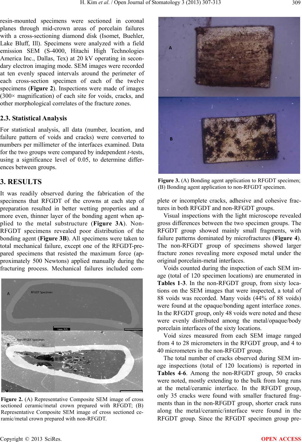 H. Kim et al. / Open Journal of Stomatology 3 (2013) 307-313 309 resin-mounted specimens were sectioned in coronal planes through mid-crown areas of porcelain failures with a cross-sectioning diamond disk (Isomet, Buehler, Lake Bluff, Ill). Specimens were analyzed with a field emission SEM (S-4000, Hitachi High Technologies America Inc., Dallas, Tex) at 20 kV operating in secon- dary electron imaging mode. SEM images were recorded at ten evenly spaced intervals around the perimeter of each cross-section specimen of each of the twelve specimens (Figure 2). Inspections were made of images (300× magnification) of each site for voids, cracks, and other morphological correlates of the fracture zones. 2.3. Statistical Analysis For statistical analysis, all data (number, location, and failure pattern of voids and cracks) were converted to numbers per millimeter of the interfaces examined. Data for the two groups were compared by independent t-tests, using a significance level of 0.05, to determine differ- ences between groups. 3. RESULTS It was readily observed during the fabrication of the specimens that RFGDT of the crowns at each step of preparation resulted in better wetting properties and a more even, thinner layer of the bonding agent when ap- plied to the metal substructure (Figure 3A). Non- RFGDT specimens revealed poor distribution of the bonding agent (Figure 3B). All specimens were taken to total mechanical failure, except one of the RFGDT-pre- pared specimens that resisted the maximum force (ap- proximately 500 Newtons) applied manually during the fracturing process. Mechanical failures included com- Figure 2. (A) Representative Composite SEM image of cross sectioned ceramic/metal crown prepared with RFGDT; (B) Representative Composite SEM image of cross sectioned ce- ramic/metal crown prepared with non-RFGDT. Figure 3. (A) Bonding agent application to RFGDT specimen; (B) Bonding agent application to non-RFGDT specimen. plete or incomplete cracks, adhesive and cohesive frac- tures in both RFGDT and non-RFGDT groups. Visual inspections with the light microscope revealed gross differences between the two specimen groups. The RFGDT group showed mainly small fragments, with failure patterns dominated by microfractures (Figure 4). The non-RFGDT group of specimens showed larger fracture zones revealing more exposed metal under the original porcelain-metal interfaces. Voids counted during the inspection of each SEM im- age (total of 120 specimen locations) are enumerated in Tables 1-3. In the non-RFGDT group, from sixty loca- tions on the SEM images that were inspected, a total of 88 voids was recorded. Many voids (44% of 88 voids) were found at the opaque/bonding agent interface zones. In the RFGDT group, only 48 voids were noted and these were evenly distributed among the metal/opaque/body porcelain interfaces of the sixty locations. Void sizes measured from each SEM image ranged from 4 to 28 micrometers in the RFGDT group, and 4 to 40 micrometers in the non-RFGDT group. The total number of cracks observed during SEM im- age inspections (total of 120 locations) is reported in Tables 4-6. Among the non-RFGDT group, 50 cracks were noted, mostly extending to the bulk from long runs at the metal/ceramic interface. In the RFGDT group, only 35 cracks were found with smaller fractured frag- ments than in the non-RFGDT group, shorter crack runs along the metal/ceramic/interface were found in the RFGDT group. Since the RFGDT specimen group pre- Copyright © 2013 SciRes. OPEN ACCESS 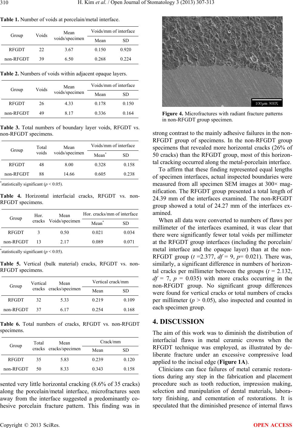 H. Kim et al. / Open Journal of Stomatology 3 (2013) 307-313 310 Table 1. Number of voids at porcelain/metal interface. Voids/mm of interface Group Voids Mean voids/specimen Mean SD RFGDT 22 3.67 0.150 0.920 non-RFGDT 39 6.50 0.268 0.224 Table 2. Numbers of voids within adjacent opaque layers. Voids/mm of interface Group Voids Mean voids/specimen Mean SD RFGDT 26 4.33 0.178 0.150 non-RFGDT 49 8.17 0.336 0.164 Table 3. Total numbers of boundary layer voids, RFGDT vs. non-RFGDT specimens. Voids/mm of interface Group Total voids Mean voids/specimen Mean* SD RFGDT 48 8.00 0.328 0.158 non-RFGDT 88 14.66 0.605 0.238 *statistically significant (p < 0.05). Table 4. Horizontal interfacial cracks, RFGDT vs. non- RFGDT specimens. Hor. cracks/mm of interface Group Hor. cracks Mean Voids/specimen Mean* SD RFGDT 3 0.50 0.021 0.034 non-RFGDT 13 2.17 0.089 0.071 *statistically significant (p < 0.05). Table 5. Vertical (bulk material) cracks, RFGDT vs. non- RFGDT specimens. Vertical crack/mm Group Vertical cracks Mean cracks/specimen Mean SD RFGDT 32 5.33 0.219 0.109 non-RFGDT 37 6.17 0.254 0.168 Table 6. Total numbers of cracks, RFGDT vs. non-RFGDT specimens. Crack/mm Group Total cracks Mean cracks/specimen Mean SD RFGDT 35 5.83 0.239 0.120 non-RFGDT 50 8.33 0.343 0.158 sented very little horizontal cracking (8.6% of 35 cracks) along the porcelain/metal interface, microfractures seen away from the interface suggested a predominantly co- hesive porcelain fracture pattern. This finding was in Figure 4. Microfractures with radiant fracture patterns in non-RFGDT group specimen. strong contrast to the mainly adhesive failures in the non- RFGDT group of specimens. In the non-RFGDT group specimens that revealed more horizontal cracks (26% of 50 cracks) than the RFGDT group, most of this horizon- tal cracking occurred along the metal-porcelain interface. To affirm that these finding represented equal lengths of specimen interfaces, actual inspected boundaries were measured from all specimen SEM images at 300× mag- nification. The RFGDT group presented a total length of 24.39 mm of the interfaces examined. The non-RFGDT group showed a total of 24.27 mm of the interfaces ex- amined. When all data were converted to numbers of flaws per millimeter of the interfaces examined, it was clear that there were significantly fewer total voids per millimeter at the RFGDT group interfaces (including the porcelain/ metal interface and the opaque layer) than at the non- RFGDT group (t =2.377, df = 9, p= 0.021). There was, similarly, a significant difference in numbers of horizon- tal cracks per millimeter between the groups (t = 2.132, df = 7, p = 0.035) with more cracks occurring in the non-RFGDT group. No significant group differences were found for vertical cracks or total numbers of cracks per millimeter (p > 0.05), also inspected and counted in each specimen group. 4. DISCUSSION The aim of this work was to diminish the distribution of interfacial flaws in metal ceramic crowns when the RFGDT technique was employed, as illustrated by de- liberate fracture under an excessive compressive load applied to the incisal edge (Figure 1A). Clinicians can face failures of metal ceramic restora- tions during any step in the fabrication and placement procedure such as tooth reduction, impression making, selection and manipulation of dental materials, labora- tory finishing, and cementation of restorations. It is speculated that the diminished presence of internal flaws Copyright © 2013 SciRes. OPEN ACCESS  H. Kim et al. / Open Journal of Stomatology 3 (2013) 307-313 311 will preserve the structural strengths of restorations, however, the current study’s intent was not to measure the failure load. Rather, it enumerates two major factors that enhance the likely clinical success of metal ceramic restorations. The first factor is the intrinsic material properties, differentiated into several categories as Ber- tolotti described [21]. The chemo-mechanical compati- bility of porcelains and casting alloys plays a key role in the success of metal ceramic crowns. It is generally ac- cepted that the three bonding mechanisms of porcelain/ casting alloys are mechanical interlocking, true covalent chemical bonding, and a physical variant of true chemi- cal bonding termed Van der Waals bonding. Thermal expansion/contraction incompatibility is an- other important factor in metal ceramic restoration fail- ures. Many studies have shown that failures occur both during fabrication and in the mouth as a result of thermal expansion incompatibility. However, thermal expansion/ contraction values alone are not sufficient to predict thermal expansion compatibility. Thermal history, ge- ometry of prosthesis, and many processing variables are equally important. Other intrinsic factors affecting the strength of metal ceramic restorations are diffusion of metal oxides into porcelain, surface roughness, and in- terface reactions such that thermodynamic driving forces result in porcelain wetting of the metal and spreading of the molten glass. In previous work, beneficial smaller contact angles have been obtained only when the metal surface was roughened, not recognizing that the lower- than-ideal degree of surface cleanliness and surface en- ergy was not actually overcome by this process. Here, RFGDT is proposed to be able to enhance the integrity of even very smooth bonding surfaces. The second factor is extrinsic surface flaws produced by machining, grinding, and other surface treatment methods. The sizes and numbers of extrinsic surface flaws, microcracks, porosities and many different in- traoral variables do affect the strength of metal ceramic restorations [22]. Dental porcelains are brittle materials, therefore, frac- tures occur in porcelain when the applied forces produce stresses at flaw tips equal to the intrinsic tensile strength of porcelain, as described by Griffith [23]. First, the frac- ture occurs at the most severe flaw present in the stressed region. In dental porcelain, surface flaws are the most important cause of fracture failures [24]. The flaws grow to critical size when placed under increasing stress. A chemical reaction between porcelain and water is re- sponsible for delayed failure in porcelains [25]. The slow extension of the crack continues until the stress intensity at the crack tip reaches a critical value for the particular material. The stress intensity at the crack tip at the time the crack becomes unstable is a material property, termed the “critical stress intensity factor” or “fracture tough- ness”. Ceramic strength is directly related to fracture toughness and inversely related to the square root of the flaw size [26]. Flaws can be introduced into a metal ce- ramic restoration during porcelain powder mixing, build- up, firing, and later contouring, or they can be inherent in the microstructures from grain size and thermal-coeffi- cient mismatches [21]. Kelly et al. analyzed failures of all-ceramic fixed partial dentures (FPDs) in clinical and laboratory situations [26]. They reported that failures originated from either the external surface of connectors of FPDs or from the core-veneer interfaces. For appro- ximately 75% of all specimens, crack initiation occur- red at the core-veneer interface, indicating that the inter- face is both a location of high tensile stress and an im- portant locus of structural flaws. Interfaces can be the sites of unique defects, boundary phases, and thermal incompatibility stresses due to the elastic modulus mis- matches across the interfaces. Scanning electron micro- scopic observations revealed numerous porous defects in both core and veneer ceramics at the interface. It can be appreciated that restoration failure was originated from the ceramo-metal interface. Kelly et al. also emphasized the importance of flaw size, indicating that calculated tensile strengths of ceramics were reduced with increases of the flaw sizes [26]. Therefore, dental porcelain is very sensitive to flaws in size, number and distribution in the area of highest tensile stress, and the flaws control the material’s fracture toughness. In the current work, the observed size of internal voids near the interface between the bonding agent and opaque/dentin porcelain ranged from 4 to 40 micrometers in the non-RFGDT group of specimens, but only from 4 to 28 micrometers in the RFGDT group. Because ceramic materials fail as a result of crack propagation and fracture, this RFGDT-imparted improvement suggests that its increased use during bond- ing of ceramic materials can lead to improve strength, fracture resistance, and improved performance due to a stronger bond between the porcelain and the metal sub- structure. When the bonding agent was applied to the metal sub- structure in this work, it was observed that RFGDT did promote a more even and thinner layer of that bonding agent. Non-RFGDT specimens showed poorer distribu- tion of the bond layer. Also, the non-RFGDT specimens showed weaker sites of the metal/ceramic restorations, when taken to total mechanical failure, where gross vis- ual and light stereomicroscopic inspection showed sub- stantially larger areas of metal-baring delamination than those of the RFGDT group which presented smaller fractured fragments with crushed failure patterns and fewer areas of metal exposure. Flaws observed at the electron microscope level were defined as follows: Mi- crofractures were radiant fracture patterns near the frac- tured surfaces or crack lines (Figure 4). Voids were Copyright © 2013 SciRes. OPEN ACCESS  H. Kim et al. / Open Journal of Stomatology 3 (2013) 307-313 312 smooth-bordered oval-shaped bubbles found on either the metal substructure or in the opaque porcelain layer (Figure 5). Cracks were divided into vertical cracks that extended from the bulk to the metal/porcelain junction and horizontal cracks were those initiated along the metal/porcelain interface (Figure 6). Cross-sectional scanning electron microscope (SEM) images revealed the existence of internal voids between metal substruc- ture/opaque/body porcelain interfaces and/or within por- celain layers. A correlation between the existence of flaw and porcelain failures has been found from the SEM im- ages (Figure 7). The total number of voids per millime- ter at the interfaces in the RFGDT group was signifi- cantly lower than in the non-RFGDT group. The non- RFGDT group demonstrated more horizontal cracks per millimeter than the RFGDT group, the apparent result of easier porcelain delamination from the metal substruc- tures. Compression-induced vertical fracture cracks were similar in the two test groups, confirming that the bulk material properties had not been modified by the RFGDT process. Thus, it was found that RFGDT decreased in- terfacial flaws in general, even though there were not Figure 5. Small voids at the boundary of the dentin and opaque porcelain layers in RFGDT group speci- men. Figure 6. Vertical and horizontal cracks generated along the metal/porcelain interface in non-RFGDT specimen group. Figure 7. Vertical fracture lines noted in bulk material, propagating to porcelain-metal interface in a non- RFGDT specimen. statistically significant differences in total numbers of cracks of all types between the two groups. Power analy- sis, using these already-obtained test results, showed that a larger number of specimens would be required to es- tablish significance in this regard (i.e. A total of 19 specimens are needed to determine differences in total number of cracks between the RFGDT and non-RFGDT groups with 80% power of achieving statistical signifi- cance at 10% level). Glow-discharge-treatment increased the integrity of the tested crowns mainly by diminishing the number of interfacial voids, and resulted in fewer horizontal cracks along the metal/porcelain interfaces when specimens were broken. Improved integrity of ce- ramic/metal crown interfaces by glow-discharge-treat- ment should lead to the better clinical performances, based on these mechanical fracture trials and SEM analyses, and should better maintain the overall integrity of porce- lain-metal interfaces during functional loading. 5. CONCLUSIONS Within the limits of this investigation, it is possible to conclude the following: 1) Mechanical fracture of ceramic/metal crowns leads to complete and incomplete cracks as well as adhesive and cohesive fractures, with differences in distribution of the initiating sites attainable by surface energy modifica- tion of the bonding faces during the preparation proc- esses. 2) Glow-discharge-treatment at each step of ceramic/ metal fabrication increases the integrity of ceramo-metal interface zones by diminishing the numbers of interfacial voids. 3) Improved integrity of ceramic/metal crown inter- faces, achieved by RFGDT, should result in better clini- cal performances under higher compressive forces. 6. ACKNOWLEDGEMENTS The authors thank Precious Chemicals Inc. for donating CaptekTM Copyright © 2013 SciRes. OPEN ACCESS  H. Kim et al. / Open Journal of Stomatology 3 (2013) 307-313 Copyright © 2013 SciRes. 313 materials for this study, and Mr. Peter J. Bush in the UB South Campus Instrument Center for his assistance with Scanning Electron Micros- copy. OPEN ACCESS REFERENCES [1] Brecker, C.S. (1956) Porcelain baked to gold: A new medium in prosthodontics. Journal of Prosthetic Den- tistry, 6, 801. doi:10.1016/0022-3913(56)90077-4 [2] Johnston, J., Dykema, R. and Cunningham, D. (1956) The use and construction of gold crowns with a fused porcelain veneer—A progress report. Journal of Pros- thetic Dentistry, 6, 811-821. doi:10.1016/0022-3913(56)90078-6 [3] Anusavice, K.J. (1985) Noble metal alloys for metal- ceramic restorations. The Dental Clinics of North Amer- ica, 29, 789-803. [4] Anusavice, K.J. (1983) Screening tests for metal-ceramic systems. In: McLean, J.W., Ed., Dental Ceramics Pro- ceedings of the First International Symposium on Ce- ramics, Quintessence, Chicago, 371-414. [5] Campbell, S.D. and Sirakian, A. (1995) Effect of firing cycle and surface finishing on distortion of metal ceramic castings. Journal of Prosthetic Dentistry, 74, 476-481. doi:10.1016/S0022-3913(05)80348-8 [6] Campbell, S.D. and Pelletier, L. (1992) Thermal cycling distortion of metal ceramics: Part I- Metal collar width. Journal of Prosthetic Dentistry, 67, 603-608. doi:10.1016/0022-3913(92)90155-4 [7] Campbell, S.D. and Pelletier, L. (1992) Thermal cycling distortion of metal ceramics: Part II- Etiology. Journal of Prosthetic Dentistry, 68, 284-289. doi:10.1016/0022-3913(92)90331-4 [8] Bridger, D.V. and Nicholls, J.I. (1981) Distortion of ce- ramometal fixed partial dentures during the firing cycle. Journal of Prosthetic Dentistry, 45, 507-514. doi:10.1016/0022-3913(81)90036-6 [9] Wiltshire, W.A., Ferreira, M.R. and Ligthelm, A.J. (1996) Allergies to Dental Materials. Quintessence International, 27, 513-520. [10] Pierce, L.H. and Goodkind, R.J. (1989) A status report of possible risks of base metal alloys and their component. Journal of Prosthetic Dentistry, 62, 234-237. doi:10.1016/0022-3913(89)90320-X [11] Della Bona, A. and Kelly, J.R. (2008) The clinical suc- cess of all-ceramic restorations. Journal of the American Dental Association, 139, 85-135. [12] Wall, J.G. and Cipra, D.L. (1992) Alternative crown sys- tems. Is the metal-ceramic crown always the restoration of choice? The Dental Clinics of North America, 36, 765-782. [13] Erpenstein, H., Borchard, R. and Kerschbaum, T. (2000) Long-term clinical results of galvano-ceramic and glass- ceramic individual crowns. Journal of Prosthetic Den- tistry, 83, 530-534. doi:10.1016/S0022-3913(00)70010-2 [14] Shoher, I. and Whiteman, A. (1995 ) Captek—A new ca- pillary casting technology for ceramometal restorations. Quintessence of Dental Technology, 18, 9-20. [15] Zappala, C., Shoher, I. and Battaini, P. (1996) Microstru- ctural aspects of the Captek alloy for Porcelain- fused-to- metal restorations. Journal of Esthetic Dentistry, 8, 151- 156. doi:10.1111/j.1708-8240.1996.tb00419.x [16] Shoher, I. (1998) Vital tooth esthetics in Captek restora- tions. The Dental Clinics of North America, 42, 713-718. [17] Baier, R.E. and DePalma, V.A. (1970) Electrodeless glow discharge cleaning and activation of high-energy sub- strates to insure their freedom from organic contamina- tion and their receptivity for adhesives and coatings. Calspan Advanced Technology Center, New York. [18] Baier, R.E. (1990) Glow-Discharge-Treatment techniques, improvement of adhesion in the intraoral environment by glow-discharge-treatment (GDT) Techniques. Transac- tions of the Academy of Dental Materials, 3, 6-29. [19] Tran, H.S., Puc, M.M., Hewitt, C.W., Soll, D.B., Marra, S.W., Simonetti, V.A., Cilley, J.H. and DelRossi, A.J. (1999) Diamond-like carbon coating and plasma or glow discharge treatment of Mechanical heart valves. Journal of Investigative Surgery, 12, 133-140. doi:10.1080/089419399272520 [20] Ramsey, W.S., Hertl, W., Nowlan, E.D. and Binkowski, N.J. (1984) Surface treatments and cell attachment, Tis- sue Culture Association, Inc. IN VITRO, 20, 802-808. doi:10.1007/BF02618296 [21] Bertolotti, R. (1983) Porcelain-to-metal bonding and com- patibility. In: McLean, J.W., Ed., Dental Ceramics Pro- ceedings of the First International Symposium on Ce- ramics, Quintessence, Chicago, 415-429. [22] Kelly, J.R. (1995) Perspectives on strength. Dental Mate- rials, 11, 103-110. doi:10.1016/0109-5641(95)80043-3 [23] Kelly, J.R., Giordano, R., Pober, R. and Cima, M.J. (1990) Fracture surface analysis of dental ceramics: Clinically failed restorations. International Journal of Prosthodon- tics, 3, 430-440. [24] Southan, D.E. (1983) The Porcelain jacket crown. In: McLean, J.W., Ed., Dental Ceramics Proceedings of the First International Symposium on Ceramics, Quintes- sence, Chicago, 207-230. [25] Kelly, J.R. (1999) Clinically relevant approach to failure testing of all-ceramic restorations. Journal of Prosthetic Dentistry, 81, 652-661. doi:10.1016/S0022-3913(99)70103-4 [26] Kelly, J.R., Tesk, J.A. and Sorensen, J.A. (1995) Failure of all-ceramic fixed partial dentures in in vitro and in vivo: Analysis and modeling. Journal of Dental Research, 74, 1253-1258. doi:10.1177/00220345950740060301 |