

Paper Menu >>
Journal Menu >>
 Vol.2, No.6, 348-350 (2013) Case Reports in Clinical Medicine http://dx.doi.org/10.4236/crcm.2013.26094 Isolated lumbar-4 vertebral cryptococcosis in an immunocompetent patient—A case report and literature review Tanya Minasian1,2,3,4,5, Omid R. Hariri1,2,3,4,5*, Casey Corsino2, Dan E. Miulli1,2, Saman Farr2, Javed Siddiqi1,2,3,4 1Department of Neurosurgery, Arrowhead Regional Medical Center, C o lton, USA; *Corresponding Author: [email protected] 2Division of Neurosurgery, Department of Surgery, College of Osteopathic Medicine, Western University of Health Sciences, Pomona, USA 3Department of Neurosurgery, Riverside County Regional Medical Center, Moreno Valley, USA 4Department of Neurosurgery, Institute of Clinical Orthopaedics & Neurosciences, Desert Regional Medical Center, Palm Springs, USA 5Department of Neurosurgery, Kaiser Permenante Medical Center, Fontana, USA Received 11 July 2013; revised 2 August 2013; accepted 8 August 2013 Copyright © 2013 Tanya Minasian et al. This is an open access article distributed under the Creative Commons Attribution License, which permits unrestricted use, distribution, and reproduction in any medium, provided the original work is properly cited. ABSTRACT Background: The purpose of this case report is to present an unusual and unique case of ver- tebral osteomyelitis due to the organism Cryp- tococcus neoformans, which was found to be isolated to the fourth lumbar vertebra in an im- munocompetent patient. Cryptotoccus neofor- mans is an encapsulated yeast which typically presents in severely immunocompromised pa- tients. Vertebral osteomyelitis is most common- ly associated with bacterial infections. Case Description: A 51-year-old male presented with severe pain localized to the lumbar region, and a high grade fever for 15 days, chills, urinary hesi- tancy, dysuria, and fatigue. The patient’s only past medical history included Type II Diabetes Mellitus. Neoplasms and HIV were ruled out. No source of entry was located upon examination and the lungs were negative for the presence of Cryptococcal pathogen. The CT scan revealed a lytic lesion located in the fourth lumb ar vertebral body. A bone biopsy confirmed the presence of Cryptococcus neoformans as the source of in- fection. A follow up visit was also conducted to examine the patient’s status of infection, and for the presence of complications. Conclusion: At this time, it is important to note Cryptococcus neoformans can be isolated to a single vertebral level. This case study is pivotal in demonstrat- ing the import ance of the comprehension of rare, and non-traumatic Cryptococcal infections in Central Nervous System, showing also that im- munocompetent patients are well at risk for this infectious process. Keywords: Vertebral Osteomyelitis; Cryptococcus; Immunocompetent; Lytic Lesion; Isolated Infection 1. INTRODUCTION Vertebral osteomyelitis is an infection of the vertebral body, which can progress to abscess formation and spread to adjacent structures hematologically. This can ultimately lead to destruction of intervertebral discs and vertebral bodies, possibly leading to eventual spinal ins- tability, vertebral body collapse, and neural compression. Because the spread of the infection is typically via the blood, multiple loci of infection are common. The most common site of infection is the lumbar spine, with a rate of 45% - 55%, followed by thoracic, cervical, and sacral regions [1]. The most common organisms found in ver- tebral osteomyelitis are Staphylococcus aureus and co- agulase negative staphylococci, and occurrences are rare- ly caused by fungal and parasitic infections [1,2]. Cryptococcus neoformans is a budding yeast sur- rounded by a polysaccharide capsule that contains anti- genic determinants permitting identification. It is distri- buted worldwide, existing in nature as a soil saprophyte, and is most commonly found in roosting sites of birds, particularly pigeons [3]. Infection results from inhalation of spores, which germinate in pulmonary tissue and may Copyright © 2013 SciRes. Openly accessible at http://www.sc irp.or g/journal/crcm/  T. Minasian et al. / Case Reports in Clinical Medicine 2 (2013) 348-3 50 349 disseminate via the bloodstream to the brain, meninges, bone marrow, and skin. Bone involvement, usually os- teolytic, is documented in 5% - 10% of patients with cryptococcal infection. Differential diagnosis must be determined to rule out neoplastic lesions or osseous tu- berculosis [4-6]. Moreover, diabetes mellitus can in- crease incidence rates of infection with Cryptococcus neoformans. This infection is common in adults with an average age of 60 to 62 years old, affecting males more so than females, with a predominance of 55% - 75% [1]. The yeast is known for its opportunistic infection, es- pecially in those who are immunosuppressed and immu- nocompromised. Infection in the United States prior to 1980 with Cryptococcus neoformans was less than one case per million persons per year. In the 1980’s during the AIDS epidemic, the incidence in creased to 5% - 10% of AIDS patients. With advances in antiretroviral therapy, the annual incidence rate has decreased to seven cases per 1000 people in the year 2000 [7]. It is very rare for immunocompetent patients to be afflicted with this disease, with amounts estimated to be 0.2 per million per year [2]. There have been few documented cases re- porting thoracic vertebral cryptococcus with cord com- pression [8]. However, to our knowledge and extensive research, there have been no previously documented presentations of an isolated lumbar vertebral crypto- coccosis [9]. We are reporting a case of an unusual presentation of a pathologically proven case of isolated cryptococcosis infection to a single level lumbar vertebral body in an immunocompetent patient. 2. CASE REPORT A 51-year-old Pakistani male presented with low back pain and fever for 15 days associated with chills and night sweats. Other symptoms reported include urinary hesitancy, dysuria, and fatigue. He described the severity of his pain as 10/10, sharp, increasing with movement, and located in the mid-lumbar region. However, he denied any weight loss, headaches, dizziness, blurred vision, cough, or nasal discharge. He also denied sore throat, neck stiffness, chest pain, hematuria, or loss of balance. The patient denied pain, paresthesias, or weak- ness radiating into the bilateral lower extremities, saddle anesthesia, or bladder or bowel incontinence. The patient’s past medical history was significant for Type II Diabetes Mellitus for ten years and he had been on Metformin. The patient reported chewing tobacco for 16 years with no use in the previous 10 years. The patient lived in Virginia and was employed as a truck driver. Patient denied illicit drug us e. Upon physical examination, the patient was awake, alert, and oriented to person, place, time, and event. Vital signs were stable except sinus tachycardia. No lympha- denopathy was noted. His exam was significant for tenderness to palpation along Lumbar 2, 3, 4 spinous processes with no tenderness to percussion. Normal muscle tone was present, and no atrophy noted. No evi- dence of trauma to the lumbar region. He had 5/5 muscle strength bilaterally in his upper and lower extremities. Normal deep tendon reflexes were noted, without any long tract signs. Rectal exam was intact. Differential diagnosis for a 51-year-old immunocom- petent male presenting with an isolated lumbar vertebral body lytic lesion must include an infectious etiology, but given the location of the lesion in addition to the pa- tient’s complaint of urinary hesitancy/urgency, a meta- static lesion from the prostate to the vertebral body must be excluded. Laboratory workup revealed a WBC count of 8.2, HgB of 14.1, Hct of 42.1, and Platelet count of 240. ESR and CRP values were elevated, 77 and 4.26, respectively. His tumor markers (AFP, CEA, CA 19-9, CA 125, B-HCG, and PSA) were all neg ative. His HIV and Hepatitis p anel were also negative. Blood cultures were negative on three separate occasions. His urinalysis was positive for 2+ glucose but urine culture was ne gat i ve. CSF was clear with WBC 61, RBC 3, Lymphocytes 100, Glucose 92 and Protein 81. His Cryptococcus anti- gen, Histoplasmosis, and India Ink all came back nega- tive. Microbiology culture was po sitive for Cryptococcus neoformans. The patient was found to have a negative protein electrophoresis for multiple myelo ma. A CT chest/abdomen/pelvis was performed and no masses or other sources of primary neoplasm were iden- tified. Other studies including dedicated spine CT were performed and a lytic lesion was identified in the L4 vertebral body. There was no evidence of canal compro- mise on MR imaging. An Interventional Radiology CT guided L4 vertebral lytic lesion biopsy was performed. Microsections demonstrated diffuse acute and chronic in- flammatory exudate with non-caseating granulomas. No normal hematopoiesis tissue was present. There were nu- merous small to intermediate size encapsulated yeast forms occurring singly and occasionally as narrow-based budding yeasts. The microorganisms were morphologi- cally suggestive of Cryptococcus by fungal stain (GMS with adequate controls) (Figure 1). 3. DISCUSSION Certain predisposing factors make the vertebral co- lumn a suitable site for infection, including the lack of epiphyseal growth, voluminous, yet slow blood supply, and the presence of bone marrow within the vertebral body. Approximately 40% of the cases of vertebral os- teomyelitis lack an obvious port of entry [1]. Cryptococcus can infect any organ in the body, but has a predilection for the lung and central nervous system Copyright © 2013 SciRes. Openly accessible at http://www.sc irp.or g/journal/crcm/ 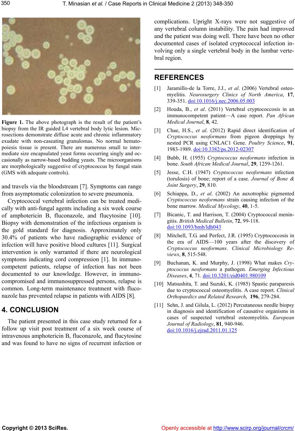 T. Minasian et al. / Case Reports in Clinical Medicine 2 (2013) 348-3 50 Copyright © 2013 SciRes. Openly accessible at http://www.sc irp.or g/journal/crcm/ 350 Figure 1. The above photograph is the result of the patient’s biopsy from the IR guided L4 vertebral body lytic lesion. Mic- rosections demonstrate diffuse acute and chronic inflammatory exudate with non-caseating granulomas. No normal hemato- poiesis tissue is present. There are numerous small to inter- mediate size encapsulated yeast forms occurring singly and oc- casionally as narrow-based budding yeasts. The microorganisms are morphologically suggestive of cryptococcus by fungal stain (GMS with adequate controls). and travels via the bloodstream [7] . S ympt oms c an ran ge from asymptomatic colonization to severe pneumonia. Cryptococcal vertebral infection can be treated medi- cally with anti-fungal agen ts including a six week course of amphotericin B, fluconazole, and flucytosine [10]. Biopsy with demonstration of the infectious organism is the gold standard for diagnosis. Approximately only 30.4% of patients who have radiographic evidence of infection will have positive blood cultures [11]. Surgical intervention is only warranted if there are neurological symptoms indicating cord compression [1]. In immuno- competent patients, relapse of infection has not been documented to our knowledge. However, in immuno- compromised and immunosuppressed persons, relapse is common. Long-term maintenance treatment with fluco- nazole has prevented relaps e in patients with AIDS [8]. 4. CONCLUSION The patient presented in this case study returned for a follow up visit post treatment of a six week course of intravenous amphotericin B, fluconazole, and flucytosine and was found to have no signs of recurrent infection or complications. Upright X-rays were not suggestive of any vertebral column instability. The pain had improved and the patient was doing well. There have been no other documented cases of isolated cryptococcal infection in- volving only a single vertebral bod y in the lumbar verte- bral region. REFERENCES [1] Jaramillo-de la Torre, J.J., et al. (2006) Vertebral osteo- myelitis. Neurosurgery Clinics of North America, 17, 339-351. doi:10.1016/j.nec.2006.05.003 [2] Houda, B., et al. (2011) Vertebral cryptococcosis in an immunocompetent patient—A case report. Pan African Medical Journal, 8, 42. [3] Chae, H.S., et al. (2012) Rapid direct identification of Cryptococcus neoformans from pigeon droppings by nested PCR using CNLAC1 Gene. Poultry Science, 91, 1983-1989. doi:10.3382/ps.2012-02307 [4] Bubb, H. (1955) Cryptococcus neoformans infection in bone. South African Medical Journal, 29, 1259-1261. [5] Jesse, C.H. (1947) Cryptococcus neoformans infection (torulosis) of bone; report of a case. Journal of Bone & Joint Surgery, 29, 810. [6] Schiappa, D., et al. (2002) An auxotrophic pigmented Cryptococcus neoformans strain causing infection of the bone marrow. Medical Mycology, 40, 1-5. [7] Bicanic, T. and Harrison, T. (2004) Cryptococcal menin- gitis. British Medical Bulletin, 72, 99-118. doi:10.1093/bmb/ldh043 [8] Mitchell, T.G. and Perfect, J.R. (1995) Cryptococcosis in the era of AIDS—100 years after the discovery of Cryptococcus neoformans. Clinical Microbiology Re- views, 8, 515-548. [9] Buchanan, K. and Murphy, J. (1998) What makes Cry- ptococcus neoformans a pathogen. Emerging Infectious Diseases, 4, 71. doi:10.3201/eid0401.980109 [10] Matsushita, T. and Suzuki, K. (1985) Spastic paraparesis due to cryptococcal osteomyelitis. A case report. Clinical Orthopaedics and Related Research, 196, 279-284. [11] Sehn, J. and Gilula, L. (2012) Percutaneous needle biopsy in diagnosis and identification of causative organisms in cases of suspected vertebral osteomyelitis. European Journal of Radiology, 81, 940-946. doi:10.1016/j.ejrad.2011.01.125 |