

Paper Menu >>
Journal Menu >>
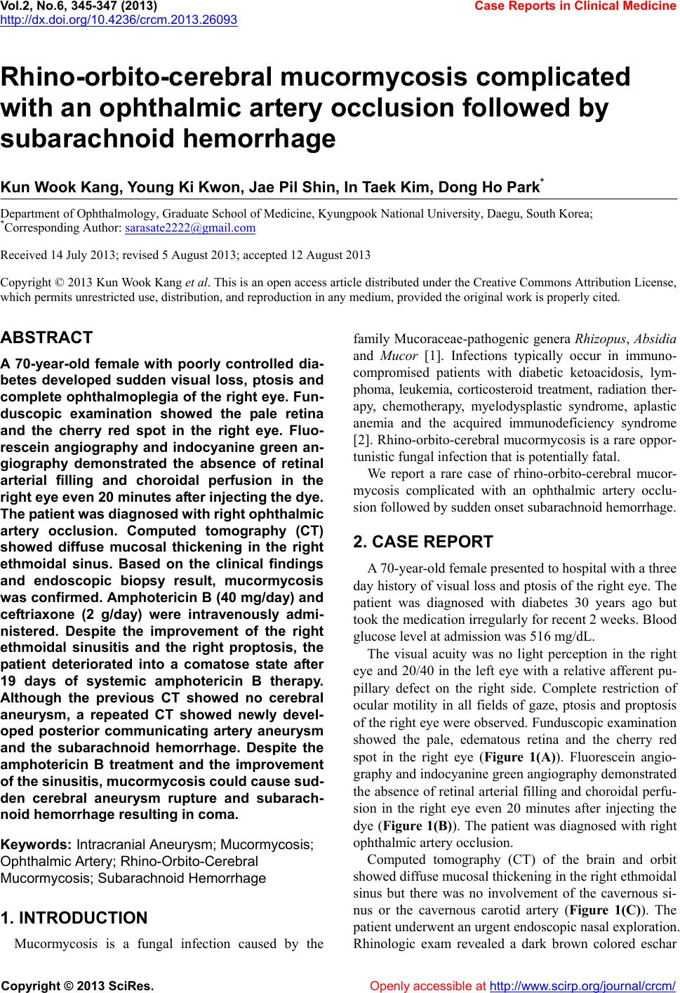 Vol.2, No.6, 345-347 (2013) Case Reports in Clinical Medicine http://dx.doi.org/10.4236/crcm.2013.26093 Rhino-orbito-cerebral mucormycosis complicated with an ophthalmic artery occlusion followed by subarachnoid hemorrhage Kun Wook Kang, Young Ki Kwon, Jae Pil Shin, In Taek Kim, Dong Ho Park* Department of Ophthalmology, Graduate School of Medicine, Kyungpook National University, Daegu, South Korea; *Corresponding Author: [email protected] Received 14 July 2013; revised 5 August 2013; accepted 12 August 2013 Copyright © 2013 Kun Wook Kang et al. This is an open access article distributed under the Creative Commons Attribution License, which permits unrestricted use, distribution, and reproduction in any medium, provided the original work is properly cited. ABSTRACT A 70-year-old female with poorly controlled dia- betes developed sudden visual loss, ptosis and complete ophthalmoplegia of the right eye. Fun- duscopic examination showed the pale retina and the cherry red spot in the right eye. Fluo- rescein angiography and indocyanine green an- giography demonstrated the absence of retinal arterial filling and choroidal perfusion in the right eye even 20 minutes after injecting the dye. The p atient was diagnosed with right ophthalmic artery occlusion. Computed tomography (CT) showed diffuse mucosal thickening in the right ethmoidal sinus. Based on the clinical findings and endoscopic biopsy result, mucormycosis was confirmed. Amphotericin B (40 mg/day) and ceftriaxone (2 g/day) were intravenously admi- nistered. Despite the improvement of the right ethmoidal sinusitis and the right proptosis, the patient deteriorated into a comatose state after 19 days of systemic amphotericin B therapy. Although the previous CT showed no cerebral aneurysm, a repeated CT showed newly devel- oped posterior communicating artery aneurysm and the subarachnoid hemorrhage. Despite the amphotericin B treatment and the improvement of the sinusitis, mucormycosis could cause sud- den cerebral aneurysm rupture and subarach- noid hemorrhage resulting in coma. Keyw ords: Intracranial Aneurysm; Mucormycosis; Ophthalmic Artery; Rhino-Orbito-Cere bral Mucormycosis; Subarach noid Hemorrhage 1. INTRODUCTION Mucormycosis is a fungal infection caused by the family Mucoraceae-pathogenic genera Rhizopus, Absidia and Mucor [1]. Infections typically occur in immuno- compromised patients with diabetic ketoacidosis, lym- phoma, leukemia, corticosteroid treatment, radiation ther- apy, chemotherapy, myelodysplastic syndrome, aplastic anemia and the acquired immunodeficiency syndrome [2]. Rhino-orbito-cerebral mucormycosis is a rare oppor- tunistic fungal infection that is potentially fatal. We report a rare case of rhino-orbito-cerebral mucor- mycosis complicated with an ophthalmic artery occlu- sion followed by sudden onset subarachnoid hemorrhage. 2. CASE REPORT A 70-year-old female presented to hospital with a three day history of visual loss and ptosis of the right eye. The patient was diagnosed with diabetes 30 years ago but took the medication irregularly for recent 2 weeks. Blood glucose level at admission was 516 mg/dL. The visual acuity was no light perception in the right eye and 20/40 in the left eye with a relative afferent pu- pillary defect on the right side. Complete restriction of ocular motility in all fields of gaze, ptosis and proptosis of the right eye were observed. Funduscopic examination showed the pale, edematous retina and the cherry red spot in the right eye (Figure 1(A)). Fluorescein angio- graphy an d indocyan ine gre en angiography demonstrated the absence of retinal arterial filling and choroidal p erfu- sion in the right eye even 20 minutes after injecting the dye (Figure 1(B)). The patient was diagnosed with right ophthalmic artery occlusion. Computed tomography (CT) of the brain and orbit showed diffuse mucosal thickening in the righ t eth moidal sinus but there was no involvement of the cavernous si- nus or the cavernous carotid artery (Figure 1(C)). The patient underwent an urgent endoscopic nasal exploration. Rhinologic exam revealed a dark brown colored eschar Copyright © 2013 SciRes. Openly accessible at http://www.sc irp.or g/journal/crcm/ 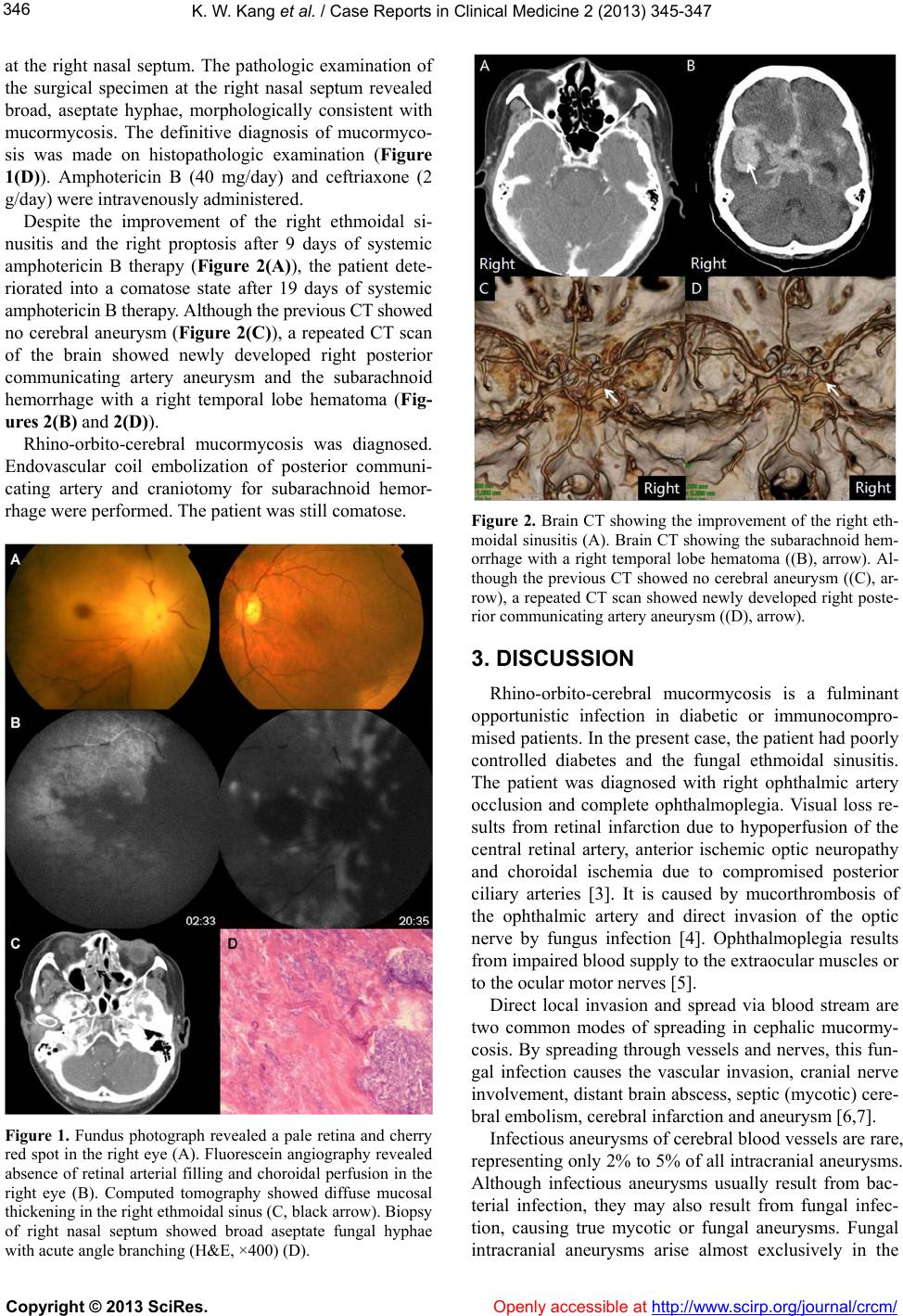 K. W. Kang et al. / Case Reports in Clinical Medicine 2 (2013) 345-347 346 at the right nasal septum. The pathologic examination of the surgical specimen at the right nasal septum revealed broad, aseptate hyphae, morphologically consistent with mucormycosis. The definitive diagnosis of mucormyco- sis was made on histopathologic examination (Figure 1(D)). Amphotericin B (40 mg/day) and ceftriaxone (2 g/day) were intravenously administered. Despite the improvement of the right ethmoidal si- nusitis and the right proptosis after 9 days of systemic amphotericin B therapy (Figure 2(A)), the patient dete- riorated into a comatose state after 19 days of systemic amphotericin B therapy. Although the previous CT show e d no cerebral aneurysm (Figure 2(C)), a repeated CT scan of the brain showed newly developed right posterior communicating artery aneurysm and the subarachnoid hemorrhage with a right temporal lobe hematoma (Fig- ures 2(B) and 2( D)). Rhino-orbito-cerebral mucormycosis was diagnosed. Endovascular coil embolization of posterior communi- cating artery and craniotomy for subarachnoid hemor- rhage were performed. The patient was still comatose. Figure 1. Fundus photograph revealed a pale retina and cherry red spot in the right eye (A). Fluorescein angiography revealed absence of retinal arterial filling and choroidal perfusion in the right eye (B). Computed tomography showed diffuse mucosal thickening in the right ethmoidal sinus (C, black arrow). Biopsy of right nasal septum showed broad aseptate fungal hyphae with acute angle branching (H&E, ×400) (D). Figure 2. Brain CT showing the improvement of the right eth- moidal sinusitis (A). Brain CT showing the subarachnoid hem- orrhage with a right temporal lobe hematoma ((B), arrow). Al- though the previous CT showed no cerebral aneurysm ((C), ar- row), a repeated CT scan showed newly developed right poste- rior communicating artery aneurysm ((D), arrow). 3. DISCUSSION Rhino-orbito-cerebral mucormycosis is a fulminant opportunistic infection in diabetic or immunocompro- mised patients. In the present case, the patient had poorly controlled diabetes and the fungal ethmoidal sinusitis. The patient was diagnosed with right ophthalmic artery occlusion and complete ophthalmoplegia. Visual loss re- sults from retinal infarction due to hypoperfusion of the central retinal artery, anterior ischemic optic neuropathy and choroidal ischemia due to compromised posterior ciliary arteries [3]. It is caused by mucorthrombosis of the ophthalmic artery and direct invasion of the optic nerve by fungus infection [4]. Ophthalmoplegia results from impaired blood supply to th e extraocu lar muscles or to the ocular motor nerves [5]. Direct local invasion and spread via blood stream are two common modes of spreading in cephalic mucormy- cosis. By spreading through vessels and nerves, this fun- gal infection causes the vascular invasion, cranial nerve involvement, distant brain ab scess, septic (mycotic) cere- bral embolism, cerebral infarction and aneurysm [6,7]. Infectious aneurysms of cerebral blood vessels are rare, representing only 2% to 5% of all intracranial aneurysms. Although infectious aneurysms usually result from bac- terial infection, they may also result from fungal infec- tion, causing true mycotic or fungal aneurysms. Fungal intracranial aneurysms arise almost exclusively in the Copyright © 2013 SciRes. Openly accessible at http://www.sc irp.org/jour nal/crcm/  K. W. Kang et al. / Case Reports in Clinical Medicine 2 (2013) 345-347 Copyright © 2013 SciRes. Openly accessible at http://www.sc irp.or g/journal/crcm/ 347 setting of an extravascular infection in an immunocom- promised host [8]. In the present case, the patient with poorly controlled diabetes complained of only ocular symptom and brain CT showed no infectious cerebral aneurysm at admission. After 9 days of systemic ampho- tericin B therapy, brain CT showed the improvement of the proptosis of the right eye and the resolution of mu- cosal thickening of ethmoidal sinus. However, 19 days after systemic amphotericin B therapy, the patient dete- riorated into a comatose state. Brain CT showed the dif- fuse subarachnoid hemorrhage with a right temporal lobe hematoma due to ruptured aneurysm. Within 19 days, new cerebral aneurysm developed and progressed rapidly. It supported our su spicion of a fungal aneur ysm. REFERENCES [1] Koc, Z., Koc, F., Yerdelen, D. and Ozdogu, H. (2007) Rhino-orbital-cerebral mucormycosis with different cere- bral involvements: Infarct, hemorrhage, and ophthalmo- plegia. International Journal of Neuroscience, 117, 1677- 1690. doi:10.1080/00207450601050238 [2] Haliloglu, N.U., Yesilirmak, Z., Erden, A. and Erden, I. (2008) Rhino-orbito-cerebral mucormycosis: Report of two cases and review of the literature. Dentomaxillofacial Radiology, 37, 161-166. doi:10.1259/dmfr/14698002 [3] Yang, S.W., Kim, S.Y., Chung, J. and Kim, K.B. (2000) Two cases of orbital infarction syndrome. Korean Journal of Ophthalmology, 14, 107-111. [4] Hussain, S., Salahuddin, N., Ahmad, I., Salahuddin, I. and Jooma, R. (1995) Rhinocerebral invasive mycosis: Oc- currence in immunocompetent individuals. European Jour- nal of Radiology, 20, 151-155. doi:10.1016/0720-048X(95)00644-6 Survival of mucormycosis with intracerebral involve- ment was thought to be virtually impossible un til the first case of clinical recovery reported in 1955 [9]. The intro- duction of amphotericin B in the 1960s reduced mortality from 70% to 40% [10]. Nevertheless, the prognosis of rhino-orbito -cerebral mucormycosis remains poo r and 20 to 50% of patients expire [11]. In the present case, the ethmoidal sinusitis and the proptosis of the right eye were improved after amphotericin B treatment. However the subarachnoid hemorrhage due to ruptured aneurysm occurred in a short period. Although endovascular coil embolization of posterior communicating artery and cra- niotomy for subarachnoid hemorrhage were performed, the patient was still comatose. [5] Zimmerman, C.F., Van Patten, P.D., Golnik, K.C., Kopit- nik Jr., T.A. and Anand, R. (1995) Orbital infarction syn- drome after surgery for intracranial aneurysms. Ophthal- mology, 102, 594-598. [6] Chimelli, L. and Mahler-Araujo, M.B. (1997) Fungal infections. Brain Pathology, 7, 613-627. d oi:1 0.1111/j .1750-3639.1997.tb01078.x [7] Peterus, T., Teguh, T. and Daofu, D. (2004) Fatal strokes in patients with rhino-orbito-cerebral mucormycosis and associated vasculopathy. Scandinavian Journal of Infec- tious Diseases, 36, 643-648. doi:10.1080/00365540410020794 In conclusion, systemic amphotericin B antifungal treatment improves the sinusitis and the proptosis. Nev- ertheless, new cerebral aneurysm can develop in a short period and it can cause subarachnoid hemorrhage. Oph- thalmologists shou ld raise the suspicion of the infectious mycotic cerebral aneurysm and the subarachnoid hem- orrhage resulting in coma, even if the sinusitis is im- proved after amphotericin B treatment. [8] Asari, S., Nishimoto, A. and Murakami, M. (1988) A rare case of cerebral aspergillus aneurysm at the site of tem- porary clip application. No Shinkei Geka, 16, 1079-1082. [9] Harris, J.S. (1955) Mucormycosis; report of a case. Pedi- atrics, 16, 857-867. [10] Shah, P.D., Peters, K.R. and Reuman, P.D. (1997) Recov- ery from rhinocerebral mucormycosis with carotid artery occlusion: A pediatric case and review of the literature Pediatric Infectious Disease Journal, 16, 68-71. doi:10.1097/00006454-199701000-00015 4. ACKNOWLEDGEMENTS This research was supported by the Basic Science Research Program through the National Research Foundation of Korea (NRF) funded by the Ministry of Education, Science and Technology (2012004585) and by the Korea Health Technology R&D Project, Ministry of Health & Welfare, Repu blic of Korea (A111345). [11] Toumi, A., Larbi Ammari, F., Loussaief, C., Hadhri, R., Ben Brahim, H., Harrathi, K., Ben Romdhane, F., Koubaa, J. and Chakroun, M. (2012) Rhino-orbito-cerebral mucor- mycosis: Five cases. Médecine et Maladies Infectieuses, 42, 591-598. doi:10.1016/j.medmal.2012.10.001 |