

Paper Menu >>
Journal Menu >>
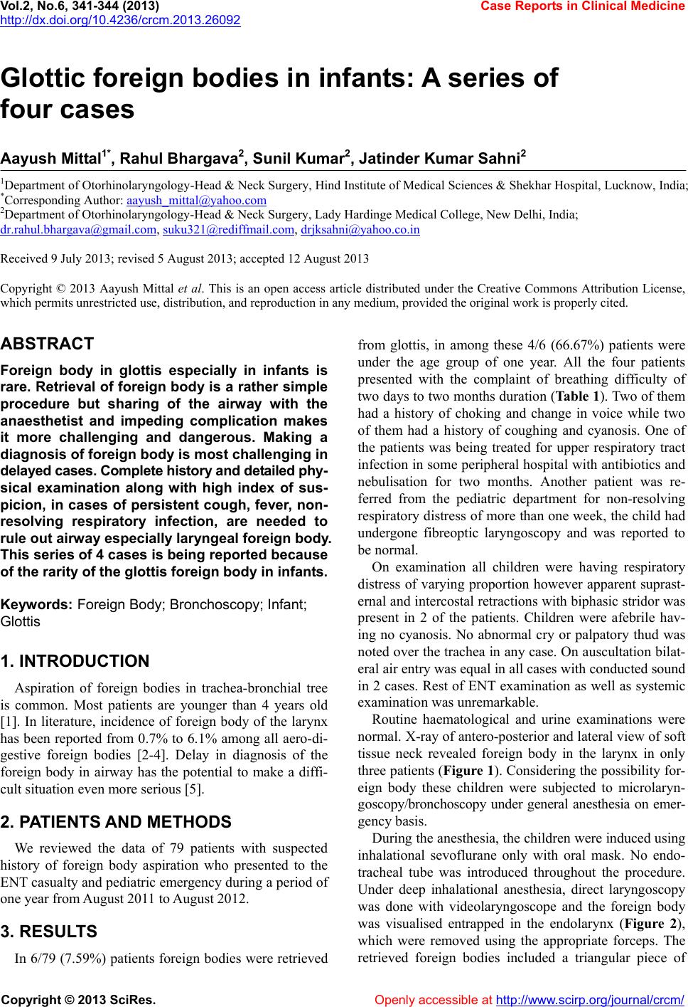 Vol.2, No.6, 341-344 (2013) Case Reports in Clinical Medicine http://dx.doi.org/10.4236/crcm.2013.26092 Glottic foreign bodies in infants: A series of four cases Aayush Mittal1*, Rahul Bhargava2, Sunil Kumar2, Jatinder Kumar Sahni2 1Department of Otorhinolaryngology-Head & Neck Surgery, Hind Institute of Medical Sciences & Shekhar Hospital, Lucknow, India; *Corresponding Author: [email protected] 2Department of Otorhinolaryngology-Head & Neck Surgery, Lady Hardinge Medical College, New Delhi, India; Received 9 July 2013; revised 5 August 2013; accepted 12 August 2013 Copyright © 2013 Aayush Mittal et al. This is an open access article distributed under the Creative Commons Attribution License, which permits unrestricted use, distribution, and reproduction in any medium, provided the original work is properly cited. ABSTRACT Foreign body in glottis especially in infants is rare. Retrieval of foreign body is a rather simple procedure but sharing of the airway with the anaesthetist and impeding complication makes it more challenging and dangerous. Making a diagnosis of foreign body is most challenging in delayed cases. Complete history and detailed phy- sical examination along with high index of sus- picion, in cases of persistent cough, fever, non- resolving respiratory infection, are needed to rule out airway especially laryngeal foreign body. This series of 4 cases is being reported because of the rarity of the glott is fore ign b od y in infants. Keyw ords: Foreign Body; Bronchoscop y ; Infant; Glottis 1. INTRODUCTION Aspiration of foreign bodies in trachea-bronchial tree is common. Most patients are younger than 4 years old [1]. In literature, incidence of foreign body of the larynx has been reported from 0.7% to 6.1% among all aero-di- gestive foreign bodies [2-4]. Delay in diagnosis of the foreign body in airway has the potential to make a diffi- cult situation even more serious [5]. 2. PATIENTS AND METHODS We reviewed the data of 79 patients with suspected history of foreign body aspiration who presented to the ENT casualty and pediatric emergency during a period of one year from August 2011 to August 2012. 3. RESULTS In 6/79 (7.59%) patients foreign bodies were retrieved from glottis, in among these 4/6 (66.67%) patients were under the age group of one year. All the four patients presented with the complaint of breathing difficulty of two days to two months duration (Table 1). Two of them had a history of choking and change in voice while two of them had a history of coughing and cyanosis. One of the patients was being treated for upper respiratory tract infection in some peripheral hospital with antibiotics and nebulisation for two months. Another patient was re- ferred from the pediatric department for non-resolving respiratory distress of more than one week, the child had undergone fibreoptic laryngoscopy and was reported to be normal. On examination all children were having respiratory distress of varying proportion however apparent suprast- ernal and intercostal retractions with biphasic stridor was present in 2 of the patients. Children were afebrile hav- ing no cyanosis. No abnormal cry or palpatory thud was noted over the trachea in any case. On auscultation bilat- eral air entry was equal in all cases with conducted sound in 2 cases. Rest of ENT examination as well as systemic examination was unremarkable. Routine haematological and urine examinations were normal. X-ray of antero-posterior and lateral view of soft tissue neck revealed foreign body in the larynx in only three patients (Figure 1). Considering the possibility for- eign body these children were subjected to microlaryn- goscopy/bronchoscopy under general anesthesia on emer- gency basis. During the anesthesia, the children were induced using inhalational sevoflurane only with oral mask. No endo- tracheal tube was introduced throughout the procedure. Under deep inhalational anesthesia, direct laryngoscopy was done with videolaryngoscope and the foreign body was visualised entrapped in the endolarynx (Figure 2), which were removed using the appropriate forceps. The retrieved foreign bodies included a triangular piece of Copyright © 2013 SciRes. Openly accessible at http://www.sc irp.or g/journal/crcm/ 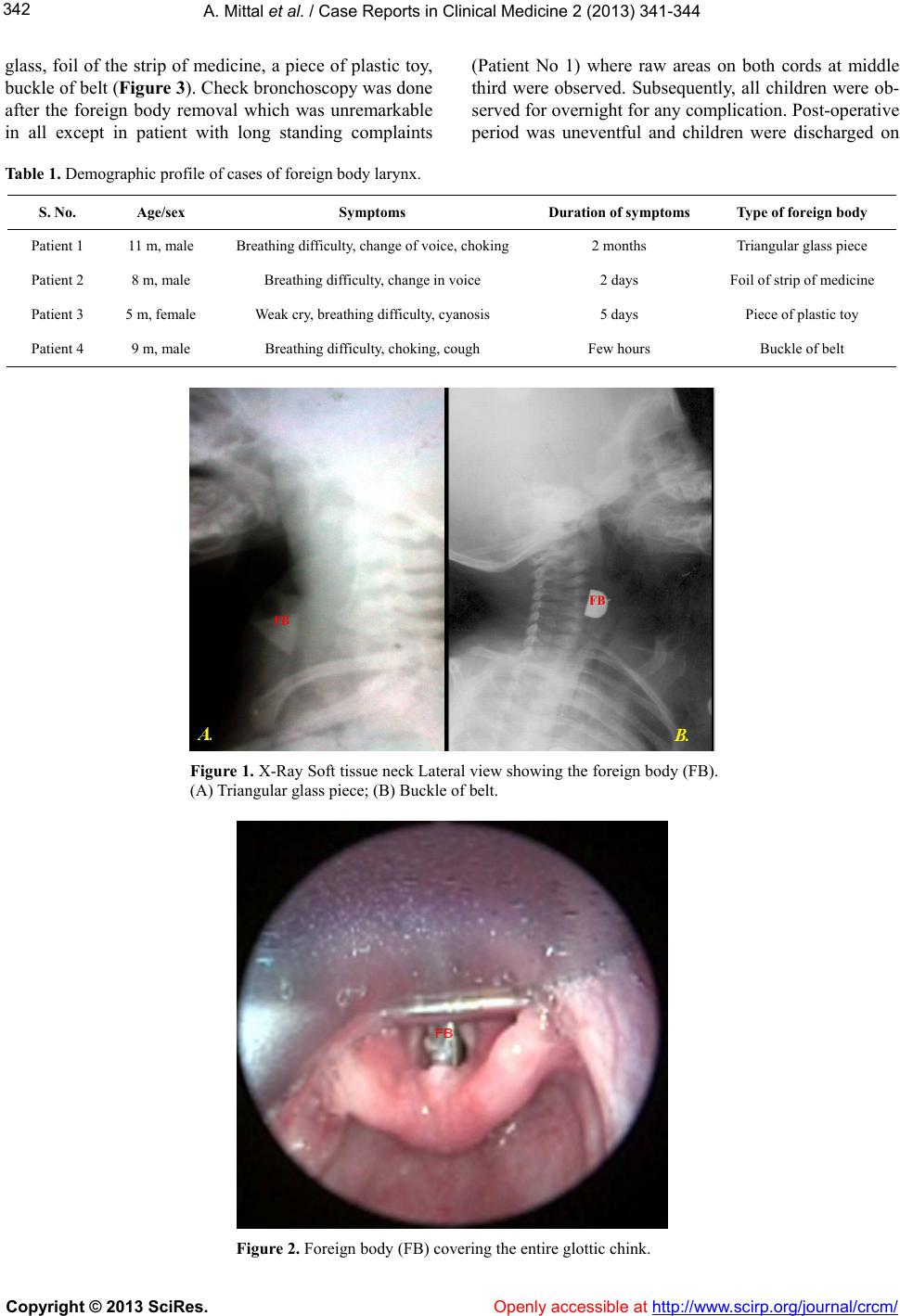 A. Mittal et al. / Case Reports in Clinical Medicine 2 (2013) 341-344 342 glass, foil of the strip of medicine, a piece of plastic toy, buckle of belt (Figure 3). Check bronchoscopy was done after the foreign body removal which was unremarkable in all except in patient with long standing complaints (Patient No 1) where raw areas on both cords at middle third were observed. Subsequently, all children were ob- served for overnight for any complication. Post-operative period was uneventful and children were discharged on Table 1. Demographic profile of cases of foreign body larynx. S. No. Age/sex Symptoms Duration of symptoms Type of foreign body Patient 1 11 m, male Breathing difficulty, change of voice, choking 2 months Triangular glass piece Patient 2 8 m, male Breathing difficulty, change in voice 2 days Foil of strip of medicine Patient 3 5 m, female Weak cry, breathing difficulty, cyanosis 5 days Piece of plastic toy Patient 4 9 m, male Breathing difficulty, choking, cough Few hours Buckle of belt Figure 1. X-Ray Soft tissue neck Lateral view showing the foreign body (FB). (A) Triangular glass piece; (B) Buckle of belt. Figure 2. Foreign body (FB) covering the entire glottic chink. Copyright © 2013 SciRes. Openly accessible at http://www.sc irp.org/jour nal/crcm/ 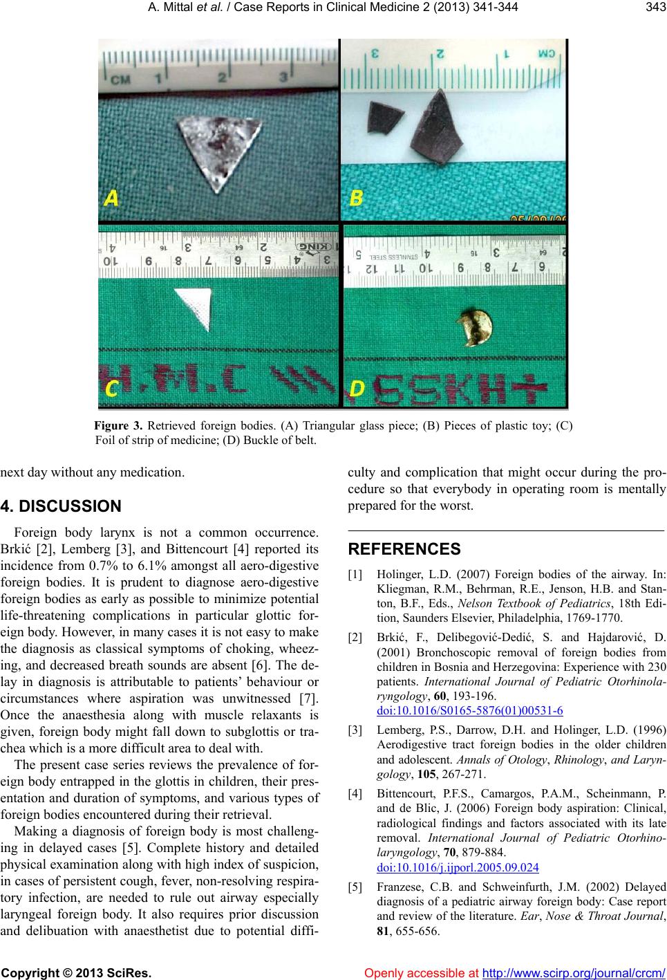 A. Mittal et al. / Case Reports in Clinical Medicine 2 (2013) 341-344 343 Figure 3. Retrieved foreign bodies. (A) Triangular glass piece; (B) Pieces of plastic toy; (C) Foil of strip of medicine; (D) Buckle of belt. next day without any medication. 4. DISCUSSION Foreign body larynx is not a common occurrence. Brkić [2], Lemberg [3], and Bittencourt [4] reported its incidence from 0.7% to 6.1% amongst all aero-digestive foreign bodies. It is prudent to diagnose aero-digestive foreign bodies as early as possible to minimize potential life-threatening complications in particular glottic for- eign body. However, in many cases it is not easy to make the diagnosis as classical symptoms of choking, wheez- ing, and decreased breath sounds are absent [6]. The de- lay in diagnosis is attributable to patients’ behaviour or circumstances where aspiration was unwitnessed [7]. Once the anaesthesia along with muscle relaxants is given, foreign body might fall down to subglottis or tra- chea which is a more difficult area to deal with. The present case series reviews the prevalence of for- eign body entrapped in the glottis in children, their pres- entation and duration of symptoms, and various types of foreign bodies encountered during their retrieval. Making a diagnosis of foreign body is most challeng- ing in delayed cases [5]. Complete history and detailed physical examination along with high index of suspicion, in cases of persistent cough, fever, non-resolving respira- tory infection, are needed to rule out airway especially laryngeal foreign body. It also requires prior discussion and delibuation with anaesthetist due to potential diffi- culty and complication that might occur during the pro- cedure so that everybody in operating room is mentally prepared for the worst. REFERENCES [1] Holinger, L.D. (2007) Foreign bodies of the airway. In: Kliegman, R.M., Behrman, R.E., Jenson, H.B. and Stan- ton, B.F., Eds., Nelson Textbook of Pediatrics, 18th Edi- tion, Saunders Elsevier, Philadelphia, 1769-1770. [2] Brkić, F., Delibegović-Dedić, S. and Hajdarović, D. (2001) Bronchoscopic removal of foreign bodies from children in Bosnia and Herzegovina: Experience with 230 patients. International Journal of Pediatric Otorhinola- ryngology, 60, 193-196. doi:10.1016/S0165-5876(01)00531-6 [3] Lemberg, P.S., Darrow, D.H. and Holinger, L.D. (1996) Aerodigestive tract foreign bodies in the older children and adolescent. Annals of Otology, Rhinology, and Laryn- gology, 105, 267-271. [4] Bittencourt, P.F.S., Camargos, P.A.M., Scheinmann, P. and de Blic, J. (2006) Foreign body aspiration: Clinical, radiological findings and factors associated with its late removal. International Journal of Pediatric Otorhino- laryngology, 70, 879-884. doi:10.1016/j.ijporl.2005.09.024 [5] Franzese, C.B. and Schweinfurth, J.M. (2002) Delayed diagnosis of a pediatric airway foreign body: Case report and review of the literature. Ear, Nose & Throat Journal, 81, 655-656. Copyright © 2013 SciRes. Openly accessible at http://www.sc irp.or g/journal/crcm/  A. Mittal et al. / Case Reports in Clinical Medicine 2 (2013) 341-344 344 [6] Reilly, J., Thompson, J., MacArthur, C., Pransky, S., Be- ste, D., Smith, M., et al. (1997) Pediatric aerodigestive foreign body injuries are complications related to timeli- ness of diagnosis. Laryngoscope, 107, 17-20. doi:10.1097/00005537-199701000-00006 [7] Esclamado, R.M. and Richardson, M.A. (1987) Laryn- gotracheal foreign bodies in children, a comparison with bronchial foreign bodies. American Journal of Diseases of Children, 141, 259-262. Copyright © 2013 SciRes. Openly accessible at http://www.sc irp.org/jour nal/crcm/ |