

Paper Menu >>
Journal Menu >>
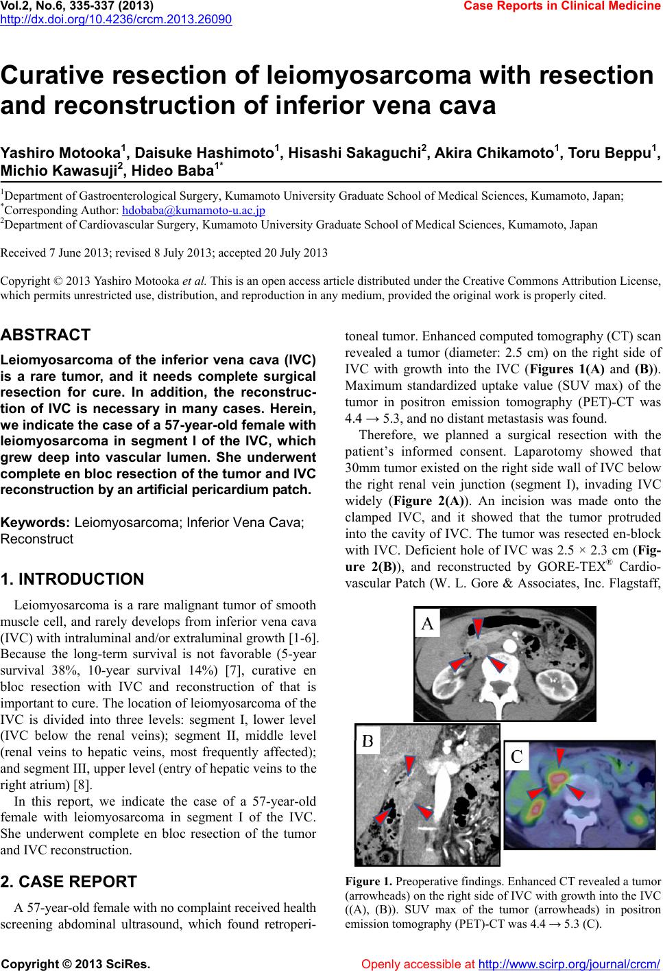 Vol.2, No.6, 335-337 (2013) Case Reports in Clinical Medicine http://dx.doi.org/10.4236/crcm.2013.26090 Curative resection of leiomyosarcoma with resection and reconstruction of inferior vena cava Yashiro Motooka1, Daisuke Hashimoto1, Hisashi Sakaguchi2, Akira Chikamoto1, Toru Beppu1, Michio Kawasuji2, Hideo Baba1* 1Department of Gastroenterological Surgery, Kumamoto University Graduate School of Medical Sciences, Kumamoto, Japan; *Corresponding Author: [email protected] 2Department of Cardiovascular Surgery, Kumamoto University Graduate School of Medical Sciences, Kumamoto, Japan Received 7 June 2013; revised 8 July 2013; accepted 20 July 2013 Copyright © 2013 Yashiro Motooka et al. This is an open access article distributed under the Creative Commons Attribution License, which permits unrestricted use, distribution, and reproduction in any medium, provided the original work is properly cited. ABSTRACT Leiomyosarcoma of the inferior vena cava (IVC) is a rare tumor, and it needs complete surgical resection for cure. In addition, the reconstruc- tion of IVC is necessary in many cases. Herein, we indicate the case of a 57-year-old female with leiomyosarcoma in segment I of the IVC, which grew deep into vascular lumen. She underwent complete en bloc resection of the tumor and IVC reconstruction by an artificial pericardium p atch. Keywords: Leiomyosarcoma; Inferior Vena Cava; Reconstruct 1. INTRODUCTION Leiomyosarcoma is a rare malignant tumor of smooth muscle cell, and rarely develops from inferior vena cava (IVC) with intraluminal and/or extraluminal growth [1-6]. Because the long-term survival is not favorable (5-year survival 38%, 10-year survival 14%) [7], curative en bloc resection with IVC and reconstruction of that is important to cure. The location of leiomyosarcoma of the IVC is divided into three levels: segment I, lower level (IVC below the renal veins); segment II, middle level (renal veins to hepatic veins, most frequently affected); and segment III, upper level (entry of hepatic veins to the right atrium) [8]. In this report, we indicate the case of a 57-year-old female with leiomyosarcoma in segment I of the IVC. She underwent complete en bloc resection of the tumor and IVC reconstruction. 2. CASE REPORT A 57-year-old female with no complaint received health screening abdominal ultrasound, which found retroperi- toneal tumor. Enhanced computed tomography (CT) scan revealed a tumor (diameter: 2.5 cm) on the right side of IVC with growth into the IVC (Figures 1(A) and (B)). Maximum standardized uptake value (SUV max) of the tumor in positron emission tomography (PET)-CT was 4.4 → 5.3, and no distant metastasis was found. Therefore, we planned a surgical resection with the patient’s informed consent. Laparotomy showed that 30mm tumor existed on the right side wall of IVC below the right renal vein junction (segment I), invading IVC widely (Figure 2(A)). An incision was made onto the clamped IVC, and it showed that the tumor protruded into the cavity of IVC. The tumor was resected en-block with IVC. Deficient hole of IVC was 2.5 × 2.3 cm (Fig- ure 2(B)), and reconstructed by GORE-TEX® Cardio- vascular Patch (W. L. Gore & Associates, Inc. Flagstaff, Figure 1. Preoperative findings. Enhanced CT revealed a tumor (arrowheads) on the right side of IVC with growth into the IVC ((A), (B)). SUV max of the tumor (arrowheads) in positron emission tomography (PET)-CT was 4.4 → 5.3 (C). Copyright © 2013 SciRes. Openly accessible at http://www.sc irp.or g/journal/crcm/ 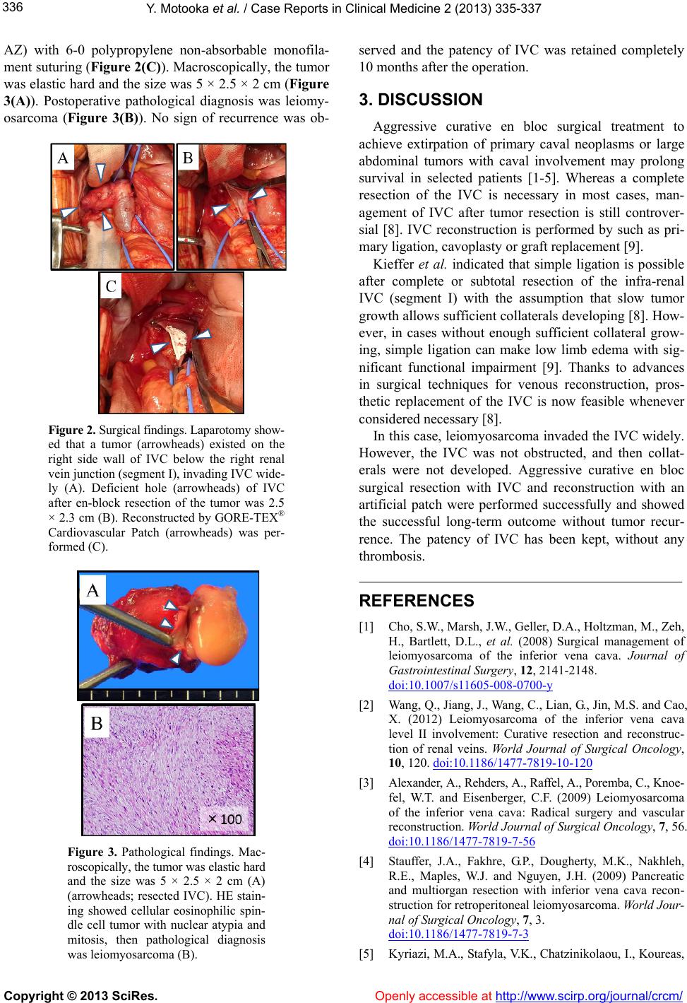 Y. Motooka et al. / Case Reports in Clinical Medicine 2 (2013) 335-337 336 AZ) with 6-0 polypropylene non-absorbable monofila- ment suturing (Figure 2(C)). Macroscopically, the tumor was elastic hard and the size was 5 × 2.5 × 2 cm (Figure 3(A)). Postoperative pathological diagnosis was leiomy- osarcoma (Figure 3(B)). No sign of recurrence was ob- Figure 2. Surgical findings. Laparotomy show- ed that a tumor (arrowheads) existed on the right side wall of IVC below the right renal vein junction (segment I), invading IVC wide- ly (A). Deficient hole (arrowheads) of IVC after en-block resection of the tumor was 2.5 × 2.3 cm (B). Reconstructed by GORE-TEX® Cardiovascular Patch (arrowheads) was per- formed (C). Figure 3. Pathological findings. Mac- roscopically, the tumor was elastic hard and the size was 5 × 2.5 × 2 cm (A) (arrowheads; resected IVC). HE stain- ing showed cellular eosinophilic spin- dle cell tumor with nuclear atypia and mitosis, then pathological diagnosis was leiomyosarcoma (B). served and the patency of IVC was retained completely 10 months after the operation. 3. DISCUSSION Aggressive curative en bloc surgical treatment to achieve extirpation of primary caval neoplasms or large abdominal tumors with caval involvement may prolong survival in selected patients [1-5]. Whereas a complete resection of the IVC is necessary in most cases, man- agement of IVC after tumor resection is still controver- sial [8]. IVC reconstruction is performed by such as pri- mary ligation, cavoplasty or graft replacement [9]. Kieffer et al. indicated that simple ligation is possible after complete or subtotal resection of the infra-renal IVC (segment I) with the assumption that slow tumor growth allows sufficient collaterals developing [8]. How- ever, in cases without enough sufficient collateral grow- ing, simple ligation can make low limb edema with sig- nificant functional impairment [9]. Thanks to advances in surgical techniques for venous reconstruction, pros- thetic replacement of the IVC is now feasible whenever considered necessary [8]. In this case, leiomyosarcoma invaded the IVC widely. However, the IVC was not obstructed, and then collat- erals were not developed. Aggressive curative en bloc surgical resection with IVC and reconstruction with an artificial patch were performed successfully and showed the successful long-term outcome without tumor recur- rence. The patency of IVC has been kept, without any thrombosis. REFERENCES [1] Cho, S.W., Marsh, J.W., Geller, D.A., Holtzman, M., Zeh, H., Bartlett, D.L., et al. (2008) Surgical management of leiomyosarcoma of the inferior vena cava. Journal of Gastrointestinal Surger y, 12, 2141-2148. doi:10.1007/s11605-008-0700-y [2] Wang, Q., Jiang, J., Wang, C., Lian, G., Jin, M.S. and Cao, X. (2012) Leiomyosarcoma of the inferior vena cava level II involvement: Curative resection and reconstruc- tion of renal veins. World Journal of Surgical Oncology, 10, 120. doi:10.1186/1477-7819-10-120 [3] Alexander, A., Rehders, A., Raffel, A., Poremba, C., Knoe- fel, W.T. and Eisenberger, C.F. (2009) Leiomyosarcoma of the inferior vena cava: Radical surgery and vascular reconstruction. World Journal of Surgical Oncology, 7, 56. doi:10.1186/1477-7819-7-56 [4] Stauffer, J.A., Fakhre, G.P., Dougherty, M.K., Nakhleh, R.E., Maples, W.J. and Nguyen, J.H. (2009) Pancreatic and multiorgan resection with inferior vena cava recon- struction for retroperitoneal leiomyosarcoma. World Jour- nal of Surgical Oncology, 7, 3. doi:10.1186/1477-7819-7-3 [5] Kyriazi, M.A., Stafyla, V.K., Chatzinikolaou, I., Koureas, Copyright © 2013 SciRes. Openly accessible at http://www.sc irp.or g/journal/crcm/  Y. Motooka et al. / Case Reports in Clinical Medicine 2 (2013) 335-337 Copyright © 2013 SciRes. Openly accessible at http://www.sc irp.or g/journal/crcm/ 337 A., Chatziioannou, A., Kondi-Paphiti, A., et al. (2010) Surgical challenges in the treatment of leiomyosarcoma of the inferior vena cava: Analysis of two cases and brief review of the literature. Annals of Vascular Surgery, 24, 826.e13-826.e17. [6] Dew, J., Hansen, K., Hammon, J., McCoy, T., Levine, E.A. and Shen, P. (2005) Leiomyosarcoma of the inferior vena cava: Surgical management and clinical results. American Journal of Surgery, 71, 497-501. [7] Mingoli, A., Cavallaro, A., Sapienza, P., Di Marzo, L., Feldhaus, R.J. and Cavallari, N. (1996) International reg- istry of inferior vena cava leiomyosarcoma: Analysis of a world series on 218 patients. Anticancer Research, 16, 3201-3205. [8] Kieffer, E., Alaoui, M., Piette, J.C., Cacoub, P. and Chi- che, L. (2006) Leiomyosarcoma of the inferior vena cava: Experience in 22 cases. Annals of Surgery, 244, 289-295. doi:10.1097/01.sla.0000229964.71743.db [9] Yoshidome, H., Takeuchi, D., Ito, H., Kimura, F., Shi- mizu, H., Ambiru, S., et al. (2005) Should the inferior vena cava be reconstructed after resection for malignant tumors? American Journal of Surgery, 189, 419-424. doi:10.1016/j.amjsurg.2005.01.010 |