

Paper Menu >>
Journal Menu >>
 World Journal of Cardiovascular Diseases, 2013, 3, 395-400 WJCD http://dx.doi.org/10.4236/wjcd.2013.36061 Published Online September 2013 (http://www.scirp.org/journal/wjcd/) Comparison of pre-dilation with a non-compliant balloon versus a dual wire scoring balloon for coronary stenting Kenji Sadamatsu*, Keiki Yoshida, Yuya Yoshidomi, Yasuaki Koga, Kaori Amari, Tomotake Tokunou Department of Cardiology, Saga-ken Medical Centre Koseikan, Saga, Japan Email: *[email protected] Received 2 July 2013; revised 2 August 2013; accepted 12 August 2013 Copyright © 2013 Kenji Sadamatsu et al. This is an open access article distributed under the Creative Commons Attribution License, which permits unrestricted use, distribution, and reproduction in any medium, provided the original work is properly cited. ABSTRACT Purpose: The aim of this study was to determine the influence of lesion preparation using the dual wire scoring balloon on stent expansion and long-term outcomes. Methods: Forty-six consecutive de novo lesions treated with a single >2.5 mm drug-eluting stent under intravascular ultrasound guidance, using two implantation strategies, were randomly assigned to: 1) pre-dilation with a non-compliant balloon (NC group; n = 23) or 2) pre-dilation with a dual wire scoring balloon (DS group; n = 23). Results: Although the balloon size and the maximal dilation pressure for pre-dilatation was larger (3.33 ± 0.28 vs 3.09 ± 0.33 mm, p = 0.01) and higher (11.6 ± 3.2 vs 8.6 ± 2.7 atm, p < 0.01) in the NC group than the DS group, there were no significant differences in the stent expansion. Quantitative coronary angiography at the follow-up demonstrated a smaller in-stent late loss (0.71 ± 0.63 mm vs 0.23 ± 0.52 mm, p = 0.03) in the DS group. During the long-term follow-up, there were no signi- ficant differences in the major adverse cardiac event rates. Conclusions: Lesion preparation with a dual wire scoring balloon prior to drug-eluting stent im- plantation might therefore be a more feasible strategy than that u si ng a non-c om pliant balloon. Keywords: Intravascular Ultrasound; Stent; Coronary Artery Disease; Restenosis; Angioplasty 1. INTRODUCTION Stent expansion remains an important predictor of re- stenosis and subacute thrombosis, even in the drug-elu- ting stent (DES) era. Plaque modification with rotational atherectomy or pre-dilation with a cutting balloon has been demonstrated to improve stent expansion [1-5]. Re- cently, pretreatment with the AngioSculpt balloon cathe- ter (AngioScore, CA) has also been reported to enhance stent expansion [6]. However, the delivery of these de- vices to stenotic lesions is occasionally difficult, and thus, the use of these devices has not become widespread. The Scoreflex balloon (Orbus Neich, Hong Kong), which is a novel semi-compliant balloon with a dual wire system, is designed for use in focused force angioplasty, and is ex- pected to allow the enhancement of luminal gain with modification of the plaque. Although the concept of both the scoring balloon catheters seems to be similar, the number of scoring elements is higher, with three in the AngioSculpt and two in the Scoreflex. Therefore, the Scoreflex balloon has the potential advantage in its de- liverability through severely stenotic lesions. This study was conducted to determine the influence of lesion pre- paration using the dual wire scoring balloon on stent ex- pansion and to examine the long-term outcomes. 2. METHODS 2.1. Study Population The institutional review board approved this study. Writ- ten informed consent was obtained from all patients. Be- tween December 2008 and September 2009, 46 consecu- tive patients who underwent elective DES implantation under intravascular ultrasound guidance were prospec- tively enrolled and randomly assigned to two implanta- tion strategies: 1) pre-dilation with a non-compliant bal- loon (Hiryu, Terumo, Tokyo) (NC group; n = 23), and 2) pre-dilation with a dual wire scoring balloon (Scoreflex) (DS group; n = 23). Eligible patients were ≥18 years old with de novo lesions in native coronary arteries ≥ 2.5 mm in angiographic diameter (by visual assessment) and who were to receive a single DES. We excluded patients with acute coronary syndrome, total occlusion, bifurca- tion lesions with a significant side branch, and severely calcified lesions that an intravascular ultrasound (IVUS) catheter could not cross, or in which rotational atherec- tomy was needed. *Corresponding author. OPEN ACCESS  K. Sadamatsu et al. / World Journal of Cardiovascular Diseases 3 (2013) 395-400 396 2.2. Te ch niques All procedures were performed using the standard tech- nique. Unfractionated heparin at 10,000 units was ad- ministered at the start of the procedure. Dual antiplatelet therapy with aspirin at 100 mg/day and clopidgrel at 75 mg/day was recommended to be continued for at least 12 months. Three different commercially available DESs were used: sirolimus-eluting stents (Cypher, Cordis, FL), paclitaxel- eluting stents (Taxus, Boston Scientific, MA) and zo- tarolimus-eluting stents (Endeavor, Medtronic Vascular, CL). The selection of the DES type, size and length were left to the discretion of the operators. The stents were deployed at the same pressure of 14 atmosphere and in- flated several times over 60 seconds in total to obtain an optimal stent expansion [7-9]. The non-compliant pre- dilation balloon was used in a diameter of 2.5 to 3.75 mm and a length of 15 mm. The dual wire scoring bal- loon catheter is a semi-compliant balloon with 2 wires that exert focused inflation forces. It can facilitate con- trolled plaque fractures, because the built-in integral wire and the coronary guide wire on the outside of the balloon create a focused force in a localized region of the plaque. The sizes used were 2.5 to 3.5 mm in a diameter, and 10 or 15 mm in a length. The pressure used for the pre- dilatation was left to the operators’ discretion. All the patients underwent baseline and post-stent de- ployment intravascular ultrasound using a commercially available catheter (Atlantis Pro, Boston Scientific, MA or Revolution, Volcano, CA) with motorized transducer pullback (0.5 mm/s). Quantitative coronary ultrasound was performed in all the lesions. Stent expansion was defined as the ratio of the intravascular ultrasound-mea- sured stent area in the lesion to the manufacturer’s pre- dicted stent area. After the post-stent deployment intra- vascular ultrasound, the addition of post-dilation was also left to the discretion of the operators. 2.3. Patient Follow-Up Patients were followed up at six months after the proce- dure and once a year routinely, unless symptoms or events required earlier consultation. Angiographic fol- low-up at eight months was encouraged for all the pa- tients. The follow-up data were collected until February 2013. Major adverse cardiac events were defined as car- diac death, non-fatal myocardial infarction, target lesion revascularization and stent thrombosis. 2.4. Statistical Analysis Quantitative data are presented as the mean values ± SD or the medians [the interquartile range], and qualitative data as frequencies. Continuous variables between two groups were compared using a Mann-Whitney test. Cate- gorical variables were compared with the chi-square test. The Kaplan-Meier method and log-rank test were used to assess the time to major adverse cardiac events. All probability values were 2-tailed and a value of p < 0.05 was considered to be statistically significant. All statisti- cal analyses were performed with the SPSS software program (SPSS, Inc, Chicago, Ill). 3. RESULTS All the procedures were successful, while an additional stent was needed in a patient in the NC group due to coronary dissection. The clinical and procedural charac- teristics are shown in Ta b l e 1 . All the patients were di- agnosed to have angina pectoris or silent myocardial is- chemia. Although the clinical background of the patients was similar, the balloon size (3.33 ± 0.28 vs 3.09 ± 0.33 mm, p = 0.01) and maximal dilation pressure (11.6 ± 3.2 vs 8.6 ± 2.7 atm, p < 0.01) for the pre-dilation were sig- nificantly different between the NC group and the DS group. The pre-interventional quantitative coronary an- giography and intravascular ultrasound, and the post- stent deployment and post-interventional quantitative data are shown in Ta bl e 2. There were no differences in the angiographic or ultrasound data. The percentage of stents that had minimum stent areas > 5.0 mm2 (a com- monly accepted criterion for adequate DES expansion) was 87% in both groups. Post-dilation was added for four lesions (17.4%) in the NC group and seven lesions (30.4%) in the DS group (p = 0.30). Clinical follow-up data at 1-year were obtained for all patients in the NC group and 20 patients in the DS group, and the durations to the final clinical follow-up were 1197 [813 - 1271] and 1206 [642 - 1301] days, respectively. During the follow- up period, 43 patients (93.5%) were assessed using some imaging modality, such as myocardial scintigraphy, com- puted tomography or coronary angiography. Follow-up coronary angiography was performed in 33 patients (71.7 %), and the periods from the index coronary intervention until angiography consisted of 258 [215 - 297] days in the NC group (n = 16) and 273 [252 - 383] days in the DS group (n = 17). Quantitative coronary angiography demonstrated a significantly lower in-stent late loss in the DS group (0.71 ± 0.63 mm vs 0.23 ± 0.52 mm, p = 0.03; Figure 1), and the other parameters including the in-stent % diameter stenosis, in-segment % diameter stenosis, and binary restenosis rate all tended to be lower in the DS group (Ta ble 3). The incidence of major ad- verse cardiovascular events was also lower in the DS group, although the difference was insignificant (Table 3, Figure 2). Non-cardiac death occurred in one patient in the NC group and four in the DS group. 4. DISCUSSION Focused-force angioplasty is a technique in which the Copyright © 2013 SciRes. OPEN ACCESS 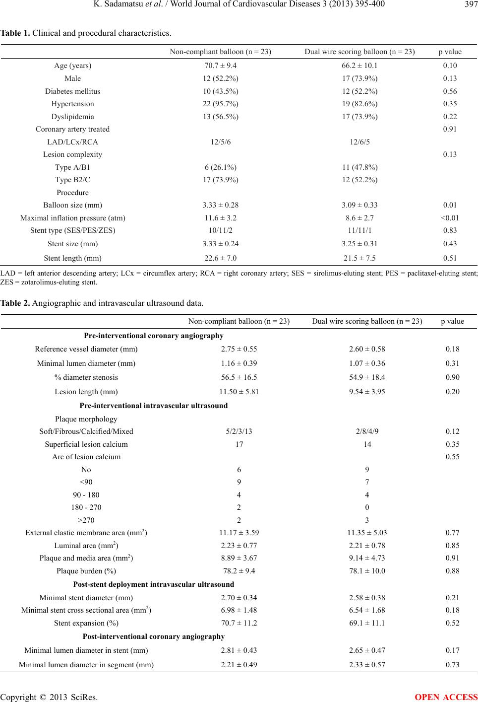 K. Sadamatsu et al. / World Journal of Cardiovascular Diseases 3 (2013) 395-400 Copyright © 2013 SciRes. 397 OPEN ACCESS Table 1. Clinical and procedural characteristics. Non-compliant balloon (n = 23) Dual wire scoring balloon (n = 23) p value Age (years) 70.7 ± 9.4 66.2 ± 10.1 0.10 Male 12 (52.2%) 17 (73.9%) 0.13 Diabetes mellitus 10 (43.5%) 12 (52.2%) 0.56 Hypertension 22 (95.7%) 19 (82.6%) 0.35 Dyslipidemia 13 (56.5%) 17 (73.9%) 0.22 Coronary artery treated 0.91 LAD/LCx/RCA 12/5/6 12/6/5 Lesion complexity 0.13 Type A/B1 6 (26.1%) 11 (47.8%) Type B2/C 17 (73.9%) 12 (52.2%) Procedure Balloon size (mm) 3.33 ± 0.28 3.09 ± 0.33 0.01 Maximal inflation pressure (atm) 11.6 ± 3.2 8.6 ± 2.7 <0.01 Stent type (SES/PES/ZES) 10/11/2 11/11/1 0.83 Stent size (mm) 3.33 ± 0.24 3.25 ± 0.31 0.43 Stent length (mm) 22.6 ± 7.0 21.5 ± 7.5 0.51 LAD = left anterior descending artery; LCx = circumflex artery; RCA = right coronary artery; SES = sirolimus-eluting stent; PES = paclitaxel-eluting stent; ZES = zotarolimus-eluting stent. Table 2. Angiographic and intravascular ultrasound data. Non-compliant balloon (n = 23) Dual wire scoring balloon (n = 23) p value Pre-interventional coronary angiography Reference vessel diameter (mm) 2.75 ± 0.55 2.60 ± 0.58 0.18 Minimal lumen diameter (mm) 1.16 ± 0.39 1.07 ± 0.36 0.31 % diameter stenosis 56.5 ± 16.5 54.9 ± 18.4 0.90 Lesion length (mm) 11.50 ± 5.81 9.54 ± 3.95 0.20 Pre-interventional intravascular ultrasound Plaque morphology Soft/Fibrous/Calcified/Mixed 5/2/3/13 2/8/4/9 0.12 Superficial lesion calcium 17 14 0.35 Arc of lesion calcium 0.55 No 6 9 <90 9 7 90 - 180 4 4 180 - 270 2 0 >270 2 3 External elastic membrane area (mm2) 11.17 ± 3.59 11.35 ± 5.03 0.77 Luminal area (mm2) 2.23 ± 0.77 2.21 ± 0.78 0.85 Plaque and media area (mm2) 8.89 ± 3.67 9.14 ± 4.73 0.91 Plaque burden (%) 78.2 ± 9.4 78.1 ± 10.0 0.88 Post-stent deployment intravascular ultrasound Minimal stent diameter (mm) 2.70 ± 0.34 2.58 ± 0.38 0.21 Minimal stent cross sectional area (mm2) 6.98 ± 1.48 6.54 ± 1.68 0.18 Stent expansion (%) 70.7 ± 11.2 69.1 ± 11.1 0.52 Post-interventional coronary angiography Minimal lumen diameter in stent (mm) 2.81 ± 0.43 2.65 ± 0.47 0.17 Minimal lumen diameter in segment (mm) 2.21 ± 0.49 2.33 ± 0.57 0.73 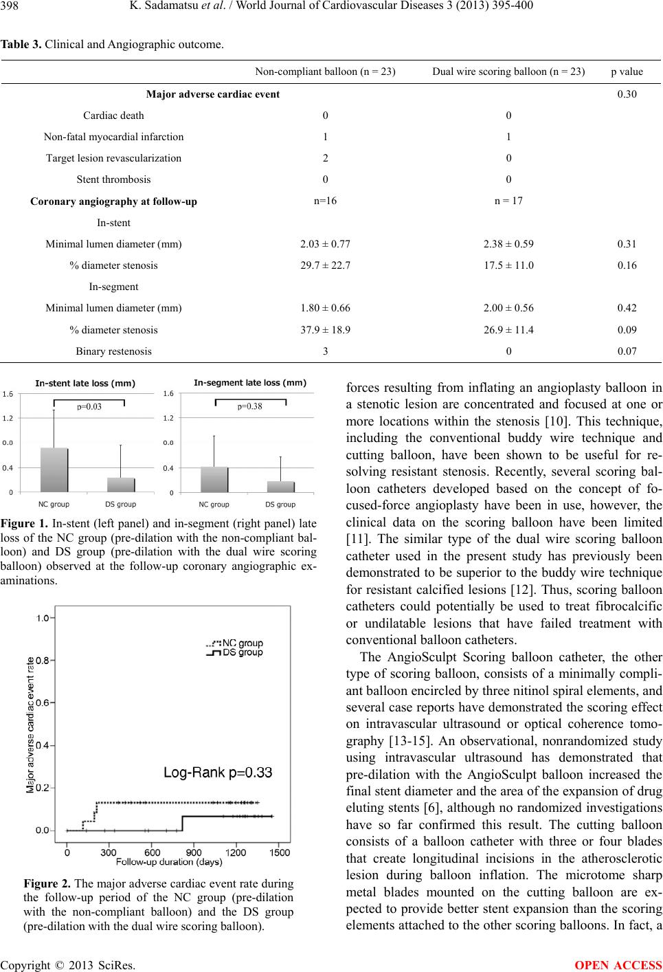 K. Sadamatsu et al. / World Journal of Cardiovascular Diseases 3 (2013) 395-400 398 Table 3. Clinical and Angiographic outcome. Non-compliant balloon (n = 23) Dual wire scoring balloon (n = 23) p value Major adverse cardiac event 0.30 Cardiac death 0 0 Non-fatal myocardial infarction 1 1 Target lesion revascularization 2 0 Stent thrombosis 0 0 Coronary angiography at follow-up n=16 n = 17 In-stent Minimal lumen diameter (mm) 2.03 ± 0.77 2.38 ± 0.59 0.31 % diameter stenosis 29.7 ± 22.7 17.5 ± 11.0 0.16 In-segment Minimal lumen diameter (mm) 1.80 ± 0.66 2.00 ± 0.56 0.42 % diameter stenosis 37.9 ± 18.9 26.9 ± 11.4 0.09 Binary restenosis 3 0 0.07 Figure 1. In-stent (left panel) and in-segment (right panel) late loss of the NC group (pre-dilation with the non-compliant bal- loon) and DS group (pre-dilation with the dual wire scoring balloon) observed at the follow-up coronary angiographic ex- aminations. Figure 2. The major adverse cardiac event rate during the follow-up period of the NC group (pre-dilation with the non-compliant balloon) and the DS group (pre-dilation with the dual wire scoring balloon). forces resulting from inflating an angioplasty balloon in a stenotic lesion are concentrated and focused at one or more locations within the stenosis [10]. This technique, including the conventional buddy wire technique and cutting balloon, have been shown to be useful for re- solving resistant stenosis. Recently, several scoring bal- loon catheters developed based on the concept of fo- cused-force angioplasty have been in use, however, the clinical data on the scoring balloon have been limited [11]. The similar type of the dual wire scoring balloon catheter used in the present study has previously been demonstrated to be superior to the buddy wire technique for resistant calcified lesions [12]. Thus, scoring balloon catheters could potentially be used to treat fibrocalcific or undilatable lesions that have failed treatment with conventional balloon catheters. The AngioSculpt Scoring balloon catheter, the other type of scoring balloon, consists of a minimally compli- ant balloon encircled by three nitinol spiral elements, and several case reports have demonstrated the scoring effect on intravascular ultrasound or optical coherence tomo- graphy [13-15]. An observational, nonrandomized study using intravascular ultrasound has demonstrated that pre-dilation with the AngioSculpt balloon increased the final stent diameter and the area of the expansion of drug eluting stents [6], although no randomized investigations have so far confirmed this result. The cutting balloon consists of a balloon catheter with three or four blades that create longitudinal incisions in the atherosclerotic lesion during balloon inflation. The microtome sharp metal blades mounted on the cutting balloon are ex- pected to provide better stent expansion than the scoring elements attached to the other scoring balloons. In fact, a Copyright © 2013 SciRes. OPEN ACCESS  K. Sadamatsu et al. / World Journal of Cardiovascular Diseases 3 (2013) 395-400 399 randomized study showed that the cutting balloon yiel- ded greater luminal diameters with less inflation pressure compared with conventional balloon angioplasty for the treatment of calcified coronary lesions [1]. However, this advantage was not demonstrated for the treatment of non- calcified lesions. The present study also did not demon- strate significant superiority of the pre-dilation with a dual wire scoring balloon in terms of the stent area or expansion, although the smaller balloon size and the lower inflation pressure used in the DS group might have affected the acute gain. Therefore, it remains unclear whether pre-dilation by a scoring balloon with the same balloon size would lead to a larger stent expansion than the pre-dilation using a conventional balloon. The less traumatic ballooning with the smaller balloon size and the lower inflation pressure associated with the DS group might therefore have led to the significantly less in-stent late loss observed in this study. A high infla- tion pressure is important with regard to the risk of ves- sel stretching and edge injuries [16]. Several animal and human studies have suggested that aggressive stent infla- tion with high pressures caused deeper injury of the ves- sel wall, with rupture of the intima or media, and might result in a long-term inflammatory response with a greater neointimal proliferative response and an increased re- stenosis rate [17,18]. Moreover, a SIRIUS substudy sug- gested that more injury to the contiguous vasoelastic nor- mal wall, coupled with a drug that delays the healing process, could contribute to late stent malapposition ow- ing to focal positive vessel remodeling [19]. Therefore, the less traumatic ballooning using the dual wire scoring balloon might be a more feasible strategy for pre-dilation prior to stent implantation than conventional ballooning in order to obtain an equivalent acute gain, while also re- sulting in less late loss. Further investigations are needed to confirm the effectiveness of this less traumatic bal- looning strategy. A large-scale randomized study investigating the use- fulness of the lesion preparation prior to coronary stent implantation is lacking, while rotational atherectomy or a cutting balloon have been demonstrated to yield a greater acute gain in selected cases [1-4]. A high-pressure pre- dilation stenting strategy was also shown to lead to supe- rior stent expansion compared to a direct or non-agres- sive pre-dilation strategy in patients with bare-metal stent restenosis in a non-randomized study [20]. These results suggest that the lesion preparation prior to coronary sten- ting may modify the vessel compliance and conesquen- tly improve the stent expansion. In fact, in some cases, lesion preparation is absolutely necessary to avoid stent thrombosis and restenosis due to stent underexpansion. In daily practice, it is difficult to differentiate the cases needing lesion preparation prior to the procedure, and stent underexpansion is often undetectable angiographi- cally. Therefore, high-pressure pre-dilation seems to be one of the most practical strategies to gain appropriate stent expansion, and the present study supports pre-dila- tion with a dual wire scoring balloon because it is an easy and feasible option. There are some important limitations asociated with our study: 1) our study involved a small number of sub- jects at a single institution; 2) the drug-eluting stents used in the present study were first-generation stents and are no longer being used as contemporary coronary stents. The improved characteristics of the stent platform might minimize the validity of the plaque modification prior to stent implantation; 3) follow-up coronary angiography was not performed in all of the patients, and thus, the follow-up results might be biased to some degree. 5. CONCLUSION In this study, pretreatment using a dual wire scoring bal- loon was found to be associated with less in-stent late loss at follow-up coronary angiography compared to that using a non-compliant balloon, although the lesion pre- paration did not demonstrate any significant superiority in acute stent expansion. 6. ACKNOWLEDGEMENTS The authors appreciate the support and collaboration of our catheteriza- tion laboratory staff, and also acknowledge Dr. Takafumi Koga and Dr. Shujiro Inoue for collecting the clinical follow-up data. The authors recevied a funding for the preparation of this article from Saga Prefec- tural Hospital Koseikan, which is the previous name of Saga-ken Medical Centre Koseikan. The authors have no conflicts of interest that are directly relevant to the content of this study. REFERENCES [1] Okura, H., Hayase, M., Shimodozono, S., Kobayashi, T., Sano, K., Matsushita, T., Kondo, T., Kijima, M., Nishi- kawa, H., Kurogane, H., Aizawa, T., Hosokawa, H., Su- zuki, T., Yamaguchi, T., Bonneau. H.N., Yock, P.G. and Fitzgerald, P.J. (2002) Mechanisms of acute lumen gain following cutting balloon angioplasty in calcified and noncalcified lesions: An intravascular ultrasound study. Catheterization & Cardiovascular Interventions, 57, 429- 436. doi:10.1002/ccd.10344 [2] Furuichi, S., Tobaru, T., Asano, R., Watanabe, Y., Taka- misawa, I., Seki, A., Sumiyoshi, T. and Tomoike, H. (2012) Rotational atherectomy followed by cutting-ba- lloon plaque modification for drug-eluting stent implan- tation in calcified coronary lesions. Journal of Invasive Cardiology, 24, 191-195. [3] Ozaki, Y., Yamaguchi, T., Suzuki, T., Nakamura, M., Ki- tayama, M., Nishikawa, H., Inoue, T., Hara, K., Usuba, F., Sakurada, M., Awano, K., Matsuo, H., Ishiwata, S., Ya- sukawa, T. and Ismail, T.F., Hishida, H. and Kato, O. (2007) Impact of cutting balloon angioplasty (CBA) prior Copyright © 2013 SciRes. OPEN ACCESS  K. Sadamatsu et al. / World Journal of Cardiovascular Diseases 3 (2013) 395-400 Copyright © 2013 SciRes. 400 OPEN ACCESS to bare metal stenting on restenosis. Circulation Journal, 71, 1-8. doi:10.1253/circj.71.1 [4] Tsuchikane, E., Otsuji, S., Awata, N., Azuma, J., Na- kaoka, Y., Uesugi, H., Kobayashi, T., Sakurai, M. and Kobayashi, T. (2001) Impact of pre-stent plaque debulk- ing for chronic coronary total occlusions on restenosis reduction. Journal of Invasive Cardiology, 13, 584-589. [5] Karvouni, E., Stankovic, G., Albiero, R., Takagi, T., Cor- vaja, N., Vaghetti, M., Di Mario, C. and Colombo, A. (2001) Cutting balloon angioplasty for treatment of calci- fied coronary lesions. Catheterization & Cardiovascular Interventions, 54, 473-481. doi:10.1002/ccd.1314 [6] de Ribamar Costa Jr., J., Mintz, G.S., Carlier, S.G., Mehran, R., Teirstein, P., Sano, K., Liu, X., Lui, J., Na, Y., Castellanos, C., Biro, S., Dani, L., Rinker, J., Moussa, I., Dangas, G., Lansky, A.J., Kreps, E.M., Collins, M., Stones, G.W., Moses, J.W. and Leon, M.B. (2007) Non- randomized comparison of coronary stenting under intra- vascular ultrasound guidance of direct stenting without predilation versus conventional predilation with a semi- compliant balloon versus predilation with a new scoring balloon. American Journal of Cardiology, 100, 812-817. doi:10.1016/j.amjcard.2007.03.100 [7] Kawasaki, T., Koga, H., Serikawa, T., Orita, Y., Ikeda, S., Mito, T., Gotou, Y., Shintani, Y., Tanaka, A., Tanaka, H., Fukuyama, T. and Koga, N. (2009) Impact of a prolonged delivery inflation time for optimal drug-eluting stent ex- pansion. Catheterization & Cardiovascular Interventions, 73, 205-211. doi:10.1002/ccd.21813 [8] Asano, T., Kobayashi, Y., Fukushima, K., Iwata, Y., Ki- tahara, H., Ishio, N., Nakayama, T., Kuroda, N. and Ko- muro, I. (2009) Effect of balloon inflation time on expan- sion of sirolimus-eluting stent. Heart and Vessels, 24, 335-339. doi:10.1007/s00380-008-1130-1 [9] Iwamoto, Y., Okamoto, M., Hashimoto, M., Fukuda, Y., Iwamoto, A., Iwasaki, T., Kinoshita, H. and Kihara, Y. (2012) Better stent expansion by two-time inflation of stent balloon and its responsible mechanism. Journal of Cardiology, 59, 160-166. doi:10.1016/j.jjcc.2011.12.003 [10] Solar, R.J. and Ischinger, T.A. (2003) Focused force angioplasty: Theory and application. Cardiovascular Ra- diation Medicine, 4, 47-50. doi:10.1016/S1522-1865(03)00119-7 [11] Kato, R., Ashikaga, T., Sakurai, K., Ito, J., Ogawa, T., Tahara, T., Yokoyama, Y. and Satoh, Y. (2012) Influ- ence of additional ballooning with a dual wire balloon after a rotational atherectomy to expand drug-eluting stent for calcified lesions. Cardiovascular Interve ntion and Th e- rapeutics, 27, 155-160. doi:10.1007/s12928-012-0102-7 [12] Otsuka, Y., Kasahara, Y. and Kawamura, A. (2007) Use of safecut balloon for treatment of in-stent restenosis of a previously underexpanded sirolimus-eluting stent with a heavily calcified plaque. Journal of Invasive Cardiology, 19, E359-E362. [13] Kanai, T., Hiro, T., Takayama, T., Fukamachi, D., Wata- nabe, Y., Ichikawa, M., Kawano, T. and Hirayama, A. (2012) Three-dimensional visualization of scoring me- chanism of “Angiosculpt” balloon for calcified coronary lesions using optical coherence tomography. Journal of Cardiology Cases, 5, e16-e19. doi:10.1016/j.jccase.2011.10.008 [14] Takano, M., Yamamoto, M., Murakami, D., Takano, H., Asai, K., Yasutake, M., Seino, Y. and Mizuno, K. (2010) Optical coherence tomography after new scoring balloon angioplasty for in-stent restenosis and de novo coronary lesions. International Journal of Cardiology, 141, e51- e53. doi:10.1016/j.ijcard.2008.11.154 [15] Hosokawa, Y., Tanaka, K. and Mizuno, K. (2012) Succe- ssful treatment for refractory coronary thrombus with scoring balloon angioplasty. Catheterization & Cardiova- scular Interventions, 79, 282-287. doi:10.1002/ccd.23073 [16] Caixeta, A.M., Brito, F.S.Jr., Rati, M., Perin, M.A., da Luz, P.L., Ramires, J.A., Ambrose, J.A. and Martinez, E.E. (2000) High versus low-pressure balloon inflation during MultilinkTM stent implantation: Acute and long- term angiographic results. Catheterization & Cardiovas- cular Interventions, 50, 398-401. doi:10.1002/1522-726X(200008)50:4<398::AID-CCD5> 3.0.CO;2-1 [17] Uretsky, B.F., Rosanio, S., Lerakis, S., Wang, F.W., Smi- ley, M., Stouffer, G.A., Tocchi, M., and Estella, P. (2000) A prospective evaluation of angiography-guided coronary stent implantation with high versus very high balloon in- flation pressure. American Heart Journal, 140, 804-812. doi:10.1067/mhj.2000.110572 [18] Hoffmann, R., Guagliumi, G., Musumeci, G., Reimers, B., Petronio, A.S., Disco, C., Amoroso, G., Moses, J.W., Fi- tzgerald, P.J., Schofer, J., Leon, M.B. and Breithardt, G. (2005) Vascular response to sirolimus-eluting stents de- livered with a nonaggressive implantation technique: Com- parison of intravascular ultrasound results from the mul- ticenter, randomized E-SIRIUS, and SIRIUS trials. Ca- theterization & Cardiovascular Interventions, 66, 499- 506. doi:10.1002/ccd.20542 [19] Ako, J., Morino, Y., Honda, Y., Hassan, A., Sonoda, S., Yock, P.G., Leon, M.B., Moses, J.W., Bonneau, H.N. and Fitzgerald, P.J. (2005) Late incomplete stent apposition after sirolimus-eluting stent implantation: A serial intra- vascular ultrasound analysis. Journal of the American College of Cardiology, 46, 1002-1005. doi:10.1016/j.jacc.2005.05.068 [20] Kalinczuk, L., Demkow, M., Mintz, G.S., Cedro, K., De- bski, A., Ciszewski, M., Ciszewski, A., Kruk, M., Karcz, M., Warminski, G., Pregowski, J., Chmielak, Z., Witkow- ski, A., Lubiszewska, B. and Ruzyllo, W. (2009) Impact of different re-stenting strategies on expansion of a drug- eluting stent implanted to treat bare-metal stent restenosis. American Journal of Cardiology, 104, 531-537. doi:10.1016/j.amjcard.2009.03.067 |