

Paper Menu >>
Journal Menu >>
 World Journal of Cardiovascular Diseases, 2013, 3, 389-393 WJCD http://dx.doi.org/10.4236/wjcd.2013.35060 Published Online August 2013 (http://www.scirp.org/journal/wjcd/) 3-D multidetector computed tomography in reoperative cardiac surgery Tsuyoshi Kaneko1, Micheal L. Steigner2, Igor Gosev1, Sary F. Aranki1* 1Department of Cardiac Surgery, Brigham and Women’s Hospital, Boston, USA 2Department of Radiology, Brigham and Women’s Hospital, Boston, USA Email: *[email protected] Received 19 May 2013; revised 27 June 2013; accepted 14 July 2013 Copyright © 2013 Tsuyoshi Kaneko et al. This is an open access article distributed under the Creative Commons Attribution License, which permits unrestricted use, distribution, and reproduction in any medium, provided the original work is properly cited. ABSTRACT Reoperative cardiac surgery is becoming more com- mon surgery although it carries significant risk due to possible injury to vital structures under the sternum. Three-dimensional multidetector computed tomogra- phic angiography (3-D MDCTA) allows identifying the relationship between the sternum and the medias- tinal structures. Evidence shows that 3-D MDCTA guides surgical strategy and enables to perform this challenging surgery safely. Keywords: 3-D MDCTA; Reoperative Cardiac Surgery 1. INTRODUCTION Reoperative cardiac surgery carries a significantly higher risk compared to first time surgery. Several reasons exist; technical difficulties include reoperative sternotomy, me- diastinal dissection and higher risk in the reoperative population. However, the cardiac surgeons are facing more and more reoperations, given the longer expectancy in the modern era. The recently published mortality rate for reoperative cardiac surgery in previous coronary ar- tery bypass graft surgery (CABG) has ranged from 2.5% to 8.3% [1-7]. The major concern during sternal reentry in these patients is the potential injury to the cardiac structures (right ventricle), previous grafts, innominate vein and Aorta. Surgical strategies, such as interposition of the lungs in the left internal mammary artery (LIMA) [8] or no dissection technique [9] have been described in the literature, but do not eliminate the chances of injury to these structures. Contrast-enhanced multidetector computed tomographic angiography (MDCTA) has emerged as a useful method to evaluate heart and mediatinal structures [10-11]. In addition, three-dimensional (3-D) volume rendering al- lows detailed identification of the previous grafts as well as the right ventricle and Aorta. The aim of this paper is to support the usage of 3-D MDCTA for preoperative evaluation for reoperative car- diac surgery. 2. METHODS 2.1. 3-D MDCTA State-of-the-art MDCTA for redo sternotomy typically is performed with 64 - 320 detector row CT scanners. Helical acquisitions are performed from above the clavi- cular heads to the base of the heart with thin slice colli- mation (0.5 - 1.0 mm) at either 100 - 120 kV and at least 350 mA. Current scanners employ z-axis tube current modulation as an option for the mA adjustment. Since small vessel CT angiography requires high contrast to noise ratios, the highest available concentration of con- trast (370 mg/dL) is injected at high flow rates (4.5 - 6 ml/sec). Volume of contrast is determined by the scan time varying from 80 - 100 ml. B-blockade for heart rate control and nitroglycerine for vasodilation are typically not required for the assessment of vessel patency and course; however, these techniques are favored if stenosis evaluation of the prior bypass grafts is paramount. Since bypass grafts don’t move with the same velocity as na- tive coronary arteries, acquisitions can typically be per- formed with prospective gating with a single phase of the cardiac cycle. Dynamic assessment of retrosternal struc- tures has recently been described as an adjunct to the proximity assessment <ref Malguria, et al. “Static and Cine CT Imaging to Identify and Characterize Mediasti- nal Adhesions as a Potential Complication Undergoing Redo Sternotomy”. AJR ACCEPTED FOR PUBLICA- TION NOT YET IN PRESS. This technique requires retrospective gating at the expense of a higher radiation exposure; however, additional studies are required to determine its value. *Corresponding author. OPEN ACCESS  T. Kaneko et al. / World Journal of Cardiovascular Diseases 3 (2013) 389-393 390 2.2. Interpretation of the 3-D MDCTA Preoperative evaluation of 3-D MDCTA allows the fol- lowing assessment. 1) Proximity of LIMA and Other Grafts in Relation to the Sternum. 2) Adhesion of the Right Ventricle (RV) to the Sternal Bed (Especially in Light of Pulmonary Hyper Tension). 3) Location of the Innominate Vein and Aorta. Distance of <1 cm from the sternum to the Aorta, RV and CABG graft crossing the midline within 1 cm of posterior to the sternumwere typically used for high risk upon sternal reentry [12-14]. A survey of 2046 significant bleeding during sternal entry showed that the most commonly injured structures were RV (39%), Saphenous vein graft (SVG: 20%), Aorta (15%), IMA (12%) and innominate vein (6%) [15]. Radiologist and Surgeon must keep this in mind to find the relationship between the sternum and these structures when 3-D MDCTA is assessed preoperatively. 2.3. Operative Strategy Based on 3-D MDCTA After assessing these structures, surgeons will consider the following for operative planning. 2.3.1. Cannul ation Str ategy 1) Direct central cannulation. 2) Guidewire placement for emergency. 3) Peripheral cannulation. a) Femoral artery cannulation. b) Axillary cannulation. First step of operative decision making is cannulation strategy. Cannulation strategy must be decided prior to incision with a backup plan. If there is good distance from the sternum, sternotomy can be performed and di- rect central cannulation can be performed following me- diastinal dissection. Mediastinal dissection should be performed around the Aorta and right atrium first so that cardiopulmonary bypass (CPB) can be established in case of emergency. It is our common practice to place guidewire in femo- ral artery and femoral vein to prepare for emergency. Guidewire is inserted under transesophageal echocardio- gram (TEE) guidance. If any structure is injured during sternal reentry, sternum is approximated and CPB is es- tablished following Seldinger technique placement of arterial and venous cannula. If adhesions present, peripheral cannulation should be considered. Femoral artery/vein can be used. Axillary artery can be used when femoral artery use is concern (peripheral vascular disease, obesity etc). Our practice is to place 8 - 10 mm dacron graft to preserve distal flow. 2.3.2. Cardio pulmonary By p ass 1) Go on cardiopulmonary bypass prior to sternotomy. 2) Deep Hypothermic circulatory arrest. 3) No cardiopulmonary bypass. In severe adhesions, consideration of establishing CPB prior to sternotomy should be made. This will allow de- compression of RV and decreases the chances of injury during sternal reentry. Luciani, et al. reported their ex- perience with 158 patients who underwent cardiopul- monary bypass prior to creating sternotomy [16]. This decision was made on the CT evidence of dense retros- ternal adhesions, depressed ejection fraction or previous mediastinitis. Use of CPB prior to sternotomy was asso- ciated with less re-entry injuries, reduced operative time, less postoperative bleeding and shorter ICU stay. Deep hypothermic circulatory arrest can be used for extreme cases with severe adhesion or injury to the aorta occurred. For low risk cases, no CPB will allow blood less dissection since no heparin is used prior to dissec- tion. 2.3.3. Sternotomy vs Non-Sternotomy 1) Reoperative sternotomy-grafts in relation to the sternal wires. 2) Anterior thoracotomy. 3) Transcatheter treatment. Access strategy can be altered depending on the op- eration being performed. If prior CABG patient with significant graft adhesion is undergoing mitral valve sur- gery, literature supports use of right thoracotomy for ac- cess [17]. Left thoracotomy can be used for reoperative CABG to circumflex artery and avoiding sternal entry [18]. If the patient has high risk comorbidity and high risk findings on 3-D MDCTA, consideration of transcatheter treatment (percutaneous coronary intervention for coro- nary disease, transcatheter valve replacements for valve disease) if indicated. 2.3.4. C anc ellation When the findings show extremely high risk, surgeon can decide to cancel the case. 3-D-CTMDA will aid this decision making. Report from Cleveland Clinic de- scribed 4% cancellation in their 167 adult reoperative cardiac surgery series [14]. Patent RIMA with additional jump graft supplying a left dominant system crossing the sternal midline at an unsafe distance in a severe valvular disease patient was one of the examples used in this se- ries. Other series report 6% - 12% cancellation rate in their series [13,19]. 2.4. Patient Images Figures 1(a) and (b) show the 3-D MDCTA on 65 years old male who underwent CABG (SVG to posterior de- scending artery) and came back for aortic valve replace- ment (AVR). Preoperative MDCTA showed SVG graft Copyright © 2013 SciRes. OPEN ACCESS 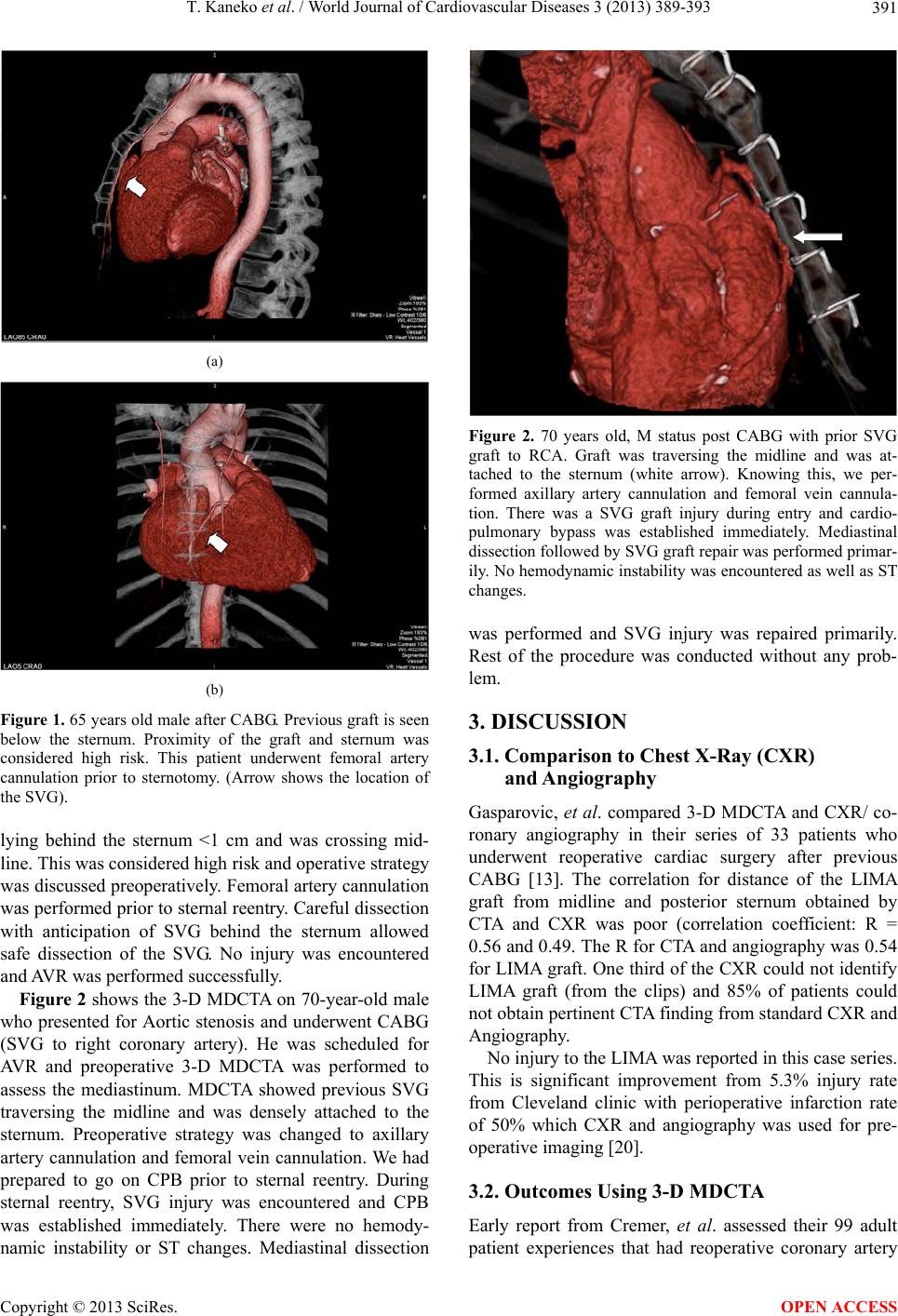 T. Kaneko et al. / World Journal of Cardiovascular Diseases 3 (2013) 389-393 391 (a) (b) Figure 1. 65 years old male after CABG. Previous graft is seen below the sternum. Proximity of the graft and sternum was considered high risk. This patient underwent femoral artery cannulation prior to sternotomy. (Arrow shows the location of the SVG). lying behind the sternum <1 cm and was crossing mid- line. This was considered high risk and operative strategy was discussed preoperatively. Femoral artery cannulation was performed prior to sternal reentry. Careful dissection with anticipation of SVG behind the sternum allowed safe dissection of the SVG. No injury was encountered and AVR was performed successfully. Figure 2 shows the 3-D MDCTA on 70-year-old male who presented for Aortic stenosis and underwent CABG (SVG to right coronary artery). He was scheduled for AVR and preoperative 3-D MDCTA was performed to assess the mediastinum. MDCTA showed previous SVG traversing the midline and was densely attached to the sternum. Preoperative strategy was changed to axillary artery cannulation and femoral vein cannulation. We had prepared to go on CPB prior to sternal reentry. During sternal reentry, SVG injury was encountered and CPB was established immediately. There were no hemody- namic instability or ST changes. Mediastinal dissection Figure 2. 70 years old, M status post CABG with prior SVG graft to RCA. Graft was traversing the midline and was at- tached to the sternum (white arrow). Knowing this, we per- formed axillary artery cannulation and femoral vein cannula- tion. There was a SVG graft injury during entry and cardio- pulmonary bypass was established immediately. Mediastinal dissection followed by SVG graft repair was performed primar- ily. No hemodynamic instability was encountered as well as ST changes. was performed and SVG injury was repaired primarily. Rest of the procedure was conducted without any prob- lem. 3. DISCUSSION 3.1. Comparison to Chest X-Ray (CXR) and Angiography Gasparovic, et al. compared 3-D MDCTA and CXR/ co- ronary angiography in their series of 33 patients who underwent reoperative cardiac surgery after previous CABG [13]. The correlation for distance of the LIMA graft from midline and posterior sternum obtained by CTA and CXR was poor (correlation coefficient: R = 0.56 and 0.49. The R for CTA and angiography was 0.54 for LIMA graft. One third of the CXR could not identify LIMA graft (from the clips) and 85% of patients could not obtain pertinent CTA finding from standard CXR and Angiography. No injury to the LIMA was reported in this case series. This is significant improvement from 5.3% injury rate from Cleveland clinic with perioperative infarction rate of 50% which CXR and angiography was used for pre- operative imaging [20]. 3.2. Outcomes Using 3-D MDCTA Early report from Cremer, et al. assessed their 99 adult patient experiences that had reoperative coronary artery Copyright © 2013 SciRes. OPEN ACCESS 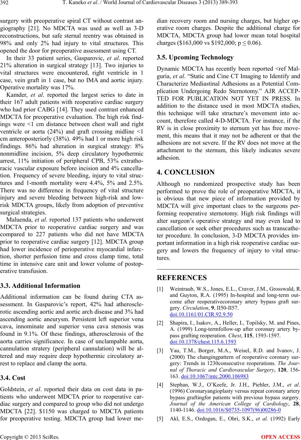 T. Kaneko et al. / World Journal of Cardiovascular Diseases 3 (2013) 389-393 392 surgery with preoperative spiral CT without contrast an- giography [21]. No MDCTA was used as well as 3-D reconstructions, but safe sternal reentry was obtained in 98% and only 2% had injury to vital structures. This opened the door for preoperative assessment using CT. In their 33 patient series, Gasparovic, et al. reported 21% alteration in surgical strategy [13]. Two injuries to vital structures were encountered, right ventricle in 1 case, vein graft in 1 case, but no IMA and aortic injury. Operative mortality was 17%. Kamder, et al. reported the largest series to date in their 167 adult patients with reoperative cardiac surgery who had prior CABG [14]. They used contrast enhanced MDCTA for preoperative evaluation. The high risk find- ings were <1 cm distance between chest wall and right ventricle or aorta (24%) and graft crossing midline <1 cm anteroposteriorly (38%). 49% had 1 or more high risk findings. 86% had alteration in surgical strategy: 8% nonmidline incision, 5% deep circulatory hypothermic arrest, 11% initiation of peripheral CPB, 53% extratho- racic vascular exposure before incision and 4% cancella- tion. Frequency of severe bleeding, injury to vital struc- tures and 1-month mortality were 4.4%, 5% and 2.5%. There was no difference in frequency of vital structure injury and severe bleeding between high-risk and low- risk MDCTA groups, likely from adoption of preventive surgical strategies. Maluenda, et al. reported 137 patients who underwent MDCTA prior to reoperative cardiac surgery and was compared to 227 patients who did not have MDCTA prior to reoperative cardiac surgery [12]. MDCTA group had lower incidence of perioperative myocardial infarc- tion, shorter perfusion time and cross clamp time, total time in intensive care unit and lower volume of postop- erative transfusion. 3.3. Additional Information Additional information can be found during CTA as- sessment. In Gasparovic’s report, 42% had atheroscle- rotic ascending aortic and aortic arch disease and 3% had ascending aortic aneurysm. Persistent left superior vena cava, innominate and superior vena cava stenosis was found in 9.1%. Of these findings, atherosclerosis of the aorta carries significance. In case of unclampable aorta, cannulation stratery (peripheral cannulation) will be al- tered and may require deep hypothermic circulatory ar- rest to replace and clamp the aorta. 3.4. Cost Goldstein, et al. reported their data on cost data in pa- tients who underwent MDCTA prior to reoperative car- diac surgery and compared to group who did not undergo MDCTA [22]. $1150 was charged to MDCTA patients for preoperative testing. MDCTA group had lower me- dian recovery room and nursing charges, but higher op- erative room charges. Despite the additional charge for MDCTA, MDCTA group had lower mean total hospital charges ($163,000 vs $192,000; p ≤ 0.06). 3.5. Upcoming Technology Dynamic MDCTA has recently been reported <ref Mal- guria, et al. “Static and Cine CT Imaging to Identify and Characterize Mediastinal Adhesions as a Potential Com- plication Undergoing Redo Sternotomy.” AJR ACCEP- TED FOR PUBLICATION NOT YET IN PRESS. In addition to the distance used in most MDCTA studies, this technique will take structure’s movement into ac- count, therefore called 4-D-MDCTA. For instance, if the RV is in close proximity to sternum yet has free move- ment, this means that it may not be adherent or that the adhesions are not severe. If the RV does not move at the attachment to the sternum, this likely indicates severe adhesion. 4. CONCLUSION Although no randomized prospective study has been performed to prove the role of preoperative MDCTA, it is obvious that new piece of information provided by MDCTA will give important clues to the surgeons per- forming reoperative sternotomy. High risk findings will alter surgeon’s operative strategy and may even lead to cancellation or seek other procedures such as transcathe- ter procedure. In conclusion, 3-D MDCTA provides im- portant information in a high risk reoperative cardiac sur- gery and lowers the frequency of injury to vital struc- tures. REFERENCES [1] Weintraub, W.S., Jones, E.L., Craver, J.M., Grosswald, R. and Guyton, R.A. (1995) In-hospital and long-term out- come after reoperativecoronary artery bypass graft sur- gery. Circulation, 9, II50-II57. doi:10.1161/01.CIR.92.9.50 [2] Shapira, I., Isakov, A., Heller, I., Topilsky, M. and Pines, A. (1999) Long-termfollow-up after coronary artery by- pass grafting reoperation. Chest, 115, 1593-1597. doi:10.1378/chest.115.6.1593 [3] Yau, T.M., Borger, M.A., Weisel, R.D. and Ivanov, J. (2000) The changingpattern of reoperative coronary sur- gery: Trends in 1230consecutive reoperations. The Jour- nal of Thoracic and Cardiovascular Surgery, 120, 156- 163. doi:10.1067/mtc.2000.106983 [4] Stephan, W.J., O’Keefe, Jr. J.H., Piehler, J.M., et al. (1996) Coronaryangioplasty versus repeat coronary artery bypass graftingfor patients with previous bypass surgery. Journal of the American College of Cardiology, 28, 1140-1146. doi:10.1016/S0735-1097(96)00286-0 [5] Akl, E.S., Ozdogan, E., Ohri, S.K., et al. (1992) Early Copyright © 2013 SciRes. OPEN ACCESS  T. Kaneko et al. / World Journal of Cardiovascular Diseases 3 (2013) 389-393 Copyright © 2013 SciRes. 393 OPEN ACCESS and long termresults of re-operation for coronary artery disease. British Heart Journal, 68, 176-180. doi:10.1136/hrt.68.8.176 [6] Fitzgibbon, G.M., Kafka, H.P., Leach, A.J., Keon, W.J., Hooper, G.D. and Burton, J.R. (1996) Coronary bypass graft fate and patient outcome: Angiographic follow-up of 5065 grafts related to survival andreoperation in 1388 patients during 25 years. Journal of the American College of Cardiology, 28, 616-626. doi:10.1016/0735-1097(96)00206-9 [7] Shimada, Y., Dixit, A., Fermanis, G. and Horton, D. (1998) Reoperation forrecurrent coronary artery disease: Results of 200 consecutivecases. Australian and New Zealand Journal of Surgery, 68, 729-734. doi:10.1111/j.1445-2197.1998.tb04661.x [8] Fullerton, D.A., St Cyr, J.A., Fall, S.M. and Whitman, G.J. (1994) Protection of the patent internalmammary ar- tery bypass graft from subsequent sternotomy. The Jour- nal of Cardiovascular Surgery (Torino), 35, 499-501. [9] Kaneko, T., Nauta, F., Borstlap, W., McGurk, S., Couper, G. and Cohn, L. (2012) The “no-dissection” technique is safe for reoperative aortic valvereplacement with a patent left internal thoracic artery graft. The Journal of Thoracic and Cardiovascular Surgery, 144, 1036-1041. doi:10.1016/j.jtcvs.2012.07.057 [10] Song, M.H., Ito, T., Watanabe, T., Nakamura, H. (2005) Multidetectorcomputed tomography versus coronary an- giogram in evaluationof coronary artery bypass grafts. The Annals of Thoracic Surgery, 79, 585-588. doi:10.1016/j.athoracsur.2004.08.014 [11] Anders, K., Baum, U., Schmid, M., et al. (2006) Coro- nary artery bypassgraft (CABG) patency: Assessment with high-resolution submillimeter16-slice multidetector- row computed tomography (MDCT) versus coronary an- giography. European Journal of Radiology, 57, 336-344. doi:10.1016/j.ejrad.2005.12.018 [12] Maluenda, G., Goldstein, M.A., Lemesle, G., Weissman, G., Weigold, G., Landsman, M.J., Hill, P.C., Pita, F., Corso, P.J., Boyce, S.W., Pichard, A.D., Waksman, R. and Taylor, A.J. (2010) Perioperative outcomes in reop- erative cardiac surgery guided by cardiac multidetector computed tomographic angiography. American Heart Journal, 159, 301-306. doi:10.1016/j.ahj.2009.11.005 [13] Gasparovic, H., Rybicki, F.J., Millstine, J., Unic, D., Byrne, J.G., Yucel, K. and Mihaljevic, T. (2005) Three dimensional computedtomographic imaging in planning the surgical approach for redo cardiac surgery after coro- nary revascularization. European Journal Cardio-Tho- racic Surgery, 28, 244-249. doi:10.1016/j.ejcts.2005.03.024 [14] Kamdar, A.R., Meadows, T.A., Roselli, E.E., Gorodeski, E.Z., Curtin, R.J., Sabik, J.F., Schoenhagen, P., White, R.D., Lytle, B.W., Flamm, S.D. and Desai, M.Y. (2008) Multidetector computed tomographic angiography in plan- ning of reoperative cardiothoracic surgery. The Annals of Thoracic Surgery, 85, 1239-1245. doi:10.1016/j.athoracsur.2007.11.075 [15] Follis, F.M., Pett, Jr. S.B., Miller, K.B., Wong, R.S., Temes, R.T. and Wernly, J.A. (1999) Catastrophic hem- orrhage on sternal reentry: Still a dreaded complication? The Annals of Thoracic Surgery, 68, 2215-2219. doi:10.1016/S0003-4975(99)01173-X [16] Luciani, N., Anselmi, A., De Geest, R., Martinelli, L., Perisano, M. and Possati, G. (2008) Extracorporeal circu- lation by peripheral cannulation before redo sternotomy: Indications and results. The Journal of Thoracic and Car- diovascular Surgery, 136, 572-577. doi:10.1016/j.jtcvs.2008.02.071 [17] Byrne, J.G., Aranki, S.F., Adams, D.H., Rizzo, R.J., Cou- per, G.S. and Cohn, L.H. (1999) Mitral valve surgery af- ter previous CABG with functioning IMA grafts. The Annals of Thoracic Surgery, 68, 2243-2247. doi:10.1016/S0003-4975(99)01120-0 [18] Byrne, J.G., Aklog, L., Adams, D.H., Cohn, L.H. and Aranki, S.F. (2001) Reoperative CABG using left thora- cotomy: A tailored strategy. The Annals of Thoracic Sur- gery, 71, 196-200. doi:10.1016/S0003-4975(00)02182-2 [19] Aviram, G., Sharony, R., Kramer, A., Nesher, N., Lober- man, D., Ben-Gal, Y., Graif, M., Uretzky, G. and Mohr, R. (2005) Modification of surgical planning based on car- diac multidetector computed tomography in reoperative heart surgery. The Annals of Thoracic Surgery, 79, 589- 595. doi:10.1016/j.athoracsur.2004.07.012 [20] Gillinov, A.M., Casselman, F.P., Lytle, B.W., et al. (1999) Injury to a patent left internal thoracic artery graft at co- ronary reoperation. The Annals of Thoracic Surgery, 67, 382-386. doi:10.1016/S0003-4975(99)00009-0 [21] Cremer, J., Teebken, O.E., Simon, A., Hutzelmann, A., Heller, M. and Haverich, A. (1998) Thoracic computed tomography prior to redo coronary surgery. European Journal Cardio-Thoracic Surgery, 13, 650-654. doi:10.1016/S1010-7940(98)00087-6 [22] Goldstein, M.A., Roy, S.K., Hebsur, S., Maluenda, G., Weissman, G., Weigold, G., Landsman, M.J., Hill, P.C., Pita, F., Corso, P.J., Boyce, S.W., Pichard, A.D., Waks- man, R. and Taylor, A.J. (2013) Relationship between routine multi-de- tector cardiac computed tomographic angiogra- phy prior to reoperative cardiac surgery, length of stay, and hospital charges. The International Journal of Cardiovascular Imaging, 29, 709-717. doi:10.1007/s10554-012-0133-2 |