

Paper Menu >>
Journal Menu >>
 World Journal of Cardiovascular Diseases, 2013, 3, 21-25 WJCD http://dx.doi.org/10.4236/wjcd.2013.35A004 Published Online August 2013 (http://www.scirp.org/journal/wjcd/) Dual effect of pre-ischemic administration of TNF-alph a on myocardial infarct size Thuy Tran Quang1, Raja Hatem2, Guy Rousseau1,3, Audrey-Anne Gosselin1, Erick Schampaert1,2, Thierry Charron1,2 1Centre de Recherche, Hôpital du Sacré-Cœur de Montréal, Montréal, Canada 2Département de Cardiologie, Hôpital du Sacré-Cœur de Montréal, Montréal, Canada 3Département de Pharmacologie, Université de Montréal, Montréal, Canada Email: [email protected] Received 26 June 2013; revised 28 July 2013; accepted 8 August 2013 Copyright © 2013 Thuy Tran Quang et al. This is an open access article distributed under the Creative Commons Attribution License, which permits unrestricted use, distribution, and reproduction in any medium, provided the original work is properly cited. ABSTRACT Tumour necrosis factor-α is a cytokine released dur- ing myocardial infarction. According to the literature, the effect of TNFα on myocardial infarction is contro- versial, especially when administered before the is- chemic period. The deleterious effects of TNFα seem to be related to the triggering of apoptosis. This study has been designed to determine if different doses of TNFα, administered before the ischemic period, have the same effect on infarct size and on activation of ca- spase-3 and -8, two enzymes involved in apoptosis. Four groups, using a porcine model of myocardial in- farction, have been used: placebo and TNFα (0.1 µg/kg; 1 µg/kg and 3 µg/kg). All administered 15 minutes before a 50 minutes occlusion of the left anterior de- scending artery. Myocardial infarct size has been de- termined at 3 hours of reperfusion. In a subgroup of animals, reperfusion period has been limited to 15 min to determine the activity of caspase-3 and -8 by spectrofluorometry. Results indicated that infarct size is significantly smaller in groups 0.1 µg/kg and 1 µg/ kg as compared to the placebo group. In contrast, the 3 µg/kg group presented an infarct size similar to the placebo group. Activity of caspase-3 and -8 is reduc- ed in the ischemic region in groups 0.1 and 1 µg/ kg as compared to the placebo group whereas activity in the 3 µg/kg group was similar to the placebo. The re- sults obtained indicated that a low dose of TNFα ad- ministered before the ischemic period reduces infarct size, whereas the cardioprotection is lost with the high dose. Keywords: TNF-Alpha; Myocardial Infarct Size; Protection; Apoptosis; Caspase-8 1. INTRODUCTION TNFα is a multifunctional 26 kDa cytokine involved as a mediator of diverse physiologic and pathophysiologic events including inflammation, growth, differentiation and apoptosis. TNFα is thought to contribute to the deve- lopment of myocardial disease, with direct correlation between plasmatic concentrations of TNFα and the se- verity and progression of heart failure [1-3]. However, recent clinical trials of anti-TNFα therapy for cardiovas- cular disease have been disappointing [4] possibly due to the dual role of TNFα in attenuation and aggravation of cardiac injury [5]. Indeed, some studies have shown that TNFα was cardioprotective [6,7] whereas others have shown that TNFα has either no effect or possibly dele- terious effects [8,9]. One of the best known effect of TNFα is its invol- vement in apoptosis [1]. When activated by TNFα, the TNF receptor 1 can activate the formation of a complex that leads to the activation of caspase-8. Nonetheless, it seems also possible that the stimulation of the TNFR1 induces the activation of enzymes involved in different cardioprotective pathways [10]. We have speculated that depending on the dose of TNFα administered before the onset of ischemia, the ef- fect on myocardial infarct size could be altered. To deter- mine the mechanism by which the different doses of TNFα can modulate the myocardium damage, activation of the caspase-3 and -8 was measured. 2. MATERIALS AND METHODS 2.1. Experimental Model Experiments were performed in accordance with the Ca- nadian Council for Animal Care guidelines and were ap- proved by the Institutional Animal Care Committee. OPEN ACCESS  T. T. Quang et al. / World Journal of Cardiovascular Diseases 3 (2013) 21-25 22 Male pigs weighing from 35 to 40 kg were used to create a porcine model of myocardial infarction. Animals were anaesthetized with intramuscular ketamine-xylazine (20 mg/kg - 2 mg/kg; Wyeth Pharmaceutical, Montreal, Ca- nada), intubated and ventilated with positive pressure ventilation (Ohmeda 7800 anaesthesia ventilator, DRE Medical, Louisville, KY) at a fraction of inspired oxygen maintained constant at 20% - 30%. Anaesthesia was main- tained with 2% isoflurane (Abbott Laboratory, Montreal, Canada) mixed in O2:N2O (2:1). The left internal jugular vein was cannulated with a 9F vascular sheath and used as an access for intravenous fluid. Fluid repletion was maintained by an I.V. infusion of 0.9% sodium chloride solution at a rate of 10 ml/kg/h. The right carotid artery was cannulated with an 8F vascular sheath and used for systemic artery pressure monitoring (8-channel poly- graph; Nihon Kohden America, Foothill Ranch, CA). 2.2. Induction of Infarct Size The procedure was performed via the right femoral ar- tery with an 8F vascular sheath using an 8F AL1 coro- nary guiding catheter. An IQ angioplasty guide wire (Bo- ston Scientific, Natick, Massachusetts, USA) was advan- ced in the left anterior descending (LAD), distally to the second diagonal, followed by the insertion of a 3.0 × 15 mm balloon catheter (ApexTM, over-the-wire) (Boston Sci- entific). The balloon catheter was inflated at 4 atm for a total period of 50 min simulating a myocardial infarction. After this period, the balloon was deflated to allow a period of reperfusion. After the reperfusion period (15 min or 3 hours), the catheter balloon was reinflated. The coronary angiogra- phy was performed to ensure complete occlusion of the LAD. We then injected Evan Blue via the 8F AL1 coro- nary guiding catheter in order to stain the heart in vivo. All pigs were euthanized, with a solution of saturated KCl, at the end of procedures. Four different groups were performed during that study. In the placebo group (P), normal saline was injec- ted 15 minutes before creating the myocardial infarction period in the coronary artery just at the level of occlusion. Three different doses (0.1 µg/kg, 1 µg/kg and 3 µg/kg) of TNF were also given 15 minutes before the myocardial infarction period in the coronary artery just at the level of occlusion. During the period of occlusion, if ventricular fibrilla- tion, detected by ECG, occurred, we would immediately defibrillate (up to a maximum of 10 shocks) with an au- tomated external defibrillator. 2.3. Angiography (TIMI Frame Count) Angiography was performed after every important step of the procedure. Cine digital image sequences of every angiography were recorded and treated at 30 frames/s. 2.4. Myocardial Infarct Size The heart was removed after 3 hours of reperfusion, fro- zen (–80˚C for 10 min), and then sliced into about 7 transverse sections of 1 cm thick, and placed in 2,3,5- triphenyltetrazolium chloride at 37˚C for 10 min. The area of necrosis (I) was expressed as a percentage of the area at risk (I/AR). AR was represented as a percentage of the left ventricular (LV), area (AR/LV). This method was described previously [11,12]. 2.5. Biochemical Analysis For the biochemical analysis, a subgroup of pigs was performed with only 15 min of reperfusion. Their heart tissues were not stained, to avoid alteration of enzymatic activity under biochemical analysis. Instead, the ische- mic area (AR) was separated from the nonischemic area of the myocardium (area not at risk, ANR). Each region was divided into epi-(Epi) and subendocardial (Endo) areas, defined as the upper and lower part of the myocar- dium. Heart tissues were snap frozen in liquid nitrogen and stored at –80˚C. Caspase-3 and -8 activities were measured according to the caspase-3 protocol described previously [13]. Brie- fly, tissues were homogenized by sonification in lysis buffer and incubated for 30 min on ice. The tissue homo- genates were centrifuged at 4˚C for 10 min. Enzymatic reactions were undertaken in reaction buffer with 25 µg protein and attested by the Bradford method and fluo- rescent substrate (Ac-DEVD-AMC or Ac-IETD-AMC, re- spectively, for caspase-3 and -8) (40 mmol/L). Reactions were studied after incubation in the dark for 3 h at 37˚C and stopped with the addition of 0.4 mol/L NaOH and 0.4 mol/L glycine buffer. Fluorescence was quantified by spectrofluorometry (Photon Technology International, Lawrenceville, N.J., USA) at an excitation wavelength of 365 nm and emission wavelength of 465 nm for caspase- 3 or 430 nm for caspase-8. 2.6. Statistical Analysis Results are reported as mean ± standard error of the mean (S.E.M). Comparisons were made using analysis of variance followed by Dunnett’s t test correction. p < 0.05 was considered statistically significant. All statistics was performed with SPSS 13.0 (SPSS, Chicago, IL). 3. RESULTS Infarct size, estimated in percent of the area at risk, showed a significant reduction in the presence of the lower doses of TNFα (0.1 and 1 µg/kg) as compared to Copyright © 2013 SciRes. OPEN ACCESS 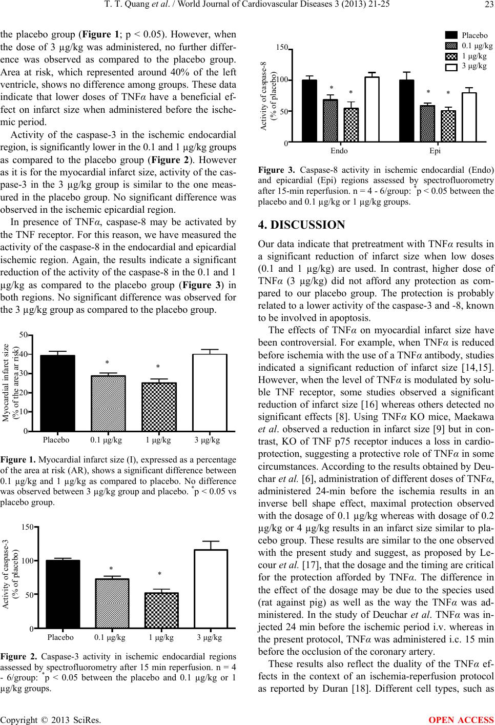 T. T. Quang et al. / World Journal of Cardiovascular Diseases 3 (2013) 21-25 23 the placebo group (Figure 1; p < 0.05). However, when the dose of 3 µg/kg was administered, no further differ- ence was observed as compared to the placebo group. Area at risk, which represented around 40% of the left ventricle, shows no difference among groups. These data indicate that lower doses of TNFα have a beneficial ef- fect on infarct size when administered before the ische- mic period. Activity of the caspase-3 in the ischemic endocardial region, is significantly lower in the 0.1 and 1 µg/kg groups as compared to the placebo group (Figure 2). However as it is for the myocardial infarct size, activity of the cas- pase-3 in the 3 µg/kg group is similar to the one meas- ured in the placebo group. No significant difference was observed in the ischemic epicardial region. In presence of TNFα, caspase-8 may be activated by the TNF receptor. For this reason, we have measured the activity of the caspase-8 in the endocardial and epicardial ischemic region. Again, the results indicate a significant reduction of the activity of the caspase-8 in the 0.1 and 1 µg/kg as compared to the placebo group (Figure 3) in both regions. No significant difference was observed for the 3 µg/kg group as compared to the placebo group. Placebo 0.1 μg/kg 1 μg/kg 3 μg/kg 50 40 30 20 10 0 Myocardial infarct size (% of the area ar risk) * * Figure 1. Myocardial infarct size (I), expressed as a percentage of the area at risk (AR), shows a significant difference between 0.1 µg/kg and 1 µg/kg as compared to placebo. No difference was observed between 3 µg/kg group and placebo. *p < 0.05 vs placebo group. Placebo 0.1 μg/kg 1 μg/kg 3 μg/kg 150 100 50 0 Activity of caspase-3 (% of placebo) * * Figure 2. Caspase-3 activity in ischemic endocardial regions assessed by spectrofluorometry after 15 min reperfusion. n = 4 - 6/group: *p < 0.05 between the placebo and 0.1 µg/kg or 1 µg/kg groups. 150 100 50 0 Activity of caspase-8 (% of placebo) * * Endo Epi * * Placebo 0.1 μg/kg 1 μg/kg 3 μg/kg Figure 3. Caspase-8 activity in ischemic endocardial (Endo) and epicardial (Epi) regions assessed by spectrofluorometry after 15-min reperfusion. n = 4 - 6/group: *p < 0.05 between the placebo and 0.1 µg/kg or 1 µg/kg groups. 4. DISCUSSION Our data indicate that pretreatment with TNFα results in a significant reduction of infarct size when low doses (0.1 and 1 µg/kg) are used. In contrast, higher dose of TNFα (3 µg/kg) did not afford any protection as com- pared to our placebo group. The protection is probably related to a lower activity of the caspase-3 and -8, known to be involved in apoptosis. The effects of TNFα on myocardial infarct size have been controversial. For example, when TNFα is reduced before ischemia with the use of a TNFα antibody, studies indicated a significant reduction of infarct size [14,15]. However, when the level of TNFα is modulated by solu- ble TNF receptor, some studies observed a significant reduction of infarct size [16] whereas others detected no significant effects [8]. Using TNFα KO mice, Maekawa et al. observed a reduction in infarct size [9] but in con- trast, KO of TNF p75 receptor induces a loss in cardio- protection, suggesting a protective role of TNFα in some circumstances. According to the results obtained by Deu- char et al. [6], administration of different doses of TNFα, administered 24-min before the ischemia results in an inverse bell shape effect, maximal protection observed with the dosage of 0.1 µg/kg whereas with dosage of 0.2 µg/kg or 4 µg/kg results in an infarct size similar to pla- cebo group. These results are similar to the one observed with the present study and suggest, as proposed by Le- cour et al. [17], that the dosage and the timing are critical for the protection afforded by TNFα. The difference in the effect of the dosage may be due to the species used (rat against pig) as well as the way the TNFα was ad- ministered. In the study of Deuchar et al. TNFα was in- jected 24 min before the ischemic period i.v. whereas in the present protocol, TNFα was administered i.c. 15 min before the occlusion of the coronary artery. These results also reflect the duality of the TNFα ef- fects in the context of an ischemia-reperfusion protocol as reported by Duran [18]. Different cell types, such as Copyright © 2013 SciRes. OPEN ACCESS  T. T. Quang et al. / World Journal of Cardiovascular Diseases 3 (2013) 21-25 24 myocytes or vascular smooth muscle cells, can synthe- size TNFα. In normal conditions the level of synthesis of TNFα is low but increases rapidly during ischemia/re- perfusion [19]. These levels then diminish during the first hours of reperfusion [20]. TNFα exerts its effects through 2 different receptors: TNFR1 and TNFR2. The binding of the TNFα to the TNFR1 induces the trimeri- zation of the receptor and promotes the dissociation of the silencer death domain from the receptor. The death domain of the activated receptor recruits the TNFR1-as- sociated death domain (TRADD), RIP1 and TRAF2, which then becomes modified and dissociate from TNFR1. The released death domain of TRADD is now able to bind to Fas-associated protein with death domain, result- ing in the activation of the caspase-8 and then caspase-3 [1]. In contrast, TNFR1 can also activate the NF-kB pathway, through the phosphorylation and ubiquity-na- tion of different proteins that bind the TNFR1-TRADD- TRAF-RIP1 complex. When NF-kB is activated, its trans- location to the nucleus mediates the transcription of dif- ferent proteins such as TNFα. TNFR1 can also activate other signaling pathway in- cluding c-Jun N-terminal kinase (JNK), p38 mitogen- activated protein kinase and PI3k/Akt [10]. These en- zymes have been identified to be members of two im- portant signaling pathways that can induce cardiopro- tection: the RISK [21] and SAFE pathways [22]. Our da- ta indicate that the activity of both caspase-3 and -8 is elevated in the control and 3 µg/kg groups whereas the activation is significantly less in the 0.1 µg/kg and 1 µg/ kg groups. This difference in the activation suggests that the lower doses of TNFα can induce the activation of the protective pathways, when activated before the ischemic period, whereas other pathways are activated with the higher doses. The difference in the myocardial infarct size between placebo and the other groups (0.1 µg/kg and 1 µg/kg) can be due to the activation of the protec- tive pathway induced by the activation of the TNFR1 before the ischemic period, but other experiments are needed to confirm this hypothesis. We have measured the activity of the caspase-3 and -8 after 15 minutes of reperfusion because previous data indicate that this activity is high during the first minute of reperfusion to reduce, and it is also basal at 5 hours of reperfusion [23]. One major limitation in this protocol is the availability of antibodies to perform biochemical analysis that is compatible with a porcine model. For this reason, we have been limited in the number of measures that can be performed. In conclusion, we have documented that low doses of TNFα can induce cardioprotection when administered before the ischemic period. However, with high doses, this protection is lost probably due to the activation of the caspase-3 and -8. 5. ACKNOWLEDGEMENTS This work was supported by the Centre de recherche, Hôpital du Sacré- Coeur de Montréal and Fonds de la recherche du Québec-Santé (FRQS). We thank Louis Chiocchio and Caroline Bouchard for their help in animal care. REFERENCES [1] Kleinbongard, P., Schulz, R. and Heusch, G. (2011) TNF- alpha in myocardial ischemia/reperfusion, remodeling and heart failure. Heart Failure Reviews, 16, 46-69. [2] Levine, B., et al. (1990) Elevated circulating levels of tumor necrosis factor in severe chronic heart failure. The New England Journal of Medicine, 323, 236-241. doi:10.1056/NEJM199007263230405 [3] Przyklenk, K., et al. (1993) Regional ischemic “precondi- tioning” protects remote virgin myocardium from subse- quent sustained coronary occlusion. Circulation, 87, 893- 899. doi:10.1161/01.CIR.87.3.893 [4] Mann, D.L. (2002) Inflammatory mediators and the fail- ing heart: Past, present, and the foreseeable future. Cir- culation Research, 91, 988-998. doi:10.1161/01.RES.0000043825.01705.1B [5] Sack, M.N. and Yellon, D.M. (2003) Insulin therapy as an adjunct to reperfusion after acute coronary ischemia: A proposed direct myocardial cell survival effect inde- pendent of metabolic modulation. Journal of the Ameri- can College of Cardiology, 41, 1404-1407. doi:10.1016/S0735-1097(03)00164-5 [6] Deuchar, G.A., Opie, L.H. and Lecour, S. (2007) TNFal- pha is required to confer protection in an in vivo model of classical ischaemic preconditioning. Life Science, 80, 1686-1691. doi:10.1016/j.lfs.2007.01.040 [7] Lecour, S., Rochette, L. and Opie, L. (2005) Free radicals trigger TNF alpha-induced cardioprotection. Cardiovas- cular Research, 65, 239-243. doi:10.1016/j.cardiores.2004.10.003 [8] Kaloustian, S., et al. (2009) Tumor necrosis factor-alpha participates in apoptosis in the limbic system after myo- cardial infarction. Apoptosis, 14, 1308-1316. doi:10.1007/s10495-009-0395-x [9] Maekawa, N., et al. (2002) Improved myocardial ische- mia/reperfusion injury in mice lacking tumor necrosis factor-alpha. Journal of the American College of Cardi- ology, 39, 1229-1235. doi:10.1016/S0735-1097(02)01738-2 [10] Bradley, J.R. (2008) TNF-mediated inflammatory disease. Journal of Pathology, 214, 149-160. doi:10.1002/path.2287 [11] Boucher, M., et al. (2002) Alterations of beta-adrenocep- tor responsiveness in postischemic myocardium after 72 h of reperfusion. European Journal of Pharmacology, 495, 185-191. doi:10.1016/j.ejphar.2004.05.040 [12] Wann, B.P., et al. (2006) Apoptosis detected in the amyg- dala following myocardial infarction in the rat. Biological Copyright © 2013 SciRes. OPEN ACCESS  T. T. Quang et al. / World Journal of Cardiovascular Diseases 3 (2013) 21-25 Copyright © 2013 SciRes. 25 OPEN ACCESS Psychiatry, 59, 430-433. doi:10.1016/j.biopsych.2005.07.018 [13] Boucher, M., et al. (2006) Reduction of apoptosis in the amygdala by an A2A adenosine receptor agonist follow- ing myocardial infarction. Apoptosis, 11, 1067-1074. doi:10.1007/s10495-006-6313-6 [14] Belosjorow, S., et al. (2003) TNF-a antibodies are as ef- fective as ischemic preconditioning in reducing infarct size in rabbits. American Journal of Physiology, 284, H927-H930. [15] Gu, Q., et al. (2006) Inhibition of TNF-alpha reduces myo- cardial injury and proinflammatory pathways following ischemia-reperfusion in the dog. Journal of Cardiovas- cular Pharmacology, 48, 320-328. doi:10.1097/01.fjc.0000250079.46526.38 [16] Sugano, M., et al. (2004) In vivo transfer of soluble TNF- alpha receptor 1 gene improves cardiac function and re- duces infarct size after myocardial infarction in rats. FASEB Journal, 18, 911-913. [17] Lecour, S., et al. (2002) Identification of a novel role for sphingolipid signaling in TNF alpha and ischemic pre- conditioning mediated cardioprotection. Journal of Mole- cular and Cellular Cardiology, 34, 509-518. doi:10.1006/jmcc.2002.1533 [18] Duran, W.N. (2008) The double-edge sword of TNF-al- pha in ischemia-reperfusion injury. American Journal of Physiology—Heart and Circulatory Physiology, 295, H2221-H2222. doi:10.1152/ajpheart.01050.2008 [19] Reil, J.C., et al. (2007) Insights from knock-out models concerning postischemic release of TNFalpha from isola- ted mouse hearts. Journal of Molecular and Cellular Car- diology, 42, 133-141. doi:10.1016/j.yjmcc.2006.09.020 [20] McVey, M., Perrone, M.H. and Clark, K.L. (1999) Does tumor necrosis factor-alpha (TNF-alpha) contribute to my- ocardial reperfusion injury in anesthetized rats? General Pharmacology, 32, 41-45. doi:10.1016/S0306-3623(98)00050-0 [21] Hausenloy, D.J., Tsang, A. and Yellon, D.M. (2005) The reperfusion injury salvage kinase pathway: A common target for both ischemic preconditioning and postcondi- tioning. Trends in Cardiovascular Medicine, 15, 69-75. doi:10.1016/j.tcm.2005.03.001 [22] Lecour, S. (2009) Multiple protective pathways against re- perfusion injury: A SAFE path without Aktion? Journal of Molecular and Cellular Cardiology, 46, 607-609. doi:10.1016/j.yjmcc.2009.01.003 [23] Boucher, M., et al. (2004) Post-ischemic cardioprotection by A2A adenosine receptors: Dependent of phosphatidy- linositol 3-kinase pathway. Journal of Cardiovascular Pharmacology, 43, 416-422. doi:10.1097/00005344-200403000-00013 |