

Paper Menu >>
Journal Menu >>
 Vol.2, No.5, 332-334 (2013) Case Reports in Clinical Medicine http://dx.doi.org/10.4236/crcm.2013.25089 Posterior reversible encephalopathy syndrome induced after blood transfusion for severe anemia Kei-ichiro Wada1, Masayoshi Kano1, Yutaka Machida1, Nobutaka Hattori2, Hideto Miwa1* 1Department of Neurology, Juntendo University Nerima Hospital, Tokyo, Japan; *Corresponding Author: [email protected] 2Department of Neurology, Juntendo University School of Medicine, Tokyo, Japan Received 6 June 2013; revised 30 June 2013; accepted 10 July 2013 Copyright © 2013 Kei-ichiro Wada et al. This is an open access article distributed under the Creative Commons Attribution License, which permits unrestricted use, distribution, and reproduction in any medium, provided the original work is properly cited. ABSTRACT Posterior reversible encephalopathy syndrome (PRES) is a clinicoradiological syndrome char- acterized by headache, confusion, seizures, and cortical visual loss, as well as subcortical ede- ma without infarction on neuroimaging. We re- port a 56-year-old woman who developed typi- cal manifestations of PRES, 6 days after blood transfusi on for severe anemia. Acu te volume over- loads by transfusion may exceed the capacity of autoregulation of perfusion pressure, possibly resulting in vasogenic edema. We propose that it is clinically important to recognize that rapid correction of anemia by blood transfusion may carry the risk of inducing PRES. Keyw ords: Posterior Reversibl e Enceph alopathy Syndrome; Anemia; Blood Transfusion 1. INTRODUCTION Posterior reversible encephalopathy syndrome (PRES), or reversible posterior leukoencephalopathy syndrome, is a clinicoradiological syndrome characterized by head- ache, confusion, seizures, and cortical visual loss, as well as subcortical edema without infarction on neuroimaging [1-3]. It was first described by Hinchey et al. [1] in 1995 based on 15 patients who had subcortical edema without cerebral infarction. To date, a wide variety of clinical con- ditions have been reported to cause PRES, such as hy- pertension, exposure to toxic agents, infection, eclampsia, autoimmune disease, and other conditions [1-3]. Recently, we encountered a patient with PRES that was induced after blood transfusion for severe anemia. 2. CASE REPORT A 56-year-old Japanese woman was admitted to the gynecology section in our hospital because of genital bleeding and abdominal pain. Her past medical and fam- ily histories were unremarkable, and in particular, she had no past history of hypertension. After admission, she was diagnosed with cancer of the corpus uteri. She had severe anemia with a hemoglobin level of 2 g/dL, and received blood transfusion with a total of 2 L packed red blood cells. After transfusion, her hemoglobin level re- covered to 9.2 g/dL for 4 days. Six days after the last blood transfusion, she suddenly developed status epilep- ticus, and was referred to our neurological section. She was immediately treated with general anesthesia by intravenous administration of midazolam with pheny- toin. Two days later, she recovered from anesthesia, but a drowsy state continued for a few days. On neurological examination, her pupils were equal, and promptly reac- tive to light. She could open her eyes if verbal stimuli were given, but she could not recognize any objects. She could not maintain her gaze on any objects, and just stared into space, suggesting that she had cortical blind- ness. Oculocephalic reflexes, towards either horizontal or vertical directions, were not disturbed. She was mute. No obvious motor paresis was noted in both upper and lower extremities. Deep tendon reflexes were all normal in both limbs. There were no signs of meningeal irritation. La- boratory examination revealed mild anemia (9.2 g/dL), leukocytosis (36,500/L) and elevated C-reactive protein (4.5 mg/dL). Other routine blood tests, blood coagulation tests, autoantibodies, and neoplastic markers were within normal limits. Cerebrospinal fluid examination revealed a mild increase in protein level (52 mg/dL). Brain mag- netic resonance imaging (MRI), which included T1-, T2- and diffusion-weighted images, fluid-attenuated inver- sion recovery (FLAIR) images, and apparent diffusion coefficient mapping, were performed. The FLAIR im- ages demonstrated high-intensity signal areas in bilateral parietal lobes (Figure 1). Within 10 days, her conscious- Copyright © 2013 SciRes. OPEN ACCESS 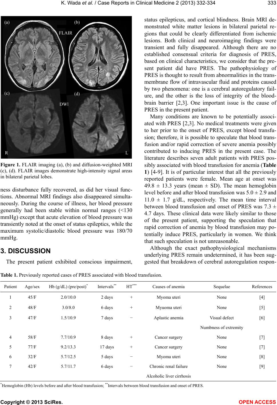 K. Wada et al. / Case Reports in Clinical Medicine 2 (2013) 332-334 333 Figure 1. FLAIR imaging (a), (b) and diffusion-weighted MRI (c), (d). FLAIR images demonstrate high-intensity signal areas in bilateral parietal lobes. ness disturbance fully recovered, as did her visual func- tions. Abnormal MRI findings also disappeared simulta- neously. During the course of illness, her blood pressure generally had been stable within normal ranges (<130 mmHg) except that acute elevation of blood pressure was transiently noted at the onset of status epileptics, while the maximum systolic/diastolic blood pressure was 180/70 mmHg. 3. DISCUSSION The present patient exhibited conscious impairment, status epilepticus, and cortical blindness. Brain MRI de- monstrated white matter lesions in bilateral parietal re- gions that could be clearly differentiated from ischemic lesions. Both clinical and neuroimaging findings were transient and fully disappeared. Although there are no established consensual criteria for diagnosis of PRES, based on clinical characteristics, we consider that the pre- sent patient did have PRES. The pathophysiology of PRES is thought to result from abnormalities in the trans- membrane flow of intravascular fluid and proteins caused by two phenomena: one is a cerebral autoregulatory fail- ure, and the other is the loss of integrity of the blood- brain barrier [2,3]. One important issue is the cause of PRES in the present patient. Many conditions are known to be potentially associ- ated with PRES [2,3]. No medical treatments were given to her prior to the onset of PRES, except blood transfu- sion; therefore, it is possible to speculate that blood trans- fusion and/or rapid correction of severe anemia possibly contributed to inducing PRES in the present case. The literature describes seven adult patients with PRES pos- sibly associated with blood transfusion for anemia (Table 1) [4-9]. It is of particular interest that all the previously reported patients were female. Mean age at onset was 49.8 ± 13.3 years (mean ± SD). The mean hemoglobin level before and after blood transfusion was 5.0 ± 2.9 and 11.0 ± 1.7 g/dL, respectively. The mean time interval between blood transfusion and onset of PRES was 7.3 ± 4.7 days. These clinical data were likely similar to those of the present patient, supporting the speculation that rapid correction of anemia by blood transfusion may po- tentially induce PRES, particularly in women. We think that such speculation is not unreasonable. Although the exact pathophysiological mechanisms underlying PRES remain undetermined, it has been sug- gested that breakdown of cerebral autoregulation respon- Table 1. Previously reported cases of PRES associated with blood transfusion. Patient Age/sex Hb (g/dL) (pre/post)* Intervals** HT*** Causes of anemia Sequelae References 1 45/F 2.0/10.0 2 days + Myoma uteri None [4] 2 48/F 3.0/8.0 6 days + Myuoma uteri None [5] 3 47/F 1.5/10.9 7 days − Aplastic anemia Visual defect [6] Numbness of extremity 4 58/F 7.7/10.9 8 days + Cancer surgery None [7] 5 77/F 9.2/13.3 17 days + Cancer surgery None [7] 6 32/F 5.7/12.5 5 days − Myoma uteri None [8] 7 42/F 5.7/11.7 6 days − Chronic renal failure None [9] Alcoholic liver cirrhosis *Hemoglobin (Hb) levels before and after blood transfusion; **Intervals between blood transfusion and onset of PRES. Copyright © 2013 SciRes. OPEN ACCESS  K. Wada et al. / Case Reports in Clinical Medicine 2 (2013) 332-334 334 sible for an increase in cerebral blood flow may play a pivotal role [2,3]. Blood transfusion may result in a rapid increase in total blood volume, leading to rapid overload of cerebral blood flow. Cerebral hyperperfusion, if it is acutely induced and exceeds the capacity for autoregula- tion of perfusion pressure in cerebral capillaries, could result in the huge vasogenic edema responsible for PRES. In addition, the possibility should not be excluded that severe anemia per se also might be the predisposing fac- tor contributing to the induction of PRES, because in- sufficient oxygen supply due to severe anemia might exert an unfavorable influence on endothelial cell func- tions, possibly causing a breakdown of the integrity of the bloodbrain barrier in capillary circulation. Another issue is whether a rapid increase in blood pressure, which is frequently associated with PRES [1-3, 10], simultaneously contributed to inducing PRES in the present study. In this regard, we think that hypertension might not have played a prominent role in the mecha- nism underlying PRES in the present case. During the course of illness, hypertension was only transiently ob- served, and an extreme increase in blood pressure (>180 mmHg) was not noted. Second, three of the seven previ- ously reported patients (Table 1) had no hypertension, suggesting that an acute increase in blood volume, but not acute hypertension, may play a predominant role in inducing PRES, if it occurs after blood transfusion for correction of anemia. Finally, we propose that it is clinically important to recognize that rapid correction of anemia by blood trans- fusion may carry a risk of inducing PRES. REFERENCES [1] Hinchey, J., et al. (1996) A reversible posterior leukoen- cephalopathy syndrome. The New England Journal of Medicine, 2, 494-500. doi:10.1056/NEJM199602223340803 [2] Legriel, S., Pico, F. and Azoulay, E. (2011) Understand- ing posterior reversible encephalopathy syndrome. In: Vincent, J.L., Ed., Annual Update in Intensive Care and Emergency Medicine, Springer, Berlin, 631-653. [3] Feske, S.K. (2011) Posterior reversible encephalopathy syndrome: A review. Seminars in Neurology, 31, 202-215. doi:10.1055/s-0031-1277990 [4] Ito, Y., et al. (1997) Post-transfusion reversible posterior leukoencephalopathy syndrome with cerebral vasocon- striction. Neurology, 49, 1174-1175. doi:10.1212/WNL.49.4.1174 [5] Boughammoura, A., Touzé, E., Oppenheim, C., Trystram, D. and Mas, J.L. (2003) Reversible angiopathy and en- cephalopathy after blood transfusion. Journal of Neurol- ogy, 250, 116-118. doi:10.1007/s00415-003-0940-4 [6] Heo, K., Park, S., Lee, J.Y., Lee, B.I. and Lee, S.K. (2003) Post-transfusion posterior leukoencephalopathy with cy- totoxic and vasogenic edema precipitated by vasospasm. Cerebrovascular Diseases, 15, 230-233. doi:10.1159/000068825 [7] Kawano, H., Suga, T., Terasaki, T., Hashimoto, Y., Baba, K. and Uchino, M. (2004) Posterior encephalopathy syn- drome in two patients after cancer surgery with transfu- sion. Rinsho Shinkeigaku, 44, 427-431. [8] Huang, Y.C., Tsai, P.L., Yeh, J.H. and Chen, W.H. (2008) Reversible posterior leukoencephalopathy syndrome caused by blood transfusion: A case report. Acta Neurologica Taiwanica, 17, 258-262. [9] Sato, T., et al. (2011) Reversible posterior leukoencepha- lopathy syndrome after blood transfusion in a patient with end-stage renal disease. Clinical and Experimental Ne- phrology, 15, 942-947. doi:10.1007/s10157-011-0515-0 [10] Li, Y., Gor, D., Walicki, D., Jenny, D., Jones, D., Barbour, P. and Castaldo, J. (2012) Spectrum and potential patho- genesis of reversible posterior leukoencephalopathy syn- drome. Journal of Stroke and Cerebrovascular Diseases, 21, 873-882. doi:10.1016/j.jstrokecerebrovasdis.2011.05.010 Copyright © 2013 SciRes. OPEN ACCESS |