

Paper Menu >>
Journal Menu >>
 Open Journal of Obstetrics and Gynecology, 2013, 3, 528-535 OJOG http://dx.doi.org/10.4236/ojog.2013.37095 Published Online September 2013 (http://www.scirp.org/journal/ojog/) Non-invasive prediction of endometriosis revisited; 3 biomarkers as Angiopoietin-2, Interleukin-1β and Vascular Endothelial Growth Factor Hoda Abdel Moety1*, Gihan I. Khalil1, Rania M. El Sharkawy1, Marwa K. El Ghandour1, Fady S. Moiety2, Hesham A. F. Salem2 1Department of Chemical Pathology, Medical Research Institute, Alexandria University, Alexandria, Egypt 2Department of Obstetrics and Gynecology, Faculty of Medicine, Alexandria University, Alexandria, Egypt Email: *[email protected] Received 2 July 2013; revised 1 August 2013; accepted 9 August 2013 Copyright © 2013 Hoda Abdel Moety et al. This is an open access article distributed under the Creative Commons Attribution Li- cense, which permits unrestricted use, distribution, and reproduction in any medium, provided the original work is properly cited. ABSTRACT Introduction: Endometriosis affects up to 1 every 5 women at their reproductive age, with variable and complex symptomatology. Patients may be asympto- matic but may have pain episodes or subfertility. Its negative impact is on patients’ health and quality of life. Objective: it was to investigate the serum and peritoneal fluid (PF) concentrations of Angiopoietin- 2, Interleukin-1β, and Vascular Endothelial Growth Factor, aiming to evaluate their diagnostic perform- ance in endometriosis. Methods: Serum and perito- neal fluid samples were taken from 112 women un- dergoing laparoscopy for infertility, pelvic pain or adnexal masses. 61 diagnosed with endometriosis and 51 controlled. Primary outcome was to estimate se- rum and PF concentrations of Angio-2, IL-1β and VEGF and secondarily correlate these concentrations to disease stages thus assuming their diagnostic po- tential. Results: Significant differences were found between patients and control as regards serum and PF concentration of all studied markers except serum IL-1β. Serum Angio-2 and PF VEGF showed a sig- nificantly higher level in more advanced stages of en- dometriosis. PF VEGF showed a positively significant correlation with the stage of the disease, spearman coefficient t = 0.442 p = 0.014. PF concentrations of Angio-2 and Serum VEGF did not show significant pattern changes with stage-related levels. Diagnostic potential of serum and PF concentrations of the 3 markers were assessed by the ROC curve. Angio-2 proved an excellent diagnostic ability for endometrio- sis. PF and serum VEGF proved an equal diagnostic performance, whereas, PF IL-1β was the least effi- cient. Based on the results, we suggested preliminary serum threshold values for these markers to be used as diagnostic or follow-up landmarks with relatively acceptable sensitivity, specificity, positive and nega- tive predictive values. Conclusion: Non-invasive pre- dictive biomarkers for endometriosis were Serum Angio-2, IL-1β, and VEGF independently or in com- bination with the estimated threshold values. Serum Angio-2 merit is considered as a novel marker for en- dometriosis due to its diagnostic power. Keywords: Endometriosis; Angiopoietin-2; VEGF; IL-1β 1. INTRODUCTION Endometriosis affects up to 1 every 5 women at their reproductive age group, with variable and often complex symptomatology. Patients may be asymptomatic but may have pain episodes, and/or subfertility [1]. Unlike its ae- tiopathogenesis, its negative impact is on patients’ health and quality of life is clear [2,3]. The stage of endome- triosis can be morphologically classified by using the revised American Fertility Society (rAFS) staging system according to the localization and the size of nodes in the peritoneum and ovaries, and the presence of adhesions in the ovary and the fallopian tube [3]. This rAFS classifi- cation includes the following stages: I = Minimal (1 - 5 points); II = Mild (6 - 15 points); III = Moderate (16 - 40 points); IV = Severe (<40 points) [4]. Effective treatment could be achieved; however, di- agnosis of the disease remains a problem, simply, be- cause accurate diagnosis can only be attained via laparo- scopic inspection, preferably with histopathologic con- firmation of suspected lesions. This remains an invasive technique, with possible complications and health care *Corresponding author. OPEN ACCESS  H. A. Moety et al. / Open Journal of Obstetrics and Gynecology 3 (2013) 528-535 529 costs [5]. However, ovarian endometriomas, and deep infiltrating endometriotic nodules can be detected ade- quately by vaginal ultrasound, but it does not rule out peritoneal endometriosis or endometriosis-associated ad- hesions [1]. This situation has led the researchers re- cently to refocus on the possible mechanism by which endometriosis occurs. The fact is that an altered immune mechanism might be involved in the development of the condition is now widely accepted, which explains why some women are more prone than others to develop the disease. The concentration of some immunological mar- kers in the peritoneal fluid largely correlates with the disease occurrence, progression, and symptoms [6]. There is substantial evidence that immunologic factors play a role in the pathogenesis of endometriosis and en- dometriosis-associated infertility. Decreased natural kil- ler cell cytotoxicity leads to an increased likelihood of implantation of endometriotic tissue and modulation of growth and inflammatory behavior of ectopic endo- metrial implants. The alteration in cytokines level and other angiogenic factors as Vascular Endothelial Growth Factor (VEGF) may explain the adhesion, implantation and the progres- sion of the transported fragment of endometrium in the different site of endometriosis. Many cytokines have been studied to prove the immunological theory of en- dometriosis as Interleukin-1β (IL-1β). It has been gener- ally accepted that the establishment of new biomarker as angiopoietin-2 (Angio-2) played a key part in the pro- gression of endometriosis, however this role still needs further confirmation to suggest a relation to endometrio- sis already proven for many pivotal angiogenesis stimu- lators such as VEGF [7,8]. Setting a non-invasive biochemical diagnostic and prognostic test for endometriosis with an acceptable level of accuracy and sensitivity, would fundamentally change those patients’ quality of life, save their costs and risks of the more invasive tools, and eventually make it smoother for health care providers to manage the condition with ease. The current study was designed to evaluate the serum and peritoneal fluid concentrations of Angiopoietin-2, Interleukin-1β, and Vascular Endothelial Growth Factor in patients with endometriosis as well as controls, thus adding some insight to the formulation of a non-invasive test to screen, detect, or follow up patients with endome- triosis. The ultimate objective was to prove any of these mar- kers independently or in combination as non-invasive diagnostic biomarker in patients with endometriosis. 2. PATIENTS AND METHODS Blood and (PF) samples were taken from 112 women undergoing laparoscopy for infertility, pelvic pain, and/or adnexal masses in their first half of the menstrual cycle. Procedures were done at a specialized unit of Gyneco- logic endoscopy at a University hospital. Sixty one were histologically confirmed to have en- dometriosis and 51 were controls. No cases were ex- cluded. A full informed consent was taken from each subject. Enrolment took place after formal approval of the Ethical committee of the Faculty of Medicine, Alex- andria University was obtained. PF samples were collected by aspiration from the per- itoneal cavity during laparoscopy. Serum samples: Five ml of venous blood were asepti- cally collected and the clear serum was stored at –70˚C until analysis. Measurement of IL-1β, VEGF, and Angio-2 in serum and PF were done using a commercially available, en- zyme-linked immunosorbent assays (ELISA) using the reagents and protocol supplied with the ELISA kit (R&D Systems, Minneapolis, Minnesota, United States) Frozen serum and PF samples were thawed and then analyzed. 3. STATISTICAL ANALYSIS Data distribution was analyzed by using the Statistical Packages for the Social Sciences (SPSS version 10.0, Chicago, IL) and the Sigma-Stat software package (ver- sion 3.5; SPSS, Chicago, IL). Normally distributed data were presented as mean, standard deviation, whereas skewed data were expressed as median and range. Pear- son’s correlation was also used for evaluation of the lin- ear relationship between different clinical variables. Pair- wise comparisons between groups were performed using the Wilcoxon-Mann-Whitney test. In order to determine which serum marker could best represent patients with endometriosis, sensitivity and specificity were assessed with receiver operating characteristic ROC curves. The area under the curve was approximately the percentage correctly classified if the test was used as a diagnostic tool. The same analyses were performed with the PF measures and statistical significance was assessed using two-tailed tests and an alpha level of p < 0.05. 4. RESULTS A total of 112 women underwent laparoscopy for infer- tility (n = 49), pelvic pain (n = 22), and/or adnexal masses (n = 41). Sixty one were diagnosed with endome- triosis and 51 were controls. No cases were excluded. Out of the 61 patients with endometriosis, 25 (40.98%) had early disease (stages I and II) and 36 (59.02%) had late disease (stages III and IV). The median age of the study group was 33.3 years, median body mass index (BMI) was 27.0, and median parity was 1. No significant differences were found as regards age, parity and BMI among the studied groups. Copyright © 2013 SciRes. OPEN ACCESS 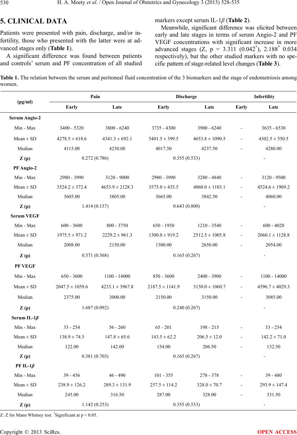 H. A. Moety et al. / Open Journal of Obstetrics and Gynecology 3 (2013) 528-535 Copyright © 2013 SciRes. 530 OPEN ACCESS 5. CLINICAL DATA Patients were presented with pain, discharge, and/or in- fertility, those who presented with the latter were at ad- vanced stages only (Table 1). A significant difference was found between patients and controls’ serum and PF concentration of all studied markers except serum IL-1β (Table 2). Meanwhile, significant difference was elicited between early and late stages in terms of serum Angio-2 and PF VEGF concentrations with significant increase in more advanced stages (Z, p = 3.311 (0.042*), 2.188* 0.034 respectively), but the other studied markers with no spe- cific pattern of stage-related level changes (Table 3). Table 1. The relation between the serum and peritoneal fluid concentration of the 3 biomarkers and the stage of endometriosis among women. Pain Discharge Infertility (pg/ml) Early Late Early Late Early Late Serum Angio-2 Min - Max 3400 - 5320 3800 - 6240 3735 - 4300 3900 - 6240 - 3635 - 6530 Mean ± SD 4278.5 ± 618.6 4341.3 ± 692.1 5401.5 ± 399.5 4653.8 ± 1090.5 - 4302.5 ± 550.5 Median 4115.00 4230.00 4017.50 4237.50 - 4280.00 Z (p) 0.272 (0.786) 0.355 (0.533) - PF Angio-2 Min - Max 2980 - 3990 3120 - 9000 2980 - 3990 3280 - 4840 - 3120 - 9500 Mean ± SD 3524.2 ± 372.4 4653.9 ± 2128.3 3575.0 ± 435.5 4060.0 ± 1103.1 - 4524.6 ± 1969.2 Median 3605.00 3805.00 3665.00 3842.50 - 4060.00 Z (p) 1.414 (0.157) 0.643 (0.800) - Serum VEGF Min - Max 600 - 3600 800 - 3750 650 - 1950 1210 - 3540 - 600 - 4020 Mean ± SD 1975.5 ± 971.2 2229.2 ± 961.3 1300.0 ± 919.2 2512.5 ± 1005.8 - 2060.1 ± 1128.8 Median 2008.00 2150.00 1300.00 2650.00 - 2054.00 Z (p) 0.571 (0.568) 0.165 (0.267) - PF VEGF Min - Max 650 - 3600 1100 - 14000 850 - 3600 2400 - 3900 - 1100 - 14000 Mean ± SD 2047.5 ± 1059.6 4233.1 ± 3967.8 2187.5 ± 1141.9 3150.0 ± 1060.7 - 4596.7 ± 4029.3 Median 2375.00 3000.00 2150.00 3150.00 - 3085.00 Z (p) 1.687 (0.092) 0.240 (0.267) - Serum IL-1β Min - Max 33 - 254 56 - 260 65 - 201 198 - 215 - 33 - 254 Mean ± SD 138.9 ± 74.5 147.8 ± 65.6 143.5 ± 62.2 206.5 ± 12.0 - 142.2 ± 71.0 Median 122.00 142.00 154.00 206.50 - 132.50 Z (p) 0.381 (0.703) 0.165 (0.267) - PF IL-1β Min - Max 39 - 456 46 - 490 101 - 355 278 - 378 - 39 - 480 Mean ± SD 238.9 ± 126.2 289.3 ± 131.9 257.5 ± 114.2 328.0 ± 70.7 - 293.9 ± 147.4 Median 245.00 316.50 287.00 328.00 - 331.50 Z (p) 1.142 (0.253) 0.355 (0.533) - Z: Z for Mann Whitney test. *Significant at p < 0.05. 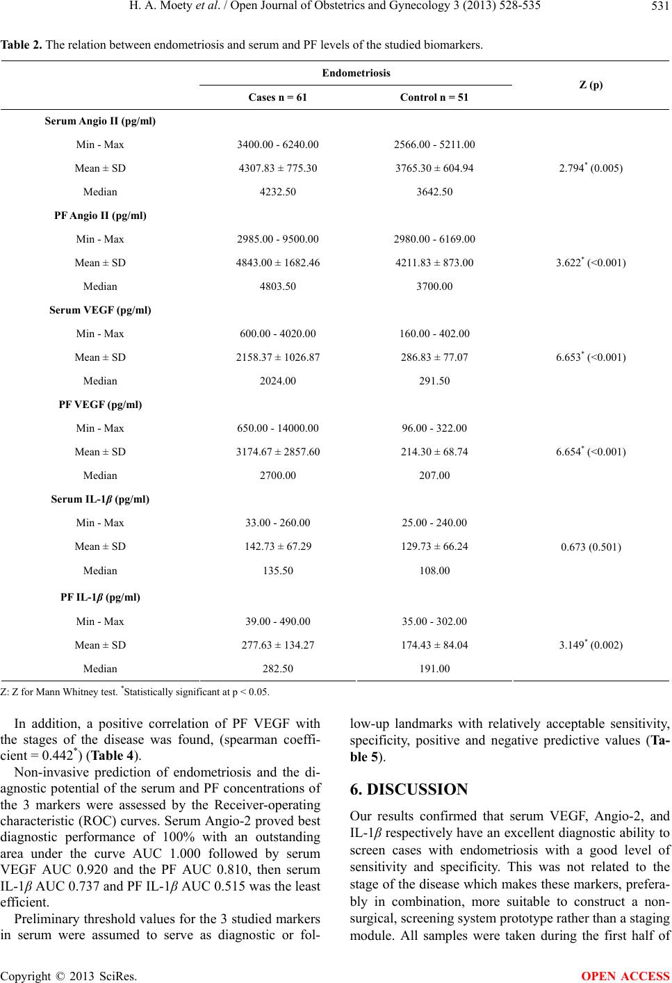 H. A. Moety et al. / Open Journal of Obstetrics and Gynecology 3 (2013) 528-535 531 Table 2. The relation between endometriosis and serum and PF levels of the studied biomarkers. Endometriosis Cases n = 61 Control n = 51 Z (p) Serum Angio II (pg/ml) Min - Max 3400.00 - 6240.00 2566.00 - 5211.00 Mean ± SD 4307.83 ± 775.30 3765.30 ± 604.94 Median 4232.50 3642.50 2.794* (0.005) PF Angio II (pg/ml) Min - Max 2985.00 - 9500.00 2980.00 - 6169.00 Mean ± SD 4843.00 ± 1682.46 4211.83 ± 873.00 Median 4803.50 3700.00 3.622* (<0.001) Serum VEGF (pg/ml) Min - Max 600.00 - 4020.00 160.00 - 402.00 Mean ± SD 2158.37 ± 1026.87 286.83 ± 77.07 Median 2024.00 291.50 6.653* (<0.001) PF VEGF (pg/ml) Min - Max 650.00 - 14000.00 96.00 - 322.00 Mean ± SD 3174.67 ± 2857.60 214.30 ± 68.74 Median 2700.00 207.00 6.654* (<0.001) Serum IL-1β (pg/ml) Min - Max 33.00 - 260.00 25.00 - 240.00 Mean ± SD 142.73 ± 67.29 129.73 ± 66.24 Median 135.50 108.00 0.673 (0.501) PF IL-1β (pg/ml) Min - Max 39.00 - 490.00 35.00 - 302.00 Mean ± SD 277.63 ± 134.27 174.43 ± 84.04 Median 282.50 191.00 3.149* (0.002) Z: Z for Mann Whitney test. *Statistically significant at p < 0.05. In addition, a positive correlation of PF VEGF with the stages of the disease was found, (spearman coeffi- cient = 0.442*) (Table 4). Non-invasive prediction of endometriosis and the di- agnostic potential of the serum and PF concentrations of the 3 markers were assessed by the Receiver-operating characteristic (ROC) curves. Serum Angio-2 proved best diagnostic performance of 100% with an outstanding area under the curve AUC 1.000 followed by serum VEGF AUC 0.920 and the PF AUC 0.810, then serum IL-1β AUC 0.737 and PF IL-1β AUC 0.515 was the least efficient. Preliminary threshold values for the 3 studied markers in serum were assumed to serve as diagnostic or fol- low-up landmarks with relatively acceptable sensitivity, specificity, positive and negative predictive values (Ta- ble 5). 6. DISCUSSION Our results confirmed that serum VEGF, Angio-2, and IL-1β respectively have an excellent diagnostic ability to screen cases with endometriosis with a good level of sensitivity and specificity. This was not related to the stage of the disease which makes these markers, prefera- bly in combination, more suitable to construct a non- surgical, screening system prototype rather than a staging module. All samples were taken during the first half of Copyright © 2013 SciRes. OPEN ACCESS  H. A. Moety et al. / Open Journal of Obstetrics and Gynecology 3 (2013) 528-535 532 Table 3. Comparison between the two stages of endometriosis according to serum and PF of the 3 biomarkers. Stages of e n dometriosis Early Late Z (p) Serum Angio-2 (pg/ml) Min - Max 3400 - 5320 3800 - 6240 Mean ± SD 4285.6 ± 559.5 4341.3 ± 692.1 Median 4115.00 4550.00 3.311 (0.042*) PF Angio-2 (pg/ml) Min-max 2980 - 3990 3120 - 9500 Mean ± SD 3524.2 ± 372.4 4670.3 ± 2045.2 Median 3605.00 3842.50 1.566 (0.117) Serum VEGF (pg/ml) Min - Max 600 - 3750 800 - 4020 Mean ± SD 2111.2 ± 109.31 2229.2 ± 961.3 Median 2024 2150 0.233 (0.816) PF VEGF (pg/ml) Min - Max 650 - 1400 1100 - 3600 Mean ± SD 2047.5 ± 1059.6 3926.1 ± 3421.8 Median 2375.00 3005.0 2.188 (0.034*) Serum IL-1β (pg/ml) Min - Max 33 - 254 56 - 260 Mean ± SD 139.4 ± 65.3 147.8 ± 68.6 Median 132.50 142.00 0.381 (0.703) PF IL-1β (pg/ml) Min - Max 39 - 480 46 - 490 Mean ± SD 269.8 ± 131.1 289.3 ± 139.9 Median 273.00 316.50 0.381 (0.703) *Statistically significant at p < 0.05. Table 4. Correlation between serum and PF levels of Ang-2, VEGF and IL-1β with the stages of the disease. Serum PF Angio-2 VEGF IL-1B Angio-2 VEGF IL-1B r 0.152 0.112 0.011 0.062 Angio-2 p 0.422 0.557 0.954 0.744 r 0.241 0.032 VEGF p 0.199 0.865 r IL-1β p rho 0.102 0.034 0.036 0.296 0.442* 0.034 Stages Early & Late p 0.592 0.858 0.850 0.112 0.014 0.859 r: Pearson coefficient. rho: Spearman coefficient. *Statistically significant at p ≤ 0.05. Copyright © 2013 SciRes. OPEN ACCESS  H. A. Moety et al. / Open Journal of Obstetrics and Gynecology 3 (2013) 528-535 533 Table 5. Diagonstic performance of the studied biomarkers with suggested threshold values. Serum Ang-2 (pg/ml) Seru m VEGF (pg/ml) Serum IL-1β (pg/ml) Threshold (cut of values) 3700 500 120 Sensitivity 100.00 90.00 60.00 Specificity 100.00 60.00 53.33 Positive predictive value (PPV) 100.00 69.23 56.25 Negative predictive value (NPV) 100.00 85.71 57.14 Accuracy 100.00 75.00 56.67 the menstrual cycle to neglect any effect of the sampling time on the results. Bedaiwy et al. [5] made the sampling time all through the menstrual cycle and found differ- ences related to sampling time that they claimed not sig- nificant, so we preferred to avoid those differences. PF laparoscopic sampling; especially for VEGF, and IL-1β can be more supportive in establishing diagnostic tests in subtle or atypically-presenting cases, where evi- dent endometriosis lesions would be hardly present to assess histologically. We did not, however find any sig- nificant correlation between symptoms such as pain with the levels of PF studied markers including the cytokine; IL-1β. This contradicts what Scholl et al. [9] stated about an increased level of PF cytokine with the severe pain and dysmenorrhea in endometriosis. We were also able to estimate a preliminary cut-off values, based on the number of patients studied, for the serum markers in this cohort of patients, where Angio-2 showed the best sensitivity, and specificity followed by VEGF and IL-1β respectively. There were relatively ac- ceptable positive and negative predictive values, as well, at the estimated cut-off levels. We assumed a promising role for these makers that could be extended to similar substances as diagnostic or follow-up landmarks in cases destinated to develop endometriosis. Similarly, Mihalyi et al. [10] concluded a diagnostic role of a panel of six selected plasma biomarkers in the diagnosis of different stages of endometriosis with good sensitivity and spe- cificity. They studied, however, different biomarkers than those in the present study, including some cytokines, and obtained serum samples during the secretory phase or during menstruation. Bedaiwy et al. [5] also assumed a non- surgical diagnosis of endometriosis be possible by measuring serum IL-6 and PF TNF-α. VEGF is a heparin-binding glycoprotein with potent angiogenic, endothelial cell-specific mitogenic and vas- cular permeability activities. Studies have demonstrated that VEGF is involved in both the etiology and maintenance of peritoneal endome- triosis [11]. Moreover, in normal endometrium the ex- pression of VEGF is potentiated by a variety of cytokines, especially Interleukin-1β (IL-1β) [12]. In this study we confirmed a good diagnostic per- formance of VEGF both in serum and PF. We found significant differences of PF VEGF and se- rum Angio-2 levels related to the stage of endometriosis, this was in accordance with Meresman et al. [13] who reported same results stating that VEGF-A levels in the serum and PF of patients with severe endometriosis (stages III-IV) were significantly higher than in those with minimal endometriosis owing to the modulation of this marker by the disease itself. Angio-2 is a cytokine that can amplify the effects of other angiogenic factors, mainly VEGF but it can not independently activate angiogenesis. Angio-2 is the en- dogenous antagonist of Angiopoietin-1, binding the same receptor. Angio-1 has been demonstrated in endometrial and periendothelial cells and in glandular and stromal cells of endometriotic lesions. Zhang et al. [14] suggested overexpression of Angio-2 gene responsible for development and/or progression of endometriosis. Data on Angio-1 and Angio-2 remain too scarce to clarify any role or mechanism in the aetiopathogenesis of endometriosis. We assumed a role for serum Angio-2 in non-invasive prediction of endometriosis based on our results which demonstrated an out-standing diagnostic ability for serum Angio-2 of 100% with (AUC 1.000), however It was much less efficient for PF Angio-2 (AUC = 0.937), this results were in accordance with Di Carlo et al. [15] who stated that patients with endometriosis had higher levels of angiogenic factors including Angio-2 compared with normal controls. Meanwhile, those an- giogenic factors’ concentrations in serum and PF, as re- ported by Bourlev et al. [16] normalize within a week after surgical removal of endometriotic lesions in women with advanced endometriosis. IL-1β, a proinflammaory cytokine, was produced main- ly by monocytes macrophages, and dendritic cells. In health, circulating human blood monocytes or bone mar- row aspirate do not constitutively express IL-1 , Stimu- lants such as the complement component C5a, hypoxia, and adherence to surfaces or clotting of blood induce the synthesis of large amounts of IL-1 mRNA in monocytic Copyright © 2013 SciRes. OPEN ACCESS  H. A. Moety et al. / Open Journal of Obstetrics and Gynecology 3 (2013) 528-535 534 cells [17-19]. A predictive performance of IL-1 was also suggested in the light of this work. Based on our analysis, the PF IL-1 was significantly higher in patients than controls wit—h better diagnostic potential compared to serum IL- 1 . This could be assumed to the fact that, being a proin- flammatory cytokine, locally released in the vicinity of active endometriotic lesions. Menstrual pain scores with endometriosis were assessed in relation to PF cytokine concentration in a study by Scholl et al. [9] where a sig- nificant difference was found according to the pain score. Cytokine profiles were also tackled in a study by Kalu et al. [20] where an elevated level of PF interleukins but not serum confirmed a role in the pathogenesis of endo- metriosis. Hou et al. [21] suggested a possible clinical strategy for the treatment of endometriosis by neutralization of IL-1 , thus counteracting its effect on endometrial stro- mal cells. Serum Angio-2 and VEGF markers with the estimated peritoneal fluid levels would be an addition to the diag- nostic potential in endometriosis. 7. CONCLUSION Non-invasive predictive biomarkers for endometriosis as Serum Angio-2, VEGF and IL-1β independently or in combination were estimated with threshold values. Se- rum angio-2 merit is considered as a novel marker for endometriosis due to its diagnostic power. REFERENCES [1] Kennedy, S., Bergqvist, A., Chapron, C., D’Hooghe, T., Dunselman, G., Greb, R., Hummelshoj, L., Prentice, A., Saridogan, E. on Behalf of the ESHRE Special Interest Group for Endometriosis Endometrium Guideline De- velopment Group (2005) ESHRE guideline for the diag- nosis and treatment of endometriosis. Human Reproduc- tion, 20, 2698-2704. doi:10.1093/humrep/dei135 [2] Vercellini, P., Trespidi, L., De Giorgi, O., Cortesi, I., Parazzini, F. and Crosignani, P.G. (1996) Endometriosis and pelvic pain: Relation to disease stage and localiza- tion. Fertility and Sterility, 65, 299-304. [3] Leng, J.H., Lang, J.H., Dai, Y., Li, H.J. and Li, X.Y. (2007) Relationship between pain symptoms and clinico- pathological features of pelvic endometriosis. Zhonghua Fu Chan, 42, 165-168. [4] American Society for Reproductive Medicine (1997) The revised American Society of endometriosis: 1996. Fertil- ity and Sterility, 67, 817-821. doi:10.1016/S0015-0282(97)81391-X [5] Bedaiwy, M.A., Falcone, T., Sharma, R.K., Goldberg, J.M., Attaran, M., Nelson, D.R. and Agarwal, A. (2008) Prediction of endometriosis with serum and peritoneal fluid markers: A prospective controlled trial. Human Re- production, 17, 426-431. doi:10.1093/humrep/17.2.426 [6] Ramey, J.W. and Archer, D.F. (1993) Fertil Steril. Peri- toneal fluid: its relevance to the development of endome- triosis. Fertility and Sterility, 60, 1-14. [7] Cai, J.T., Zhang, Y.D., Zhang, Y., Yu, R., Li, M.X., Zhang, Y., Peng, G.Q. and Peng, L.X. (2008) Expression of heparanase and angiopoietin-2 in patients with endo- metriosis. European Journal of Obstetrics & Gynecology and Reproductive Biology, 136, 199-209. doi:10.1016/j.ejogrb.2006.09.018 [8] Wang, H., Gorpudolo, N., Li, Y., Feng, D., Wang, Z. and Zhang, Y. (2009) Elevated vascular endothelial growth factor-A in the serum and peritoneal fluid of patients with endometriosis. Journal of Huazhong University of Sci- ence and Technology—Medical Sciences, 29, 637-641. doi:10.1007/s11596-009-0520-7 [9] Scholl, B., Bersinger, N.A., Kuhn, A. and Mueller, M.D. (2009) Correlation between symptoms of pain and peri- toneal fluid inflammatory cytokine concentrations in en- dometriosis. Gynecological Endocrinology, 25, 701-706. doi:10.3109/09513590903159680 [10] Mihalyi, A., Gevaert, O., Kyama, C.M., Simsa, P., Po- chet, N., De Smet, F., De Moor, B., Meuleman, C., Billen, J., Blanckaert, N., Vodolazkaia, A., Fulop, V. and D’Ho- oghe, T.M. (2010) Non-invasive diagnosis of endome- triosis based on a combined analysis of six plasma bio- markers. Human Reproduction, 25, 654-664. doi:10.1093/humrep/dep425 [11] Donnez, J., Smoes, P., Gillerot, S., Casanas-Roux, F. and Nisolle, M. (1998) Vascular endothelial growth factor (VEGF) in endometriosis. Human Reproduction, 13, 1686- 1690. doi:10.1093/humrep/13.6.1686 [12] Lebovic, D.I., Shifren, J.L., Ryan, I.P., Mueller, M.D., Korn, A.P., Darney, P.D. and Taylor, R.N. (2000) Ovar- ian steroid and cytokine modulation of human endome- trial angiogenesis. Human Reproduction, 15, 67-77. doi:10.1093/humrep/15.suppl_3.67 [13] Meresman, G., Buquet, R., Bilotas, M., Sueldo, C. and Baranao, R.I. (2010) Effect of vascular endothelial grow- th factor and interleukin-1 on apoptosis in endometrial cell cultures from patients with endometriosis and con- trols. Journal of Reproductive Immunology, 84, 193-198. doi:10.1016/j.jri.2009.12.002 [14] Zhang, Y., Cai, J.T., Li, M.X., Zhang, Y., Liu, H.N. and Yu, R. (2006) Expression of heparanase and angiopoi- etin-2 mRNA in endometriosis. Zhonghua Fu Chan Ke Za Zhi, 41, 291-294. [15] Di Carlo, C., Bonifacio, M., Tommaselli, G.A., Bifulco, G., Guerra, G. and Nappi, C. (2009) Metalloproteinases, vascular endothelial growth factor, and angiopoietin 1 and 2 in eutopic and ectopic endometrium. Fertility and Sterility, 91, 2315-2323. doi:10.1016/j.fertnstert.2008.03.079 [16] Bourlev, V., Iljasova, N., Adamyan, L., Larsson, A. and Olovsson, M. (2010) Signs of reduced angiogenic activity after surgical removal of deeply infiltrating endometrio- sis. Fertility and Sterility, 94, 52-57. doi:10.1016/j.fertnstert.2009.02.019 Copyright © 2013 SciRes. OPEN ACCESS  H. A. Moety et al. / Open Journal of Obstetrics and Gynecology 3 (2013) 528-535 Copyright © 2013 SciRes. 535 OPEN ACCESS [17] Rein, D.T., Schmidt, T., Bauerschmitz, G., Hampl, M., Beyer, I.M., Paupoo, A.V., Curiel, D.T. and Breidenbach, M. (2010) Vascular Endothelial Growth Factor (VEGF)- targeted gene therapy for the treatment of endometriosis. Fertility and Sterility, 93, 2687-2694. [18] Plank, M.J., Sleeman, B.D. and Jones, P.F. (2004) The role of the angiopoietins in tumour angiogenesis. Growth Factors, 22, 1-11. doi:10.1080/08977190310001643218 [19] Gescher, D.M., Berndorrf, U., Meyhoefer-Malik, A., Moubayed, P. and Malik, E. (2004) Immunolocalization of angiopoietin1 in human peritoneal endometriotic le- sions. Fertility and Sterility, 81, 857-862. doi:10.1016/j.fertnstert.2003.09.032 [20] Kalu, E., Sumar, N., Giannopoulos, T., Patel, P., Crou- cher, C., Sherriff, E. and Bansal, A. (2007) Cytokine pro- files in serum and peritoneal fluid from infertile women with and without endometriosis. Journal of Obstetrics and Gynaecology Research, 33, 490-495. doi:10.1111/j.1447-0756.2007.00569.x [21] Hou, Z., Zhou, J., Ma, X., Fan, L., Liao, L. and Liu, J. (2008) Role of interleukin-1 receptor type II in the patho- genesis of endometriosis. Fertility and Sterility, 89, 42- 51. doi:10.1016/j.fertnstert.2007.01.044 |