

Paper Menu >>
Journal Menu >>
 Vol.2, No.5, 318-321 (2013) Case Reports in Clinical Medicine http://dx.doi.org/10.4236/crcm.2013.25085 Etiology of the bifid (double) femoral head, with prior history of developmental dysplasia of the hip Leonard P. Seimon*, Christine Kohler-Ekstrand, Howard D. Dorfman Montefiore Medical Center, Albert Einstein College of Medicine, Bronx, USA; *Corresponding Author: [email protected] Received 23 April 2013; revised 26 May 2013; accepted 3 June 2013 Copyright © 2013 Leonard P. Seimon et al. This is an open access article distributed under the Creative Commons Attribution Li- cense, which permits unrestricted use, distribution, and reproduction in any medium, provided the original work is properly cited. ABSTRACT Our patient presented with a double femoral head, that is, two separate heads with individual epiphyses, but a single contiguous metaphysis. Two similar cases had been described in the literature. The only common feature in these three cases is that they had open reduction for De- velopmental Dysplasia of the Hip (DDH) through an anterior approach. No other pathology was detected in these patients. A rabbit model was created in which the cartilaginous anlage of the rabbit femoral head was surgically split. After 2 - 4 weeks a bifid femoral head developed, mim- icking that described in the literature. We sug- gest that inadvertent damage to the femoral head during surgery for DDH may in fact lead to the development of a bifid femoral head. Prior history of DDH should be considered when the isolated bifid femoral head is identified. Keywords: Bifid; Femoral Head; Etiology; Rabbit Study 1. INTRODUCTION Bifid femoral heads are an unusual and puzzling de- formity. While the etiology of the deformity is unknown, Ferguson [1], Rossman [2] and Salter [3] proposed that since increased pressure or forced positions contribute to the development of avascular necrosis of the hip, a tight iliopsoas tendon, or focal necrosis through forced posi- tions, may be responsible for the development of a bifid femoral head. However, if a tight iliopsoas muscle is the major factor in producing a bifid femoral head then the condition should be common in the cerebral palsy popu- lation. Yet it has never been described in those patients. Variants of bifid femoral heads are described in the liter- arure, such as seen in Perthe’s Disease and Meyer Dys- plasia, and in a recent article by Osuji, et al. [4]. Two patients with bifid femoral heads, with prior his- tory of surgical treatment for D.D.H. have been de- scribed in the literature [1,2]. Ours is the third similar case to be reported. 2. CASE REPORT An 8-year-old girl presented for evaluation of left hip pain. She felt this at times when walking, and also on turning over in bed. Open reduction though an anterior approach was performed for D.D.H. as an infant. She walked with a mild limp, with the limb in neutral align- ment, but ran rapidly with her limb internally rotated. Radiographs revealed a split femoral head with a larger anterior segment and a smaller posteromedial segment (Figures 1 and 2). The smaller femoral head was con- tained in the acetabulum with the hip in neutral align- ment, and the larger head with the hip internally rotated. This explained why she felt pain when walking, as the smaller head was in the acetabulum. She internally ro- tated her limb when running and had no pain, as the lar- ger head was in the acetabulum. Intraoperatively the ili- opsoas tendon was not significantly tight, and the labrum was not deformed. The smaller head was excised. The larger head was reduced by internal rotation into its most optimal position in the acetabulum, and a derotational osteotomy was performed with internal fixation (Figure 3). She made excellent progress. She had mild residual shortening of the limb. She participated in sporting ac- tivities and at 8-year follow-up, she was a cheerleader. 3. OBJECTIVE The common thread in our patient, and the two prior cases, is surgery at a very young age for developmental dysplasia of the hip. We propose that the deformity may be related to damage to the ossific nucleus of the femoral head during surgery for DDH. We therefore devised an Copyright © 2013 SciRes. OPEN A CCESS 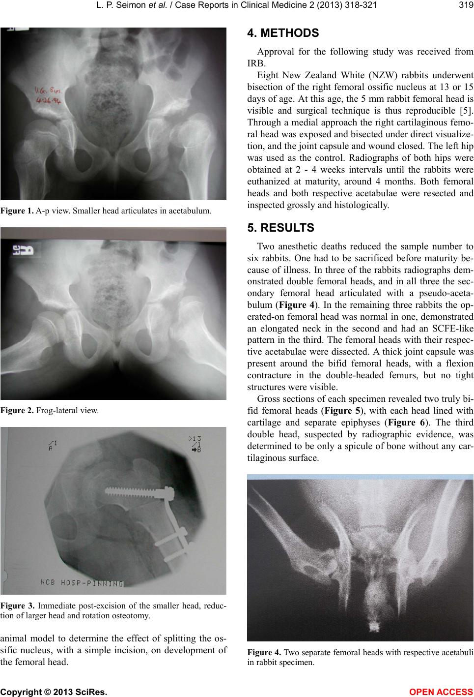 L. P. Seimon et al. / Case Reports in Clinical Medici ne 2 (2013) 318-321 319 Figure 1. A-p view. Smaller head articulates in acetabulum. Figure 2. Frog-lateral view. Figure 3. Immediate post-excision of the smaller head, reduc- tion of larger head and rotation osteotomy. animal model to determine the effect of splitting the os- sific nucleus, with a simple incision, on development of the femoral head. 4. METHODS Approval for the following study was received from IRB. Eight New Zealand White (NZW) rabbits underwent bisection of the right femoral ossific nucleus at 13 or 15 days of age. At this ag e, the 5 mm rabbit femoral head is visible and surgical technique is thus reproducible [5]. Through a medial approach the right cartilaginous femo- ral head was exposed and bisected under direct visualize- tion, and the joint capsu le and wound closed. Th e left hip was used as the control. Radiographs of both hips were obtained at 2 - 4 weeks intervals until the rabbits were euthanized at maturity, around 4 months. Both femoral heads and both respective acetabulae were resected and inspecte d grossly and histolog i c ally. 5. RESULTS Two anesthetic deaths reduced the sample number to six rabbits. One had to be sacrificed before maturity be- cause of illness. In three of the rabbits radiographs dem- onstrated double femoral heads, and in all three the sec- ondary femoral head articulated with a pseudo-aceta- bulum (Figure 4). In the remaining three rabbits the op- erated-on femoral head was normal in one, demonstrated an elongated neck in the second and had an SCFE-like pattern in the third. The femoral heads with their respec- tive acetabulae were dissected. A thick joint capsule was present around the bifid femoral heads, with a flexion contracture in the double-headed femurs, but no tight structures were visible. Gross sections of each specimen revealed two truly bi- fid femoral heads (Figure 5), with each head lined with cartilage and separate epiphyses (Figure 6). The third double head, suspected by radiographic evidence, was determined to be only a spicule of bone without any car- tilaginous surface. Figure 4. Two separate femoral heads with respective acetabuli in rabbit specimen. Copyright © 2013 SciRes. OPEN A CCESS  L. P. Seimon et al. / Case Reports in Clinical Medici ne 2 (2013) 318-321 320 Figure 5. Gross specimen of double femoral heads and aceta- buli. Figure 6. Gross section: two femoral heads with articular car- tilage, single physis. Histology showed all controls to have hemispherical heads. Two bifid femoral heads were present (Figure 7). One of these specimens showed a couple of dead bone spicules at the cleft site, however this was due to remod- eling and not consistent with osteonecrosis. Another spe- cimen showed one head with a second head-like struc- ture, which was actually composed of compact bone with- out a physis. The fourth specimen demonstracted an elongated epiphysis without any scar tissue. The fifth specimen had a widened acetabulum and the last speci- men had irregularities in the piriformis fossa and femoral neck with abundant scar tissue. 6. DISCUSSION Bifurcation of the femoral head is a rare entity, with few reports in the literature [1,2,4]. Our patient is the third to be reported who had prior surgery for DDH. In order to evaluate the hypothesis that surgical trauma during surgery for DDH may be related to the develop- Figure 7. Histology of two separate femoral heads, single phy- sis. ment of a bifid femoral head, an animal model was cre- ated. A bifid femoral head was demonstrated in 2 of 6 rabbits who underwent surgical bisection of the femoral head. Another specimen was similar on radiographic findings, howeve r the histology revealed only on e femo- ral head with a compact bone spicule similar to an exo- stosis. The question remains: why did not all of the femoral heads become bifid post-surgery? The specimens without pathology in the femoral head may have been caused by surgical error. Additionally, the ossific nucleus may not have formed in 13 - 15 days since it has been shown by Heikel [6] that the rabbit ossific nucleus presents in only 43 of 57 animals at 6 - 10 days. Alternatively, the surgi- cal insult may have healed and remodeled. 7. CONCLUSIONS Bifid femoral heads can be induced by splitting the ossific nucleus in immature rabbits. This should be kept in mind when performing surgery around the ossific nu- cleus in immature patients. Focal necrosis and tight sur- rounding structures are not the etiology of the true bifid femoral head. In children in whom a bifid femoral head is id entified, a history of surgery at a young age, for DDH, should be sought. REFERENCES [1] Ferguson, A. (1981) Orthopaedic surgery in infancy and childhood. 5th Edition, Williams and Wilkins, Baltimore, 292-294. [2] Rossman, M. and Jequier, S. (1982) The double-headed femur. Clinical Orthopaedics and Related Research, 162, 170-172. [3] Salter, R., Kostuik, J. and Dallas, S. (1969) Avascular Copyright © 2013 SciRes. OPEN A CCESS  L. P. Seimon et al. / Case Reports in Clinical Medici ne 2 (2013) 318-321 Copyright © 2013 SciRes. OPEN ACCESS 321 necrosis of the femoral head as a complication of the dis- location of the hip in young children. A clinical and ex- perimental investigation. Canadian Journal of Surgery, 12, 44. [4] Rudicel, S., Lee, K. and Pelker, R. (1985) Dimensions of the rabbit femur during growth. American Journal of Vet- erinary Research, 46, 289-299. [5] Heikel, H. (1960) On ossification and growth of certain bones of the rabbit; with a comparison of the skeletal age in the rabbit and in man. Acta Orthopaedica Scandi- navica, 39, 171-185. [6] Osuji, C.K., Chafik, D. and Moseley, C.F. (2009) Unilat- eral proximal bifurcation of the femur. Journal of Pediat- ric Orthopaedics, 29, 695-697. doi:10.1097/BPO.0b013e3181b76985 |