

Paper Menu >>
Journal Menu >>
 Open Journal of Orthopedics, 2013, 3, 208-212 http://dx.doi.org/10.4236/ojo.2013.34038 Published Online August 2013 (http://www.scirp.org/journal/ojo) Copyright © 2013 SciRes. OJO Treatment of Complex Proximal Tibial Fractures (Types V & VI of Schautzker Classification) by Double Plate Fixation with Single Anterior Incision Ebrahim Ghayem Hassankhani*, Farzad Omidi Kashani, Golnaz Ghayem Hassankhani Orthopedic and Spine Surgery, Imam Reza Hospital, Mashad University of Medical Sciences, Mashad, Iran. Email: *[email protected], *[email protected] Received June 1st, 2013; revised July 1st, 2013; accepted July 14th, 2013 Copyright © 2013 Ebrahim Ghayem Hassankhani et al. This is an open access article distributed under the Creative Commons At- tribution License, which permits unrestricted use, distribution, and reproduction in any medium, provided the original work is prop- erly cited. ABSTRACT Background: complex proximal tibial fractures (Types V & VI of Schautzker classification) are the major problems in orthopedic surgery and associated with high complication rates. There are many alternatives in treatment of these frac- tures. Aim: to evaluate the results of double plating with single anterior incision in complex proximal tibial fractures (Types V & VI of Schautzker classification). Methods and Materials: 22 patients (16 males and 6 females) with Types V and VI of Schautzker classification of proximal tibial fractures (14 cases were Type V and 8 cases were Type VI) were treated by double plating with single anterior incision method between May 2006 and May 2011. The bony and functional outcome was evaluated according to Knee Society Score. Results: According to Knee Society Score, the re- sults were as follows: excellent in 19 patients (86.4%), good in 2 patients (9.1%), fair in 1 patient (4/5%), and poor in no patient (0%). Conclusion: the double plate fixation with single anterior incision is the best, effective and simple procedure in treatment of complex proximal tibial fractures (Types V and VI of Schautzker classification). Keywords: Proximal Tibial Fractures; Types V & VI of Schautzker Classification; Double Plate Fixation; Single Anterior Incision 1. Background Complex proximal tibial fractures (Types V & VI of Schautzker classification) are the major problems in or- thopedic surgery and associated with high complication rates. These fractures include bicondylar injuries with significant articular depression; multiple, displaced con- dylar fracture lines; metadiaphyseal fracture extension and comminution; and open wounds or extensive, closed injuries [1-3]. Both the complexity and the soft-tissue disruption of this subgroup of fractures contribute to the high rate of unsatisfactory results which follow bo th non- surgical and surgical management [4,5]. The goals of operative treatment of these fractures in- clude anatomic reduction for restoration of articular con- gruity and alignment, and stable fixation to allow early motion. Dual plating is preferred to other techniques in the set- ting of a significantly displaced fracture of the articular surface, especially in cases with significantly depressed fragments [6,7]. This technique, when performed through a single, midline extensile or double incision with wide stripping of the proximal tibia, has been associated with deep in- fection, wound dehiscence, and soft tissue complications in 23% to 100% of patien ts [8-11]. The two most common complications of double plat- ing with two or single incision are; th e co mpro mised skin and soft tissue envelope, which invites a high rate of complications following attempted open reduction and internal fixation, and poor bone quality and comminuted fracture patterns creating difficulty in achieving stable fixation [3,7,9-11]. The purpose of th is study was to evaluate the resu lts of double plating with single anterior midline incision in complex proximal tibial fractures (Types V & VI of Schautzker classification) and compare this rate to other series in the literature. *Corresponding a uthor. 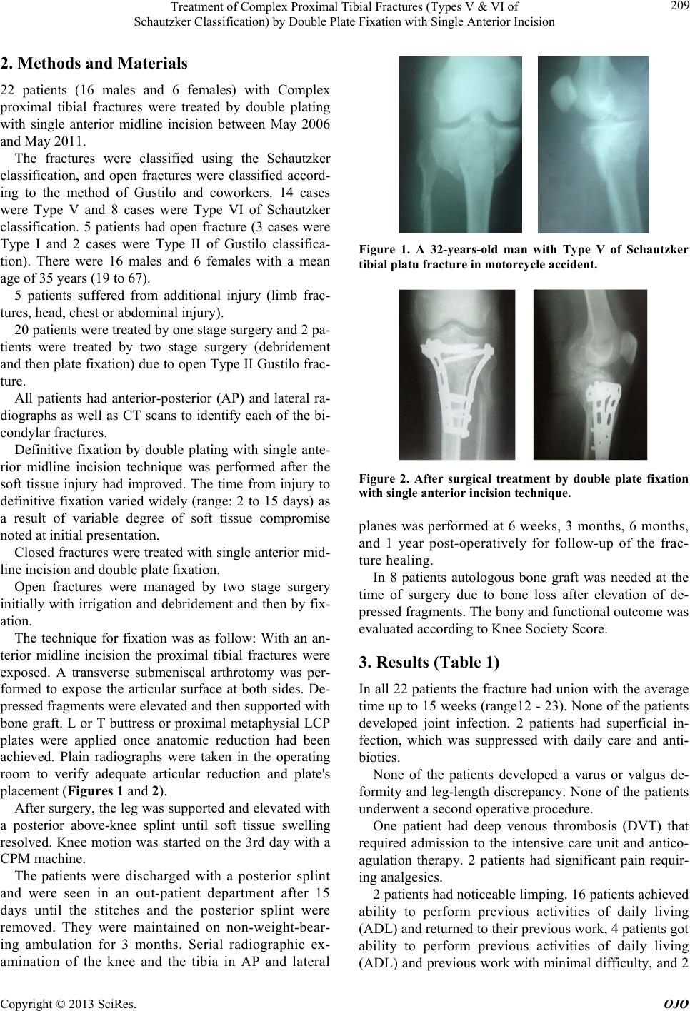 Treatment of Complex Proximal Tibial Fractures (Types V & VI of Schautzker Classification) by Double Plate Fixation with Single Anterior Incision Copyright © 2013 SciRes. OJO 209 2. Methods and Materials 22 patients (16 males and 6 females) with Complex proximal tibial fractures were treated by double plating with single anterior midline incision between May 2006 and May 2011. The fractures were classified using the Schautzker classification, and open fractures were classified accord- ing to the method of Gustilo and coworkers. 14 cases were Type V and 8 cases were Type VI of Schautzker classification. 5 patients had open fracture (3 cases were Type I and 2 cases were Type II of Gustilo classifica- tion). There were 16 males and 6 females with a mean age of 35 years (19 to 67). 5 patients suffered from additional injury (limb frac- tures, head, chest or abdomin a l injury). 20 patients were treated by one stage surgery and 2 pa- tients were treated by two stage surgery (debridement and then plate fixation) due to open Type II Gustilo frac- ture. All patients had anterior-posterior (AP) and lateral ra- diographs as well as CT scans to identify each of the bi- condylar fractures. Definitive fixation by double plating with single ante- rior midline incision technique was performed after the soft tissue injury had improved. The time from injury to definitive fixation varied widely (range: 2 to 15 days) as a result of variable degree of soft tissue compromise noted at initial presentation. Closed fractures were treated with single anterior mid- line incision and double plate fixation. Open fractures were managed by two stage surgery initially with irrigation and debridement and then by fix- ation. The technique for fixation was as follow: With an an- terior midline incision the proximal tibial fractures were exposed. A transverse submeniscal arthrotomy was per- formed to expose the articular surface at both sides. De- pressed fragments were elevated and then supported with bone graft. L or T buttress or proximal metaphysial LCP plates were applied once anatomic reduction had been achieved. Plain radiographs were taken in the operating room to verify adequate articular reduction and plate's placement (Figures 1 and 2). After surgery, the leg was supported and elevated with a posterior above-knee splint until soft tissue swelling resolved. Knee motion was started on the 3rd day with a CPM machine. The patients were discharged with a posterior splint and were seen in an out-patient department after 15 days until the stitches and the posterior splint were removed. They were maintained on non-weight-bear- ing ambulation for 3 months. Serial radiographic ex- amination of the knee and the tibia in AP and lateral Figure 1. A 32-years-old man with Type V of Schautzker tibial platu fracture in motorcycle accident. Figure 2. After surgical treatment by double plate fixation with single anterior incision te chnique . planes was performed at 6 weeks, 3 months, 6 months, and 1 year post-operatively for follow-up of the frac- ture healing. In 8 patients autologous bone graft was needed at the time of surgery due to bone loss after elevation of de- pressed fragments. The bony and functional outcome was evaluated according to Knee Society Score. 3. Results (Table 1) In all 22 patients the fracture had un ion with the average time up to 15 weeks (range12 - 23). None of the patients developed joint infection. 2 patients had superficial in- fection, which was suppressed with daily care and anti- biotics. None of the patients developed a varus or valgus de- formity and leg-length discrepancy. None of the patients underwent a second operative procedure. One patient had deep venous thrombosis (DVT) that required admission to the intensive care unit and antico- agulation therapy. 2 patients had significant pain requir- ing analgesics. 2 patients had noticeable limping. 16 patients achieved ability to perform previous activities of daily living (ADL) and returned to th eir previou s work, 4 p atient s got ability to perform previous activities of daily living (ADL) and previous work with minimal difficulty, and 2 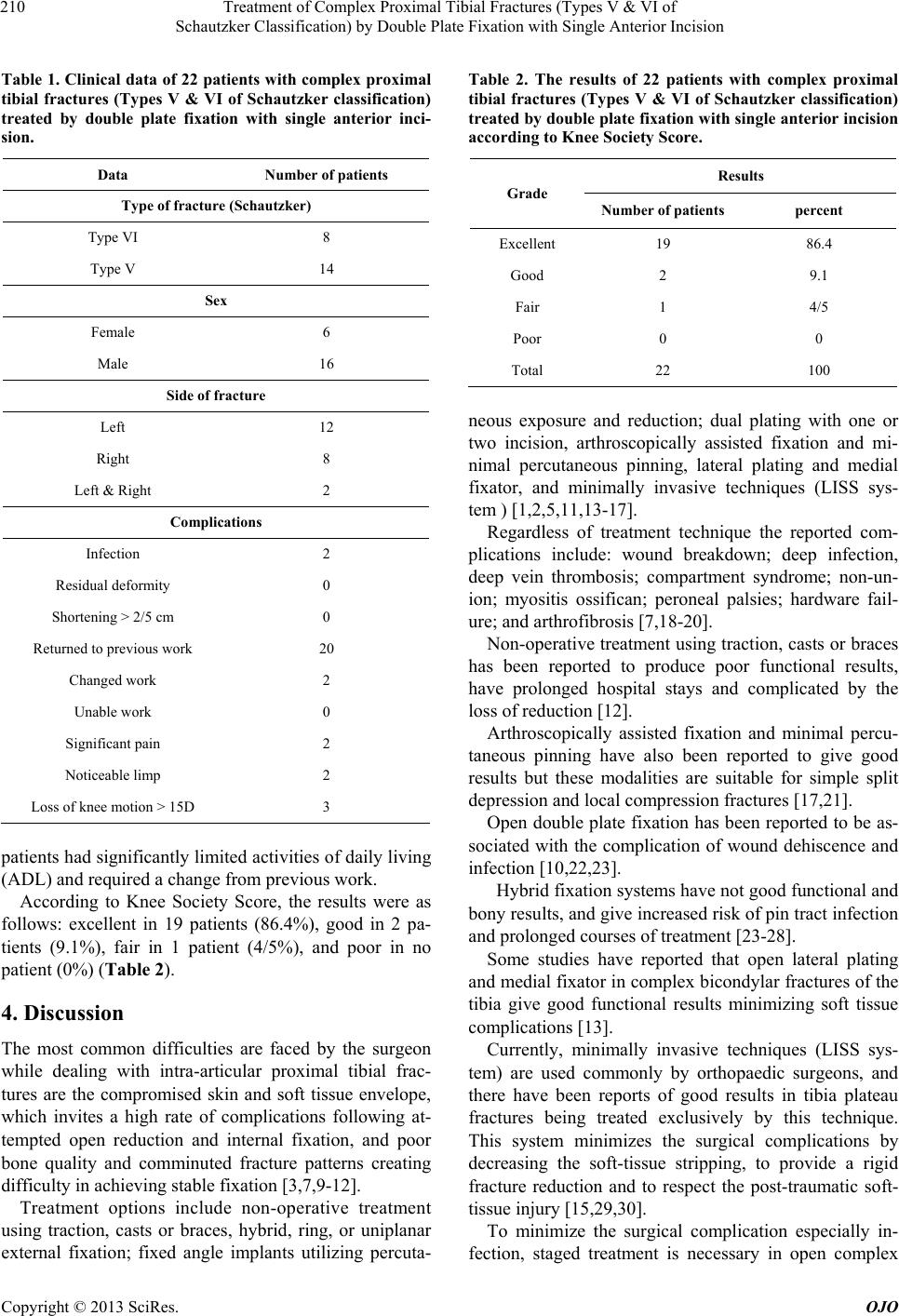 Treatment of Complex Proximal Tibial Fractures (Types V & VI of Schautzker Classification) by Double Plate Fixation with Single Anterior Incision Copyright © 2013 SciRes. OJO 210 Table 1. Clinical data of 22 patients with complex proximal tibial fractures (Types V & VI of Schautzker classification) treated by double plate fixation with single anterior inci- sion. Data Number of patients Type of fracture (Schautzker) Type VI 8 Type V 14 Sex Female 6 Male 16 Side of fracture Left 12 Right 8 Left & Right 2 Complications Infection 2 Residual deformity 0 Shortening > 2/5 cm 0 Returned to previous work 20 Changed work 2 Unable work 0 Significant pain 2 Noticeable limp 2 Loss of knee motion > 15D 3 patients had significantly limited activities of daily liv ing (ADL) and required a change from previous work. According to Knee Society Score, the results were as follows: excellent in 19 patients (86.4%), good in 2 pa- tients (9.1%), fair in 1 patient (4/5%), and poor in no patient (0%) (Table 2). 4. Discussion The most common difficulties are faced by the surgeon while dealing with intra-articular proximal tibial frac- tures are the compromised skin and soft tissue envelope, which invites a high rate of complications following at- tempted open reduction and internal fixation, and poor bone quality and comminuted fracture patterns creating difficulty in achieving stable fixation [3,7,9-12]. Treatment options include non-operative treatment using traction, casts or braces, hybrid, ring, or uniplanar external fixation; fixed angle implants utilizing percuta- Table 2. The results of 22 patients with complex proximal tibial fractures (Types V & VI of Schautzker classification) treated by double plate fixation with single anterior incision according to Knee Society Score. Results Grade Number of patients percent Excellent 19 86.4 Good 2 9.1 Fair 1 4/5 Poor 0 0 Total 22 100 neous exposure and reduction; dual plating with one or two incision, arthroscopically assisted fixation and mi- nimal percutaneous pinning, lateral plating and medial fixator, and minimally invasive techniques (LISS sys- tem ) [1,2,5,11,13-17]. Regardless of treatment technique the reported com- plications include: wound breakdown; deep infection, deep vein thrombosis; compartment syndrome; non-un- ion; myositis ossifican; peroneal palsies; hardware fail- ure; and arthrofibrosis [7,18-20]. Non-operative treatment using traction, casts or braces has been reported to produce poor functional results, have prolonged hospital stays and complicated by the loss of redu ct ion [1 2]. Arthroscopically assisted fixation and minimal percu- taneous pinning have also been reported to give good results but these modalities are suitable for simple split depression and local compression fr actures [17,21]. Open double plate fixation has been reported to be as- sociated with the complication of wound dehiscence and infection [10,22,23]. Hybrid fixation systems have not good functional an d bony results, and give increased risk of pin tract infection and prolonged courses of treatment [23-28]. Some studies have reported that open lateral plating and medial fixator in complex bicondylar fractures of the tibia give good functional results minimizing soft tissue complications [13]. Currently, minimally invasive techniques (LISS sys- tem) are used commonly by orthopaedic surgeons, and there have been reports of good results in tibia plateau fractures being treated exclusively by this technique. This system minimizes the surgical complications by decreasing the soft-tissue stripping, to provide a rigid fracture reduction and to respect the post-traumatic soft- tissue injury [15,29,30]. To minimize the surgical complication especially in- fection, staged treatment is necessary in open complex  Treatment of Complex Proximal Tibial Fractures (Types V & VI of Schautzker Classification) by Double Plate Fixation with Single Anterior Incision Copyright © 2013 SciRes. OJO 211 proximal tibial fractures [31]. In this study 2 patients with open Type II Gustilo fractures were treated by two stage surgery (debridement and then plate fixation). Anatomical knee joint reduction, the relative stability and alignment of the proximal tibia allowing the earliest knee mobilization, while keeping complications to a mi- nimum rate, are the major goals in the treatment of com- plex proximal tibial fractures [27,32]. In order to obtain stability of bicondylar and complex proximal tibial fractures, reduction and fixation of both medial and lateral columns is necessary. Dual plating successfully gives a good stability by buttressing both columns; but high rate of complications associated with this open techniqu e is reported [10,11]. Steven N., et al. reported wound dehiscence and infec- tion in proximal tibial fractures treated with double plate fixation [33]. In our study superficial infection was ob- served in 2 cases and no sof t tissue breakdown was noted. We also had good functional and bony results. In this study, our results demonstrate a lower risk for deep infection and soft tissue complications, good func- tional and bony results in complex proximal tibial frac- tures when compared to earlier reports about other tech- niques. 5. Conclusion The double plate fixation with single anterior incision is the best, effective and simple procedure in treatment of complex proximal tibial fractures (Types V and VI of Schautzker classification) with lower risk for deep infec- tion and good results when compared to other reported techniques. 6. Acknowledgements The authors thank orthopaedic research center group for their assistance. REFERENCES [1] W. J. Mills and S. E. Nork, “Open Reduction and Internal Fixation of High-Energy Tibial Plateau Fractures,” Jour- nal of Orthopaedic Trauma, Vol. 18, No. 10, 2004, pp. 649-657. doi:10.1097/00005131-200411000-00001 [2] J. T. Watson, “High-Energy Fractures of the Tibial Pla- teau,” Orthopedic Clinics of North America, Vol. 25, No. 4, 1994, pp. 723-752. [3] K. Christensen, J. Powell and R. Bucholz, “Early Results of a New Technique for Treatment of High Grade Tibial Plateau Fractures,” Journal of Orthopaedic Trauma, Vol. 4, 1990, p. 226. doi:10.1097/00005131-199004020-00059 [4] C. P. Blokker, C. H. Rorabeck and R. B. Bourne, “Tibial Plateau Fractures: An Analysis of the Results of Treat- ment in 60 Patients,” Clinical Orthopaedics and Related Research, No. 182, 1984, pp. 193-199. [5] A. M. Ali, M. Burton, M. Hashmi and M. Saleh, “Out- come of Complex Fractures of the Tibial Plateau Treated with a Beam-Loading Ring Fixation System,” The Jour- nal of Bone and Joint Surgery, Vol. 85, No. 5, 2003, pp. 691-699. [6] D. P. Barei, S. E. Nork, W. J. Mills, et al., “Complica- tions Associated with Internal Fixation of High-Energy Bicondylar Tibial Plateau Fractures Utilizing a Two-In- cision Technique,” Journal of Orthopaedic Trauma, Vol. 18, No. 10, 2004, pp. 649-657. doi:10.1097/00005131-200411000-00001 [7] S. Singh, P. R. Patel, A. K. Joshi, R. N. Naik, C. Nagaraj and S. Kumar, “Biological Approach to Treatment of Intra- Articular Proximal Tibial Fractures with Double Osteo- synthesis,” International Orthopaedics, Vol. 33, No. 1, 2009, pp. 271-274. doi:10.1007/s00264-007-0480-0 [8] M. J. Young and R. L. Barrack, “Complications of Inter- nal Fixation of Tibial Plateau Fractures,” Orthopedic Re- views, Vol. 23, No. 2, 1994, pp. 149-154. [9] D. P. Barei, S. E. Nork, W. J. Mills, et al., “Functional Outcomes of Severe Bicondylar Tibial Plateau Fractures Treated with Dual Incisions and Medial and Lateral Plates,” The Journal of Bone and Joint Surgery. Ameri- can Volume, Vol. 88, No. 8, 2006, pp. 1713-1721. doi:10.2106/JBJS.E.00907 [10] Z. Yu, L. Zheng, Y. Zhang, J. Li and B. Ma, “Functional and Radiological Evaluations of High-Energy Tibial Pla- teau Fractures Treated with Double-Buttress Plate Fixa- tion,” European Journal of Medical Research, Vol. 14, No. 5, 2009, pp. 200-205. doi:10.1186/2047-783X-14-5-200 [11] C. R. Perry and G. Evans, “New Surgical Approach to Fractures of the Lateral Tibial Plateau,” The Journal of Bone & Joint Surgery, Vol. 66, No. 8, 1984, pp. 1236- 1240. [12] A. Apley, “Fractures of the Tibial Plateau,” Orthopedic Clinics of North America, Vol. 10, 1979, pp. 61-74. [13] J. L. Marsh, S. T. Smith and T. T. Do, “External Fixation and Limited Internal Fixation for Complex Fractures of the Tibial Plateau,” The Journal of Bone and Joint Sur- gery. American Volume, Vol. 77, No. 5, 1995, pp. 661- 673. [14] A. Kumar and A. P. Whittle, “Treatment of Complex (Schautzker Type VI) Fractures of the Tibial Plateau with Circular Wire External Fixation: Retrospective Case Re- view,” Journal of Orthopaedic Trauma, Vol. 14, No. 5, 2000, pp. 339-344. doi:10.1097/00005131-200006000-00006 [15] R. Jiang, C. Luo, M. Wan g, T. Yang and B. Zeng, “A Com- parative Study of Less Invasive Stabilization System (LISS) Fixation and Two-Incision Double Plating for the Treatment of Bicondylar Tibial Plateau Fractures,” Knee, Vol. 15, No. 2, 2008, pp. 139-143. doi:10.1016/j.knee.2007.12.001 [16] E. Strauss, R. Schwarzkopf, F. Kummer and K. Egol, “The Current Status of Locked Plating: The Good, the Bad, and  Treatment of Complex Proximal Tibial Fractures (Types V & VI of Schautzker Classification) by Double Plate Fixation with Single Anterior Incision Copyright © 2013 SciRes. OJO 212 the Ugly,” Journal of Orthopaedic Trauma, Vol. 22, No. 7, 2008, pp. 479-486. doi:10.1097/BOT.0b013e31817996d6 [17] Y. S. Chan, L. J. Yuan, S. S. Hung, C. J. Wang, S. W. Yu, C. Y. Chen, E. K. Chao and M. S. Lee, “Arthroscopic- Assisted Reduction with Bilateral Buttress Plate Fixation of Complex Tibial Plateau Fractures,” Arthroscopy, Vol. 19, No. 9, 2003, pp. 974-984. doi:10.1016/j.arthro.2003.09.038 [18] S. A. Sems, B. A. Levy , K. Dajani, D. A. Herrera and D. C. Templeman, “Incidence of Deep Venous Thrombosis after Temporary Joint Spanning External Fixation for Complex Lower Extremity Injuries,” Journal of Trauma, Vol. 66, No. 4, 2009, pp. 1164-1166. doi:10.1097/TA.0b013e318183801e [19] N. Manidakis, A. Dosani, R. Dimitriou, D. Stengel, S. Matthews and P. Giannoudis, “Tibial Plateau Fractures: Functional Outcome and Incidence of Osteoarthritis in 125 Cases,” International Orthopaedics, Vol. 34, No. 4, 2010, pp. 565-570. doi:10.1007/s00264-009-0790-5 [20] M. Subasi, A. Kapukaya, H. Arslan, E. Ozkul and O. Cebesoy, “Outcome of Open Comminuted Tibial Plateau Fractures Treated Using an External Fixator,” Journal of Orthopaedic Science, Vol. 12, No. 4, 2007, pp. 347-353. doi:10.1007/s00776-007-1149-7 [21] C. A. Gaunche and A. W. Markman, “Arthroscopic Man- agement of Tibial Plateau Fractures,” Arthroscopy, Vol. 9, No. 4, 1993, pp. 467-471. doi:10.1016/S0749-8063(05)80324-X [22] G. Kumar, N. Peterson and B. Narayan, “Bicondylar Tibial Fractures: Internal or External Fixation?” Indian Journal of Orthopaedics, Vol. 45, No. 2, 2011, pp. 116- 124. doi:10.4103/0019-5413.77130 [23] T. F. Higgins, J. Klatt and K. N. Bachus, “Biomechanical Analysis of Bicondylar Tibial Plateau Fixation: How Does Lateral Locking Plate Fixation Compare to Dual Plate Fixation?” Journal of Orthopaedic Trauma, Vol. 21, No. 5, 2007, pp. 301-306. doi:10.1097/BOT.0b013e3180500359 [24] H. Barbary, H. A. Ghani, H. Misbah and K. Salem, “Complex Tibial Plateau Fractures Treated with Ilizarov External Fixator with or without Minimal Internal Fixa- tion,” International Orthopaedics, Vol. 29, No. 3, 2005, pp. 182-185. doi:10.1007/s00264-005-0638-6 [25] H. M. Ariffin, N. M. Mahdi, S. A. Rhani, A. Baharudin and M. H. Shukur, “Modified Hybrid Fixator for High- Energy Schautzker V and VI Tibial Plateau Fractures,” Strategies Trauma Limb Reconstruct, Vol. 6, No. 1, 2011, pp. 21-26. doi:10.1007/s11751-011-0105-4 [26] T. M. McLaurin, “Hybrid Ring External Fixation in the Treatment of Complex Tibial Plateau Fractures,” Tech- niques in Knee Surgery, Vol. 4, 2005, pp. 226-236. doi:10.1097/01.btk.0000187517.87625.3b [27] J. A. Hall, M. J. Beue rlein and M. D. McKee, “Open Re- duction and Internal Fixation Compared with Circular Fixator Application for Bicondylar Tibial Plateau Frac- tures,” The Journal of Bone and Joint Surgery. American Volume, Vol. 1, No. 91, 2009, pp. 74-88. [28] D. Mahadeva, M. L. Costa and A. Gaffey, “Open Reduc- tion and Internal Fixation versus Hybrid Fixation for Bi- condylar/Severe Tibial Plateau Fractures: A Systematic Review of the Literature,” Archives of Orthopaedic and Trauma Surgery, Vol. 128, No. 10, 2008, pp. 1169-1175. doi:10.1007/s00402-007-0520-7 [29] J. A. Lee, S. A. Papadakis, C. Moon and C. G. Zalavras, “Tibial Plateau Fractures Treated with the Less Invasive Stabilization System,” International Orthopaedics, Vol. 31, No. 3, 2007, pp. 415-418. doi:10.1007/s00264-006-0176-x [30] J. P. Stannard, T. C. Wilson, D. A. Volgas and J. E. Alon- so, “The Less Invasive Stabilization System in the Treat- ment of Complex Fractures of the Tibial Plateau: Short- Term Results,” Journal of Orthopaedic Trauma, Vol. 18, No. 8, 2004, pp. 552-558. doi:10.1097/00005131-200409000-00012 [31] K. A. Egol, N. C. Tejwani, E. L. Capla, P. L. Wolinsky and K. J. Koval, “Staged Management of High-Energy Proximal Tibia Fractures (OTA Types 41): The Results of a Prospective, Standardized Protocol,” Journal of Ortho- paedic Trauma, Vol. 19, No. 7, 2005, pp. 448-455. doi:10.1097/01.bot.0000171881.11205.80 [32] S. Eggli, M. J. Hartel, S. Kohl, U. Haupt, A. K. Exadak- tylos and C. Röder, “Unstable Bicondylar Tibial Plateau Fractures: A Clinical Investigation,” Journal of Ortho- paedic Trauma, Vol. 22, No. 10, 2008, pp. 673-679. doi:10.1097/BOT.0b013e31818b1452 [33] S. N. Shah and M. A. Karunakar, “Early Wound Compli- cations after Operative Treatment of High Energy Tibial Plateau Fractures through Two Incisions,” Bulletin of the NYU Hospital for Joint Diseases, Vol. 65, No. 2, 2007, pp. 115-119. |