Paper Menu >>
Journal Menu >>
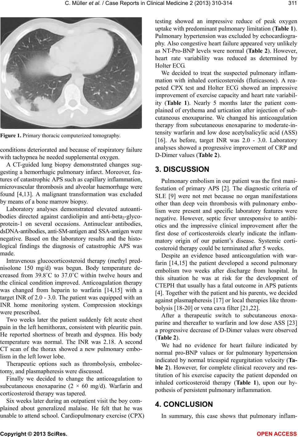
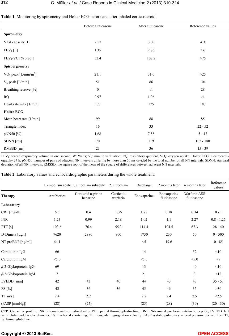
![]() Vol.2, No.5, 310-314 (2013) Case Reports in Clinical Medicine http://dx.doi.org/10.4236/crcm.2013.25083 Recurrent pulmonary embolism in a boy with antiphospholipid syndrome Christian Müller1*, Christian Willaschek1, Mathias Borst2, Reiner Buchhorn1 1Department of Pediatrics, Caritas Krankenhaus, Bad Mergentheim, Germany; *Corresponding Author: [email protected] 2Department of Medicine, Caritas Krankenhaus, Bad Mergentheim, Germany Received 2 October 2012; revised 16 November 2012; accepted 8 December 2012 Copyright © 2013 Christian Müller et al. This is an open access article distributed under the Creative Commons Attribution License, which permits unrestricted use, distribution, and reproduction in any medium, provided the original work is properly cited. ABSTRACT We report a 14-year-old patient with recurrent pulmonary embolism due to catastrophic anti- phospholipid syndrome (APS) with severe pul- monary inflammation. We considered elevated antibodies against cardiolipin and anti-beta2- glycoprotein-1, but no clinical nor laboratory manifest atio ns of syst emic lupus erythem atosus (SLE). Pulmonary embolism had been the first manifestation of catastrophic APS in this patient. We prescribed warfarin and systemic corticos- teroids. A second embolism appeared during anticoagulation with warfarin. This event led to a marked decrease of his physical performance due to his obstructive and restrictive lung dis- ease. Anticoagulation was changed from enteral warfarin to subcutaneous enoxaparine. We also prescribed inhaled corticosteroid which led to an improvement of his respiratory symptoms and overall poor physical condition. Keywords: Pulmonary Embolism; Antiphospholipid Syndrome; APS; Inhaled Corticosteroid 1. INTRODUCTION APS is a rare disease, especially in children [1]. As opposed to the secondary form of APS occurring in pa- tients with autoimmune disorders, predominately SLE. APS is said to be primary if there are no features of SLE or other autoimmune disorders [1,2]. Clinical manifestations of primary APS are variable [3]: one feature is pulmonary embolism [4] which in one case study occurred in 2.1% of patients with APS [5]. Especially in children, pulmonary embolism associ- ated with APS is rare [6], but may be its first manifesta- tion [2]. Chronic thromboembolic pulmonary hypertension (CTE- PH) due to APS is a severe disease with high morbidity and mortality [2,7]. Histopathologically, there may be signs of catastrophic APS such as alveolar hemorrhage, microvascular thrombosis and pulmonary capillaritis, which is the most severe course of high mortality due to multi- ple vascular occlusive events [8]. Pulmonary embolism in APS associated with fever and elevated autoantibodies are usually caused by SLE. Diagnosis of SLE is based on clinical and laboratory features according to the classifi- cation criteria [9-12]. Because there was no evidence of autoimmune disorders the present case was classified to primary APS. 2. CASE REPORT We report a 14-year-old male in good overall physical condition (BMI: 19.6 kg/m2) with no history of severe diseases. He did not consume any medication, nicotine, alcohol, or other drugs. The familial history was unre- markable. He presented with acute chest pain and fever up to 40˚C having lasted for one week. There was no trauma recalled by the patient or his family. He presented with pain, which was located in the right hemithorax and increased in intensity when in supine position. During physical examination a reduced respiratory sound over his right lower pulmonary lobe was found. Chest x-ray showed a consolidation in the right lower pulmonary lobe. C-reactive protein (CRP) was elevated to 9.37 mg/dL. An initial diagnosis of pneumonia was made and antibiotic therapy (Cefuroxim, Doxycycline, Gentamicin) was initiated. Five days later there was no clinical improvement. A computed tomography (CT) scan of the thorax (Figure 1) showed signs of past pulmonary embolism in the right lower lobe. We started effective anticoagulation with intravenous heparin and enhanced the antibiotic therapy (Cefuroxim, Doxycycline, Gentamicin and Vancomycin). An ultrasound examination showed a deep venous throm- bosis of the left lower extremity. The patient’s clinical Copyright © 2013 SciRes. OPEN ACCESS ![]() C. Müller et al. / Case Reports in Clinical Medicine 2 (2013) 310-314 311 Figure 1. Primary thoracic computerized tomography. conditions deteriorated and because of respiratory failure with tachypnea he needed supplemental oxygen. A CT-guided lung biopsy demonstrated changes sug- gesting a hemorrhagic pulmonary infarct. Moreover, fea- tures of catastrophic APS such as capillary inflammation, microvascular thrombosis and alveolar haemorrhage were found [4,13]. A malignant transformation was excluded by means of a bone marrow biopsy. Laboratory analyses demonstrated elevated autoanti- bodies directed against cardiolipin and anti-beta2-glyco- protein-1 on several occasions. Antinuclear antibodies, dsDNA-antibodies, anti-SM-antigen and SSA-antigen were negative. Based on the laboratory results and the histo- logical findings the diagnosis of catastrophic APS was made. Intravenous glucocorticosteroid therapy (methyl pred- nisolone 150 mg/d) was begun. Body temperature de- creased from 39.8˚C to 37.0˚C within twelve hours and the clinical condition improved. Anticoagulation therapy was changed from heparin to warfarin [14,15] with a target INR of 2.0 - 3.0. The patient was equipped with an INR home monitoring system. Compression stockings were prescribed. Two weeks later the patient suddenly felt acute chest pain in the left hemithorax, consistent with pleuritic pain. He reported shortness of breath and dyspnea. His body temperature was normal. The INR was 2.18. A second CT scan of the thorax showed a new pulmonary embo- lism in the left lower lobe. Therapeutic options such as thrombolysis, embolec- tomy, and plasmapheresis were discussed. Finally we decided to change the anticoagulation to subcutaneous enoxaparine (2 × 60 mg/d). Warfarin and corticosteroid therapy was tapered. Six weeks later during an outpatient visit the boy com- plained about generalized malaise. He felt that he was unable to attend school. Cardiopulmonary exercise (CPX) testing showed an impressive reduce of peak oxygen uptake with predominant pulmonary limitation (Table 1). Pulmonary hypertension was excluded by echocardiogra- phy. Also congestive heart failure appeared very unlikely as NT-Pro-BNP levels were normal (Ta bl e 2). However, heart rate variability was reduced as determined by Holter ECG. We decided to treat the suspected pulmonary inflam- mation with inhaled corticosteroids (fluticasone). A rea- peted CPX test and Holter ECG showed an impressive improvement of exercise capacity and heart rate variabil- ity (Table 1). Nearly 5 months later the patient com- plained of erythema and urtication after injection of sub- cutaneous enoxaparine. We changed his anticoagulation therapy from subcutaneous enoxaparine to moderate-in- tensity warfarin and low dose acetylsalicylic acid (ASS) [16]. As before, target INR was 2.0 - 3.0. Laboratory analyses showed a progressive improvement of CRP and D-Dimer values (Table 2). 3. DISCUSSION Pulmonary embolism in our patient was the first mani- festation of primary APS [2]. The diagnostic criteria of SLE [9] were not met because no organ manifestations other than deep vein thrombosis with pulmonary embo- lism were present and specific laboratory features were negative. However, septic fever unresponsive to antibi- otics and the impressive clinical improvement after the first dose of corticosteroids clearly indicate the inflam- matory origin of our patient’s disease. Systemic corti- costeroid therapy could be terminated after 5 weeks. Despite an evidence based anticoagulation with war- farin [14,15] the patient developed a second pulmonary embolism two weeks after discharge from hospital. In this situation he was at risk for the development of CTEPH that usually has a fatal outcome in APS patients [4]. Together with the patient and his parents, we decided against plasmapheresis [17] or local therapies like throm- bolysis [18-20] or vena cava filter [21,22]. After a therapeutic switch to subcutaneous enoxa- parine and thereafter to warfarin and low dose ASS [23] a progressive decrease of D-Dimer values were observed (Table 2). We had no evidence for heart failure indicated by normal pro-BNP values or for pulmonary hypertension indicated by normal tricuspid regurgitation velocity (Ta- ble 2). However, for complete clinical recovery and res- titution of his exercise capacity the patient depended on inhaled corticosteroid therapy (Table 1), upon our hy- pothesis of persistent pulmonary inflammation. 4. CONCLUSION In summary, this case shows that pulmonary inflam- Copyright © 2013 SciRes. OPEN ACCESS ![]() C. Müller et al. / Case Reports in Clinical Medicine 2 (2013) 310-314 Copyright © 2013 SciRes. OPEN ACCESS 312 Table 1. Monitoring by spirometry and Holter ECG before and after inhaled corticosteroid. Before fluticasone After fluticasone Reference values Spirometry Vital capacity [L] 2.57 3.09 4.3 FEV1 [L] 1.35 2.76 3.6 FEV1/VC [% pred.] 52.4 107.2 >75 Spiroergometry VO2 peak [L/min/m2] 21.1 31.0 >25 VE peak [L/min] 51 86 104 Breathing reserve [%] 0 11 28 RQ 0.97 1.06 >1 Heart rate max [1/min] 173 175 187 Holter ECG Mean heart rate [1/min] 99 88 85 Triangle index 16 33 22 - 52 pNN50 [%] 1,68 7,58 5 - 47 SDNN [ms] 70 119 102 - 180 RMSSD [ms] 23 36 15 - 39 FEV1: forced exspiratory volume in one second; W: Watts; VE: minute ventilation; RQ: respiratory quotient; VO2: oxygen uptake. Holter ECG: electrocardi- ography 24 h; pNN50: number of pairs of adjacent NN intervals differing by more than 50 ms divided by the total number of all NN intervals; SDNN: standard deviation of all NN intervals; RMSSD: the square root of the mean of the square of differences between adjacent NN intervals. Table 2. Laboratory values and echocardiographic parameters during the whole treatment. 1. embolism acute 1. embolism subacute2. embolism Discharge 2 months later 4 months later Reference values Therapy Antibiotics Corticoid aspirine heparine Corticoid warfarin Enoxaparine Enoxaparine fluticasone Warfarin ASS fluticasone Laboratory CRP [mg/dl] 6.3 0.4 1.36 1.78 0.18 0.34 0 - 1 INR 1.23 0.99 2.18 1.02 1.1 2.27 0.8 - 1.25 PTT [s] 103.6 76.4 55.3 114.4 104.5 67.3 28 - 40 D-Dimers [μg/l] 7620 2980 900 1730 230 50 0 - 500 NT-proBNP [pg/ml] 64.1 <5 19.6 0 - 85 Cardiolipin IgG 66 14 52 <10 Cardiolipin IgM <5.0 <5.0 <5.0 <7 β-2-Glykoprotein IgG 69 13 40 <10 β-2-Glykoprotein IgM 7 21 3 <12 LVEDD [mm] 42 43 40 44 43 43 35 - 51 FS [%] 42 36 36 45 46 35 >30 TI [m/s] 2.4 2.2 2.2 2.4 2.5 <2.5 (PASP [mmHg]) (28) (25) (25) (28) (30) (20 - 30) CRP: C-reactive protein; INR: international normalized ratio; PTT: partial thromboplastin time; BNP: N-terminal pro brain natriuretic peptide; LVEDD: left ventriclular enddiastolic diameter; FS: fractional shortening; TI: tricuspidal regurgitation velocity; PASP systolic pulmonary arterial pressure derived from TI; Ig: Immunglobuline. ![]() C. Müller et al. / Case Reports in Clinical Medicine 2 (2013) 310-314 313 mation can play an important role in manifestation of primary APS that requires consequent immunosuppres- sant treatment. Inflammation and anticoagulation have to be monitored by D-Dimer values, CRP values and spe- cific tests that depend upon the regiments. A critical cardiopulmonary monitoring including echo- cardiography, ECG, spirometry, CPX and Holter ECG is necessary to recognize pulmonary hypertension and de- creased endurance of the patient. Cardiac limitations due to pulmonary hypertension may be treated by endarter- ectomy or endothelin receptor antagonist, if patients are deemed inoperable. In patients with only pulmonary limitations like in our case inhaled corticosteroid seems to be a good course of treatment. REFERENCES [1] von Scheven, E., Glidden, D.V. and Elder, M.E. (2002) Anti-beta2-glycoprotein I antibodies in pediatric systemic lupus erythematosus and antiphospholipid syndrome. Ar- thritis & Rheumatism, 47, 414-420. doi:10.1002/art.10510 [2] Espinosa, G., Cervera, R., Font, J. and Asherson, R.A. (2002) The lung in the antiphospholipid syndrome. An- nals of the Rheumatic Diseases, 61, 195-198. doi:10.1136/ard.61.3.195 [3] Asherson, R.A., et al. (1998) Catastrophic antiphospho- lipid syndrome. Clinical and laboratory features of 50 pa- tients. Medicine (Baltimore), 77, 195-207. doi:10.1097/00005792-199805000-00005 [4] Asherson, R.A. and Cervera, R. (1995) Review: Anti- phospholipid antibodies and the lung. Journal of Rheu- matology, 22, 62-66. [5] Cervera, R., et al. (2009) Morbidity and mortality in the antiphospholipid syndrome during a 5-year period: A multicenter prospective study of 1,000 patients. Annals of the Rheumatic Diseases, 68, 1428-1432. [6] Olivier, C., Blondiaux, E., Blanc, T., Borg, J.Y. and Da- cher, J.N. (2006) Catastrophic antiphospholipid syndrome and pulmonary embolism in a 3-year-old child. Pediatric Radiology, 36, 870-873. doi:10.1007/s00247-006-0183-5 [7] Porres-Aguilar, M., Pena-Ruiz, M.A., Burgos, J.D., Por- res-Munoz, M. and Hughes, H.W. (2008) Chronic throm- boembolic pulmonary hypertension as an uncommon pre- sentation of primary antiphospholipid syndrome. Journal of the National Medical Association, 100, 734-736. [8] Espinosa, G., Bucciarelli, S., Asherson, R.A. and Cervera, R. (2008) Morbidity and mortality in the catastrophic an- tiphospholipid syndrome: Pathophysiology, causes of death, and prognostic factors. Seminars in Thrombosis and He- mostasis, 34, 290-294. doi:10.1055/s-0028-1082274 [9] Tan, E.M., et al. (1982) The 1982 revised criteria for the classification of systemic lupus erythematosus. Arthritis & Rheumatism, 25, 1271-1277. doi:10.1002/art.1780251101 [10] Hochberg, M.C. (1997) Updating the American College of Rheumatology revised criteria for the classification of systemic lupus erythematosus. Arthritis & Rheumatism, 40, 1725. doi:10.1002/art.1780400928 [11] Petri, M. (2005) Review of classification criteria for sys- temic lupus erythematosus. Rheumatic Disease Clinics of North America, 31, 245-254. doi:10.1016/j.rdc.2005.01.009 [12] Gaubitz, M. and Schotte, H. (2005) Early diagnosis in patients with systemic lupus erythematosus (SLE). Zeits- chrift für Rheumatologie, 64, 547-552. doi:10.1007/s00393-005-0795-4 [13] Gertner, E. and Lie, J.T. (1993) Pulmonary capillaritis, alveolar hemorrhage, and recurrent microvascular throm- bosis in primary antiphospholipid syndrome. Journal of Rheumatology, 20, 1224-1228. [14] Lim, W., Crowther, M.A. and Eikelboom, J.W. (2006) Management of antiphospholipid antibody syndrome: A systematic review. JAMA, 295, 1050-1057. doi:10.1001/jama.295.9.1050 [15] Crowther, M.A., et al. (2003) A comparison of two inten- sities of warfarin for the prevention of recurrent thrombo- sis in patients with the antiphospholipid antibody syn- drome. New England Journal of Medicine, 349, 1133- 1138. doi:10.1056/NEJMoa035241 [16] Ruiz-Irastorza, G., Hunt, B.J. and Khamashta, M.A. (2007) A systematic review of secondary thromboprophylaxis in patients with antiphospholipid antibodies. Arthritis & Rheu- matism, 57, 1487-1495. doi:10.1002/art.23109 [17] Castro, P., et al. (2003) Catastrophic antiphospholipid syndrome and acute heart failure. Report of a case. Re- vista Medica de Chile, 131, 1037-1041. doi:10.4067/S0034-98872003000900010 [18] Huang, H.J., Wang, J.N., Tsai, Y.C., Lin, C.S. and Wu, J.M. (2002) Intrapulmonary thrombolytic therapy in a child with acute pulmonary embolism due to primary an- tiphospholipid syndrome. Acta Paediatrica Taiwanica, 43, 351-353. [19] Kim, W.J., Hong, Y.K. and Yoo, W.H. (2008) Epidural hematoma mimicking transverse myelitis in a patient with primary antiphospholipid syndrome. Rheumatology In- ternational, 28, 709-712. doi:10.1007/s00296-007-0512-5 [20] Ordi-Ros, J., Perez-Peman, P. and Monasterio, J. (1994) Clinical and therapeutic aspects associated to phosphol- ipid binding antibodies (lupus anticoagulant and anticar- diolipin antibodies). Haemostasis, 24, 165-174. [21] Cherian, J. and Gertner, E. (2005) Recurrent pulmonary embolism despite inferior vena cava filter placement in patients with the antiphospholipid syndrome. Journal of Clinical Rheumatology, 11, 56-58. doi:10.1097/01.rhu.0000152150.01274.1b [22] Zifman, E., Rotman-Pikielny, P., Berlin, T. and Levy, Y. (2009) Insertion of inferior vena cava filters in patients with the antiphospholipid syndrome. Seminars in Arthri- tis and Rheumatism, 38, 472-477. [23] Palareti, G., et al. (2006) D-dimer testing to determine the duration of anticoagulationtherapy. New England Journal of Medicine, 355, 1780-1789. doi:10.1056/NEJMoa054444 Copyright © 2013 SciRes. OPEN ACCESS ![]() C. Müller et al. / Case Reports in Clinical Medicine 2 (2013) 310-314 314 ABBREVIATIONS APS: antiphosphorlipid syndrome ASS: acetylsalicylic acid BMI: body mass index CPX: cardiopulmonary exercise CT: computed tomography CTEPH: chronic thromboembol pulmonary hypertension ECG: electrocardiography INR: international normalized ratio SLE: systemic lupus erythematosus Copyright © 2013 SciRes. OPEN ACCESS |