

Paper Menu >>
Journal Menu >>
 World Journal of AIDS, 2013, 3, 187-191 http://dx.doi.org/10.4236/wja.2013.33024 Published Online September 2013 (http://www.scirp.org/journal/wja) 187 Graves’ Disease as a Late Manifestation of Immune Reconstitution Syndrome after Highly Active Antiretroviral Therapy in an HIV-1 Infected Patient Evelin Mingote1, Agustina Urrutia1, Alejandra Viteri2, Cristina Faingold1, Carla Musso1 1Department of Endocrinology and Metabolism, Doctor César Milstein Hospital, Buenos Aires, Argentine; 2Department of Infectol- ogy, Doctor César Milstein Hospital, Buenos Aires, Argentine. Email: [email protected] Received February 21st, 2013; revised March 21st, 2013; accepted April 21st, 2013 Copyright © 2013 Evelin Mingote et al. This is an open access article distributed under the Creative Commons Attribution License, which permits unrestricted use, distribution, and reproduction in any medium, provided the original work is properly cited. ABSTRACT Context: Highly active antiretroviral therapy (HAART) inhibits the HIV replication and consequently increases CD4 levels and decreases viral load. This immune system improvement can trigger various immunological phenomena, entity called Immune Reconstitution Syndrome (IRS). Graves’ disease is a late Immune Reconstitution consequence. Patient: We report the case of a 48 years old man with HIV infection who developed Graves’ disease three years after he was on effective HAART because of the Immune Reconstitution Syndrome. At presentation he had a very low CD4 T-cell count (17 cells/µL). When he started HAART h e presented a lipodystrophy syndrome. HAART was ch anged be- cause of the persistent low CD4-T cells count (less than 100 cell/µL). Afterwards serum lipid levels began to decrease and that was the first manifestation of Graves’ disease, which was diagnosed when CD4 T-cells increased up to 343 cell/µL. Our patient developed Graves’ disease 36 months after initiating effective HAART with protease inhibitors which was coincident with viral suppression and a rise of CD4 T cells. Conclusion: The most immunosuppressed pa- tients with a CD4 T cell count less than 100 cells/µL are at greatest risk for th e development of Immune Reconstitution Syndrome after HAART initiation. We conclude that clinicians will have to consider the importance of the early diag- nosis of thyroid disease to bring an adequate treatment. Keywords: Graves’ Disease; Immune Reconstitution Syndrome; Highly Active Antiretroviral Therapy; HIV-1; Lipodystrop hy Sy nd rome 1. Introduction Highly active antiretroviral therapy (HAART) has been shown to decrease progression and mortality rates of hu- man immunodeficiency virus (HIV) infection, leading to an increased incidence of metabolic abnormalities inclu- ding insulin resistance, type 2 diabetes, hyperlipidaemia and abnormal body fat redistribution known as lipodys- trophy syndrome [1,2]. Antiretroviral drugs association act by inhibiting the HIV replication and consequently increased CD4 levels and decreased viral load (VL). This immune system improvement can trigger various immu- nological phenomena entity called Immune Reconstitu- tion Syndrome (IRS) [3,4]. This syndrome can be ex- presse d in weeks or years after startin g antiretrov iral the- rapy and Graves’ disease (GD) is a late IRS consequence [5]. 2. Patient The patient is a 48 years old man with HIV infection who was diagnosed 7 years earlier. At presentation he had a very low CD4 T-cell count of 17 cells/µL and a viral load of 9100 copies/mL (Ta ble 1). His weight was 71 kg and his height was 169 cm, normal body mass index and normal blood pressure. Laboratory assay showed total cholesterol (TC) 150 mg/dl, alkaline phosphatase 180 U/l (0 - 240 U/l) and glucose 87 mg/dl. He was treated with lamivudine, zidovudine (nucleoside reverse transcriptase inhibitors, NRTI) and efavirenz (non-nucleoside reverse transcriptase inhibitors, NNRTI). A few months later we found an increase in TC (226 mg/dl) and a very high triglycerides (TG) levels (793 mg/dl) associated with a mild increase in CD4 T-cells count (41 cell/µL) and a decrease in VL (less than 50 copies/mL). He began life- Copyright © 2013 SciRes. WJA 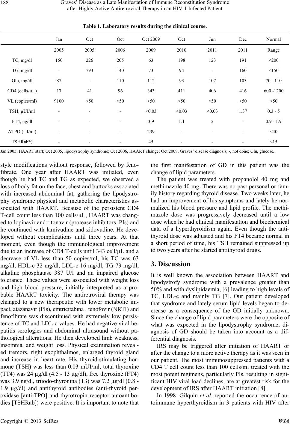 Graves’ Disease as a Late Manifestation of Immune Reconstitution Syndrome after Highly Active Antiretroviral Therapy in an HIV-1 Infected Patient 188 Table 1. Laboratory results during the clinical course. Jan Oct Oct Oct 2009 Oct Jun Dec Normal 2005 2005 2006 2009 2010 2011 2011 Range TC, mg/dl 150 226 205 63 198 123 191 <200 TG, mg/dl - 793 140 73 94 - 160 <150 Glu, mg/dl 87 - 110 112 93 107 103 70 - 110 CD4 (cells/µL) 17 41 96 343 411 406 416 600 -1200 VL (copies/m l) 9100 <50 <50 <50 <50 <50 <50 <50 TSH, µUI/ml - - - <0.03 <0.03 <0.03 1.37 0.3 - 5 FT4, ng/dl - - - 3.9 1.1 2 - 0. 9 - 1.9 ATPO (UI/ml) - - - 239 - - - <40 TSHRab% - - - 45 - - - <15 Jan 2005, HAART start; Oct 2005, lipodystrophy syndrome; Oct 2006, HAART change; Oct 200 9, Graves’ disease diagnosis; -, not done; Glu, glucose . style modifications without response, followed by feno- fibrate. One year after HAART was initiated, even though he had TC and TG as expected, we observed a loss of body fat on the face, chest and buttocks associated with increased abdominal fat, gathering the lipodystro- phy syndrome physical and metabolic characteristics as- sociated with HAART. Because of the persistent CD4 T-cell count less than 100 cells/µL, HAART was chang- ed to lopinavir and ritonavir (protease inhibitors, PIs) and he continued with lamivudine and zidovudine. He deve- loped without complications until three years. At that moment, even though the immunological improvement due to an in crease of CD4 T-cells until 343 cell/µL and a decrease of VL less than 50 copies/ml, his TC was 63 mg/dl, HDL-c 32 mg/dl, LDL-c 16 mg/dl, TG 73 mg/dl, alkaline phosphatase 387 U/l and an impaired glucose tolerance. These values were associated with weight loss and high blood pressure, initially interpreted as a pro- bable HAART toxicity. The antiretroviral therapy was changed to a new therapeutic with lower metabolic im- pact, atazanavir (PIs), emtr icitabina , tenofovir (NRTI) and fenofibrate was discontinued with extremely low persis- tence of TC and LDL-c values. He had negative viral he- patitis serologies and abdominal ultrasound without pa- thological alterations. He then developed limb weakness, insomnia, and weight loss. Physical examination reveal- ed tremors, right exophthalmos, enlarged thyroid gland and increase in heart rate. His thyroid-stimulating hor- mone (TSH) was less than 0.03 mUI/ml, total thyroxine (TT4) was 24 µg/d l (4.5 - 13 µg /dl), free th yroxine (FT4) was 3.9 ng/dl, triiodo-thyronina (T3) was 7.2 µg/dl (0.8 - 1.9 µg/dl) and antithyroid antibodies (anti-thyroid per- oxidase [anti-TPO] and thyrotropin receptor autoantibo- dies [TSHRab]) were positive. It is important to note that the first manifestation of GD in this patient was the change of lipid parameters. The patient was treated with propanolol 40 mg and methimazole 40 mg. There was no past personal or fam- ily history regarding thyroid disease. Two weeks later, he had an improvement of his symptoms and lately he nor- malized his blood pressure and lipid profile. The methi- mazole dose was progressively decreased until a low dose when he had clinical manifestation and biochemical data of a hyperthyroidism again. Even though the anti- thyroid dose was adjusted and his FT4 became normal in a short period of time, his TSH remained suppressed up to two years after he started antithyro id drugs. 3. Discussion It is well known the association between HAART and lipodystrofy syndrome with a prevalence greater than 50% and with dyslipidaemia, [6] leading to high levels of TC, LDL-c and mainly TG [7]. Our patient developed that syndrome and lately serum lipid levels began to de- crease as a consequence of the GD initially unknown. Since the change of lip id parameters were the oppo site of what was expected in the lipodystrophy syndrome, di- agnosis of GD should be taken into account as a dif- ferential diagnosis. IRS may be triggered after initiation of HAART or after the change to a more active therapy as it was seen in our patient. The most immunosuppressed patients with a CD4 T cell count less than 100 cells/ml treated with the most potent regimens, particularly PIs, resulting in signi- ficant HIV viral load declines, are at greatest risk for the development of IRS after HAART initiation [8]. In 1998, Gilquin et al. reported the occurrence of au- toimmune hyperthyroidism in 3 patients with HIV after Copyright © 2013 SciRes. WJA 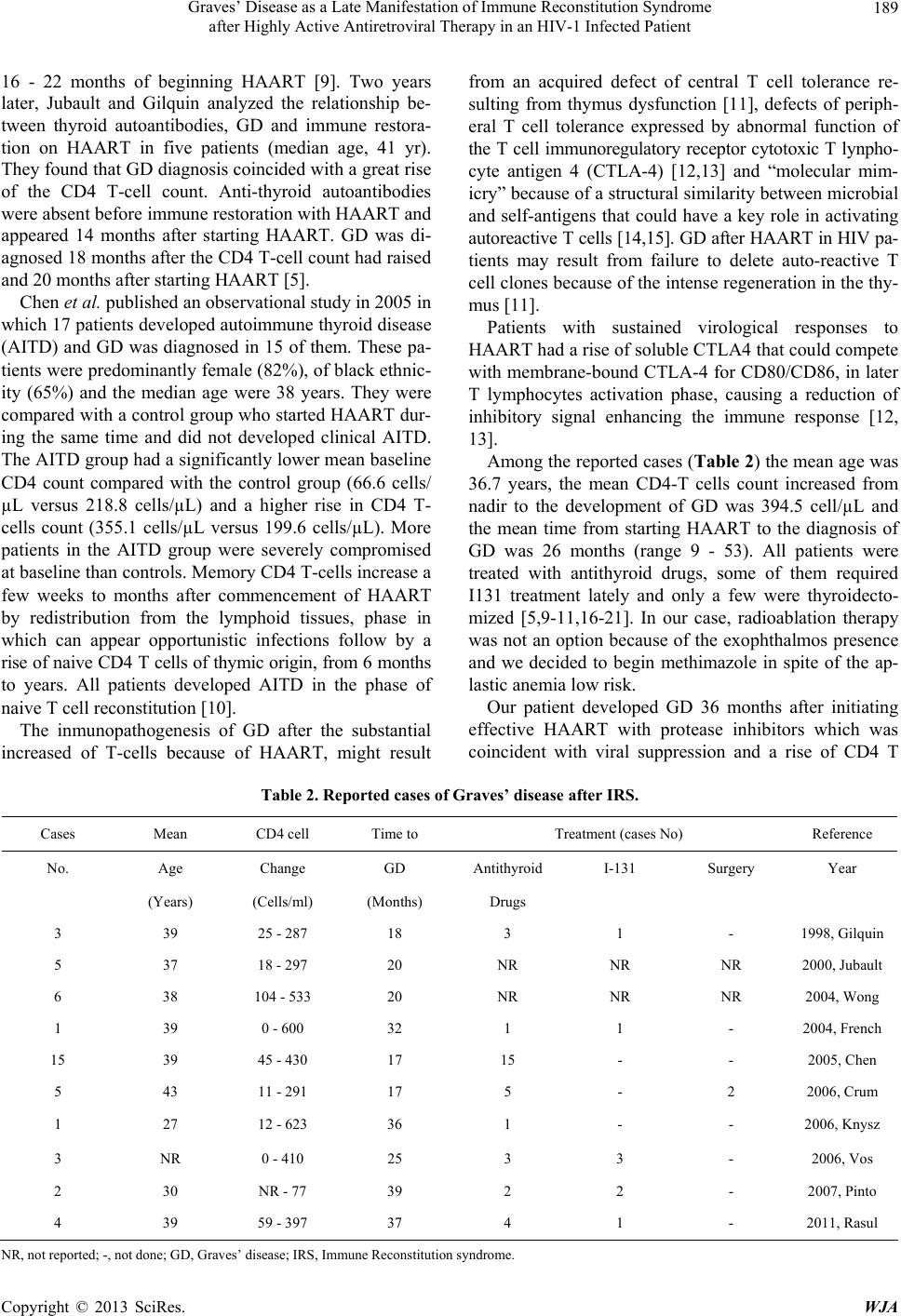 Graves’ Disease as a Late Manifestation of Immune Reconstitution Syndrome after Highly Active Antiretroviral Therapy in an HIV-1 Infected Patient 189 16 - 22 months of beginning HAART [9]. Two years later, Jubault and Gilquin analyzed the relationship be- tween thyroid autoantibodies, GD and immune restora- tion on HAART in five patients (median age, 41 yr). They found that GD diagnosis coincided with a great rise of the CD4 T-cell count. Anti-thyroid autoantibodies were absent before immune restoration with HAART and appeared 14 months after starting HAART. GD was di- agnosed 18 months after the CD4 T-cell count had raised and 20 months after starting HAART [5]. Chen et al. published an ob serv ation al study in 2005 in which 17 patients developed autoimmune thyroid disease (AITD) and GD was diagnosed in 15 of them. These pa- tients were predominantly female (82%), of black ethnic- ity (65%) and the median age were 38 years. They were compared with a contro l group who started HAART dur- ing the same time and did not developed clinical AITD. The AITD group had a significantly lower mean baselin e CD4 count compared with the control group (66.6 cells/ µL versus 218.8 cells/µL) and a higher rise in CD4 T- cells count (355.1 cells/µL versus 199.6 cells/µL). More patients in the AITD group were severely compromised at base line th an con tro ls. Memo r y CD4 T- cells in cr eas e a few weeks to months after commencement of HAART by redistribution from the lymphoid tissues, phase in which can appear opportunistic infections follow by a rise of naive CD4 T cells of thymic origin, from 6 months to years. All patients developed AITD in the phase of naive T cell reconstitution [10]. The inmunopathogenesis of GD after the substantial increased of T-cells because of HAART, might result from an acquired defect of central T cell tolerance re- sulting from thymus dysfunction [11], defects of periph- eral T cell tolerance expressed by abnormal function of the T cell immunoregulatory receptor cytotoxic T lynpho- cyte antigen 4 (CTLA-4) [12,13] and “molecular mim- icry” because of a structural similarity between microbial and self-antigens that could have a key role in activating autoreactive T cells [14,15]. GD after HAART in HIV pa- tients may result from failure to delete auto-reactive T cell clones because of the intense regeneration in the thy- mus [11]. Patients with sustained virological responses to HAART had a rise of soluble CTLA4 that could compete with membrane-bou nd CTLA-4 for CD80/CD86, in later T lymphocytes activation phase, causing a reduction of inhibitory signal enhancing the immune response [12, 13]. Among the reported cases (Table 2 ) the mean age was 36.7 years, the mean CD4-T cells count increased from nadir to the development of GD was 394.5 cell/µL and the mean time from starting HAART to the diagnosis of GD was 26 months (range 9 - 53). All patients were treated with antithyroid drugs, some of them required I131 treatment lately and only a few were thyroidecto- mized [5,9-11,16-21]. In our case, radioablation therapy was not an option because of the exophthalmos presence and we decided to begin methimazole in spite of the ap- lastic anemia low risk. Our patient developed GD 36 months after initiating effective HAART with protease inhibitors which was coincident with viral suppression and a rise of CD4 T Table 2. Reported cases of Graves’ disease after IRS. Cases Mean CD4 cell Time to Treatment (cases No) Reference No. Age Change GD Antithyroid I-131 Surgery Year (Years) (Cells/ml) (Months) Drugs 3 39 25 - 287 18 3 1 - 1998, Gilquin 5 37 18 - 297 20 NR NR NR 2000, Jubault 6 38 104 - 533 20 NR NR NR 2004, Wong 1 39 0 - 600 32 1 1 - 2004, French 15 39 45 - 430 17 15 - - 2005, Chen 5 43 11 - 291 17 5 - 2 2006, Crum 1 27 12 - 623 36 1 - - 2006, Knysz 3 NR 0 - 410 25 3 3 - 2006, Vos 2 30 NR - 77 39 2 2 - 2007, Pinto 4 39 59 - 397 37 4 1 - 2011, Rasul NR, not reported; -, not done; GD, Graves’ disease; IRS, Immune Reconstitution syndrome. Copyright © 2013 SciRes. WJA  Graves’ Disease as a Late Manifestation of Immune Reconstitution Syndrome after Highly Active Antiretroviral Therapy in an HIV-1 Infected Patient 190 cells from less than 100 to 343 cells/µL, being that an infrequent but recognized late presentation of the Im- mune Reconstitution Syndrome in mostly immune sup- pressed HIV patients. 4. Conclusion The most immunosuppressed patients with a CD4 T cell count < 100 cells/ml treated with the most potent regi- mens, particularly PIs, resulting in significant HIV viral load declines are at greatest risk for the development of IRS after HAART initiation [8]. Even though it is an uncommon manifestation of IRS, we think that Clini- cians will have to consider the importance of the early diagnosis to bring an adequate treatment and to decrease the associated morbidity of Graves’ disease. REFERENCES [1] A. Carr, K. Samaras, D. J. Chrisholm and D. A. Cooper, “Pathogenesis of HIV-1-Proteasa Inhibitor Associated Pe- Ripheral Lipodystrophy, Hyperlipidaemia and Insulin Re- sistance,” Lancet, Vol. 352, No. 9144, 1998, pp. 1881- 1883. doi:10.1016/S0140-6736(98)03391-1 [2] R. Singhania and D. Kotler, “Lipodystrophy in HIV Pa- tients: Its Challenges and Management Approaches,” HIV/AIDS-Research and Palliative Care, Vol. 3, 2011, pp. 135-143. [3] M. A. French, M. Lenzo, M. John, S. Mallal, E. J. Mc- Kinnon, I. R. James, P. Price, J. P. Flexman and M. Tay- Kearney, “Immune Restoration Disease after the Treat- ment of Immunodeficient HIV-Infected Patients with High- ly Active Antiretroviral Therapy,” HIV Medicine, Vol. 1, No. 2, 2000, pp. 107-115. doi:10.1046/j.1468-1293.2000.00012.x [4] M. A. French, “HIV/AIDS: Immune Reconstitution Inflam- matory Syndrome,” Clinical Infectious Diseases, Vol. 48, No. 1, 2009, pp. 101-107. doi:10.1086/595006 [5] V. Jubault, A. Penfornis, F. Schillo, B. Hoen, M. Izem- bart, J. Timsit, M. D. Kazatchkine, J. Gilkin and J. P. Vi- ard, “Sequential Occurrence of Thyroid Autoantibodies and Graves’ Disease after Immune Restoration in Severe- ly Immuno compromise d Human Immunode ficiency Vi rus- 1 Infected Patients,” The Journal of Clinical Endocrinol- ogy & Metabolism, Vol. 85, No. 11, 2000, pp. 4254-4257. doi:10.1210/jc.85.11.4254 [6] K. Samara s, H. Wand, M. Law, S. Emery, D. Cooper and A. Carr, “Prevalence of Metabolic Syndrome in HIV-In- fected Patients Receiving Highly Active Antiretroviral Therapy Using International Diabetes Foundation and Adult Treatment Panel III Criteria: Associations with In- sulin Resistance,” Diabetes Care, Vol. 30, No. 2, 2001, p. 455. [7] E. Fontas, F. van Leth, C. A. Sabin, N. Friis-Møller, M. Rickenbach, A. d’Arminio Monforte, O. Kirk, M. Du- pon, L. Morfeldt, S. Mateu, K. Petoumenos, W. El-Sadr, S. de Wit, J. D. Lundgren, C. Pradier and Reiss, “Profiles in HIV-Infected Patients Receiving Combination Anti- retroviral Therapy: Are Different Antiretroviral Drugs As- sociated with Different Lipid Profiles?” JID, Vol. 189, No. 6, 2004, pp. 1056-1074. doi:10.1086/381783 [8] Y. C. Manabe, J. D. Campbell, E. Sydnor and R. D. Moore, “Immune Reconstitution Inflammatory Syndrome: Risk Factors and Treatment Implications,” JAIDS Journal of Acquired Immune Deficiency Syndromes, Vol. 46, No. 4, 2007, pp. 456-462. doi:10.1097/QAI.0b013e3181594c8c [9] J. Gilquin, J. P. Viard, V. Jubault, C. Sert and M. D. Ka- zatchkine, “Delayed Occurrence of Graves’ Disease after Immune Restoration with HAART,” Lancet, Vol. 352, No. 9119, 1998, pp. 1907-1908. doi:10.1016/S0140-6736(05)60398-4 [10] F. Chen, S. L. Day, R. A. Metcalfe, G. Sethi, M. S. Ka- pembwa, M. G. Brook, D. Churchill, A. de Ruiter, S. Ro- binson, C. J. Lacey and A. P. Weetman, “Characteristics of Autoimmune Thyroid Disease Occurring as a Late Com- plication of Immune Reconstitution in Patients with Ad- vanced Human Immunodeficiency (HIV) Disease,” Medi- cine, Vol. 84, No. 2, 2005, pp. 98-106. doi:10.1097/01.md.0000159082.45703.90 [11] M. A. French, S. R. Lewin, C. Dykstra, R. Krueger, P. Price and P. J. Leedman, “Graves’ Disease during Immune Reconstitution after Highly Active Antiretroviral Therapy for HIV Infection: Evidence of Thyroid Dysfunction,” AIDS Research and Human Retroviruses, Vol. 20, No. 2, 2004, pp. 157-162. doi:10.1089/088922204773004879 [12] S. F. Stone, P. Price and M. A. French, “Dy sregulation of CD28 and CTLA-4 Expression by CD4 T Cells from Pre- viously Immunodeficient HIV-Infected Patients with Sus- tained Virological Responses to Highly Active Antiretro- viral Therapy,” HIV Medicine, Vol. 6, No. 4, 2005, pp. 278-283. doi:10.1111/j.1468-1293.2005.00307.x [13] D. Saverino, R. Brizzolara, R. Simone, A. Chiappori, F. Milintenda-Floriani, G. Pesce and M. Bagnasco, “Soluble CTLA-4 in Autoimmune Thyroid Diseases: Relationship with Clinical Status and Possible Role in the Immune Re- sponse Dysregulation,” Clinical Immunology, Vol. 123, No. 2, 2007, pp. 190-198. doi:10.1016/j.clim.2007.01.003 [14] T. Kamradt and N. A. Mitchison, “Tolerance and Autoin- munity,” The New England Journal of Medicine, Vol. 344, No. 9, 2001, pp. 655-664. doi:10.1056/NEJM200103013440907 [15] L. J. Albert and R. D. Inma n, “Molecular Mi micry and Au- toimmunity,” The New England Journal of Medicine, Vol. 341, No. 27, 1999, pp. 2068-2074. doi:10.1056/NEJM199912303412707 [16] K. H. Wong, W. S. Chow and S. S. Lee, “Clinical Hyper- thyroidism in Chinese Patients with Stable HIV Disease,” Clinical Infectious Diseases, Vol. 39, No. 8, 2004, pp. 1257-1259. doi:10.1086/424749 [17] N. F. Crum, A. Ganesan, S. T. Johns and M. R. Wallace, “Graves Disease: An Increasingly Recognized Immune Reconstitution Syndrome,” AIDS, Vol. 20, No. 3, 2006, pp. 466-469. doi:10.1097/01.aids.0000196173.42680.5f [18] B. Knysz, M. Bolanowski, M. Klimczak, A. Gladysz and K. Zwolinska, “Graves’ Disease as an Immune Recons- Copyright © 2013 SciRes. WJA  Graves’ Disease as a Late Manifestation of Immune Reconstitution Syndrome after Highly Active Antiretroviral Therapy in an HIV-1 Infected Patient 191 titution Syndrome in an HIV-1-Positive Patient Commen- cing Effective Antiretroviral Therapy,” Case Report and Literature Review, Viral Immunology, Vol. 19, No. 1, 2006, pp. 102-107. doi:10.1089/vim.2006.19.102 [19] F. Vos, G. Pie ter s , M. Keu te r and A. Va n De r Ve n, “Gr a ve s’ Disease during Immune Reconstitution in HIV-Infected Patients Treated with HAART,” Scandinavian Journal of Infectious Diseases, Vol. 38, No. 2, 2006, pp. 124-126. doi:10.1080/00365540500348960 [20] V. M. Pinto, Q. D. Iglesias, C. S. Corigliano, V. C. Ma- guiña, Z. J. Echevarria, C . S. Villena and R. C. Seas, “Graves Disease: Report of Two HIV Cases of Inmune Reconsti- tution Syndrome Following Highly Active Antiretroviral Therapy,” Revista Médica Herediana, Vol. 18, No. 4, 2007, pp. 218-221. [21] S. Rasul, R. Delape nha, F. Farhat, J. Gajjala and S. Zahra, “Graves’ Disease as a Manifestation of Immune Recon- stitution in HIV-Infected Individuals after Initiation of Highly Active Antiretroviral Therapy,” AIDS Research and Treatment, 2011, 4 p. doi:10.1155/2011/743597 Copyright © 2013 SciRes. WJA |