

Paper Menu >>
Journal Menu >>
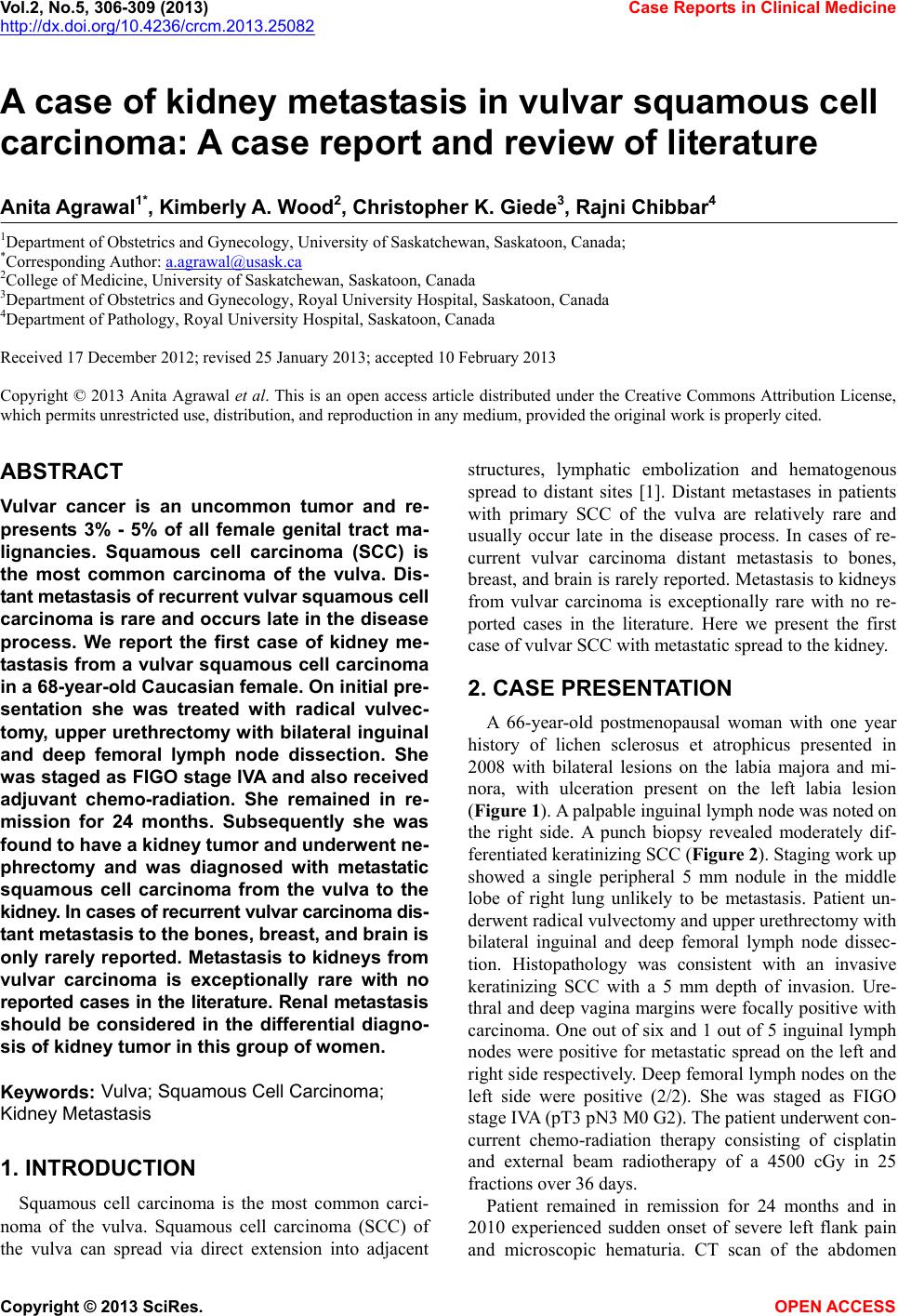 Vol.2, No.5, 306-309 (2013) Case Reports in Clinical Medicine http://dx.doi.org/10.4236/crcm.2013.25082 A case of kidney metastasis in vulvar squamous cell carcinoma: A case report and review of literature Anit a Agrawal1*, Kimberly A. Wood2, Christopher K. Giede3, Rajni Chibbar4 1Department of Obstetrics and Gynecology, University of Saskatchewan, Saskatoon, Canada; *Corresponding Author: [email protected] 2College of Medicine, University of Saskatchewan, Saskatoon, Canada 3Department of Obstetrics and Gynecology, Royal University Hospital, Saskatoon, Canada 4Department of Pathology, Royal University Hospital, Saskatoon, Canada Received 17 December 2012; revised 25 January 2013; accepted 10 February 2013 Copyright © 2013 Anita Agrawal et al. This is an open access article distributed under the Creative Commons Attribution License, which permits unrestricted use, distribution, and reproduction in any medium, provided the original work is properly cited. ABSTRACT Vulvar cancer is an uncommon tumor and re- presents 3% - 5% of all female genital tract ma- lignancies. Squamous cell carcinoma (SCC) is the most common carcinoma of the vulva. Dis- t ant met astasis of recurrent vulvar squamous cell carcinoma is rare and occurs late in the disease process. We report the first case of kidney me- tastasis from a vulvar squamous cell carcinoma in a 68-year-old Caucasi an female. On initial pre- sentation she was treated with radical vulvec- tomy, upper urethrectomy w ith bilateral inguinal and deep femoral lymph node dissection. She was staged as FIGO stage IVA and also received adjuvant chemo-radiation. She remained in re- mission for 24 months. Subsequently she was found to have a k idney tumor and underwent ne- phrectomy and was diagnosed with metastatic squamous cell carcinoma from the vulva to the kidney. In ca ses of recur rent v ulvar carci noma dis - t ant metastasis to the bo nes, breast, and brain is only rarely reported. Metastasis to kidneys from vulvar carcinoma is exceptionally rare with no reported cases in the literature. Renal meta stas is should be considered in the differential diagno- sis of kidney tumor in this group of women. Keyw ords: Vulva; Squamous Cell Carcinoma; Kidney Metastasis 1. INTRODUCTION Squamous cell carcinoma is the most common carci- noma of the vulva. Squamous cell carcinoma (SCC) of the vulva can spread via direct extension into adjacent structures, lymphatic embolization and hematogenous spread to distant sites [1]. Distant metastases in patients with primary SCC of the vulva are relatively rare and usually occur late in the disease process. In cases of re- current vulvar carcinoma distant metastasis to bones, breast, and brain is rarely reported. Metastasis to k idneys from vulvar carcinoma is exceptionally rare with no re- ported cases in the literature. Here we present the first case of vulvar SCC with metastatic spread to the kidney. 2. CASE PRESENTATION A 66-year-old postmenopausal woman with one year history of lichen sclerosus et atrophicus presented in 2008 with bilateral lesions on the labia majora and mi- nora, with ulceration present on the left labia lesion (Figure 1). A palpable inguinal lymph node was noted on the right side. A punch biopsy revealed moderately dif- ferentiated keratinizin g SCC (Figure 2). Staging wo rk up showed a single peripheral 5 mm nodule in the middle lobe of right lung unlikely to be metastasis. Patient un- derwent radical vulvectomy and upper urethrectomy with bilateral inguinal and deep femoral lymph node dissec- tion. Histopathology was consistent with an invasive keratinizing SCC with a 5 mm depth of invasion. Ure- thral and deep vagina margins were focally positive with carcinoma. One out of six and 1 out of 5 inguinal lymph nodes were positive for metastatic spread on the left and right side respectively. Deep femoral lymph nodes on the left side were positive (2/2). She was staged as FIGO stage IVA (pT3 pN3 M0 G2). The patient underwent con- current chemo-radiation therapy consisting of cisplatin and external beam radiotherapy of a 4500 cGy in 25 fractions o ve r 3 6 day s. Patient remained in remission for 24 months and in 2010 experienced sudden onset of severe left flank pain and microscopic hematuria. CT scan of the abdomen Copyright © 2013 SciRes. OPEN A CCESS 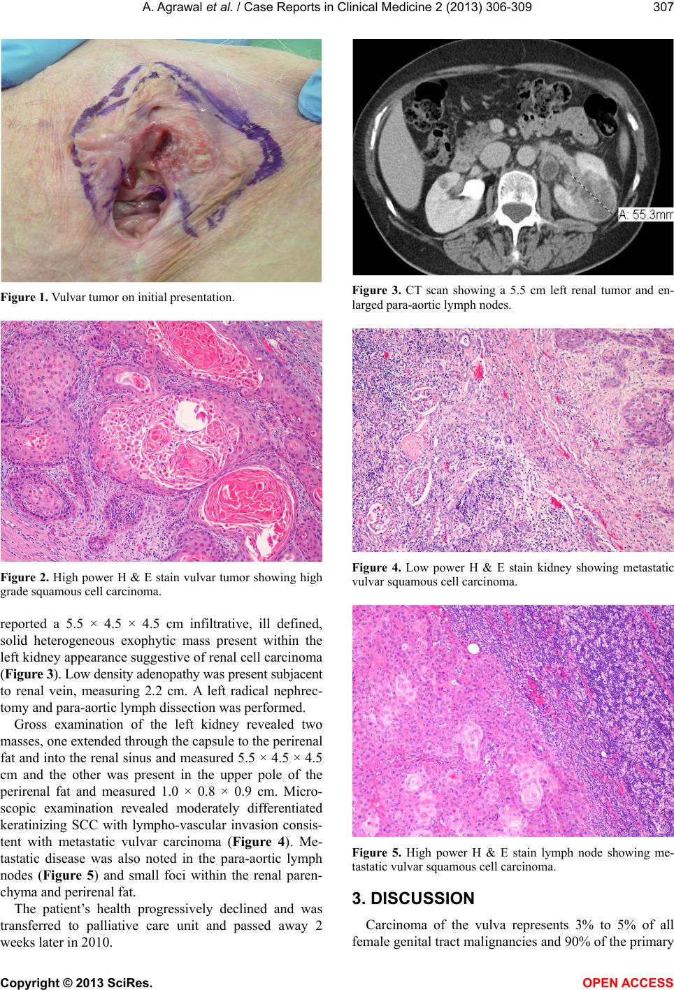 A. Agrawal et al. / Case Reports in Clinical Medicine 2 (2013) 306-309 307 Figure 1. Vulvar tumor on initial presentation. Figure 2. High power H & E stain vulvar tumor showing high grade squamous cell carcinoma. reported a 5.5 × 4.5 × 4.5 cm infiltrative, ill defined, solid heterogeneous exophytic mass present within the left kidney appearance suggestive of renal cell carcinoma (Figure 3). Low density adenopathy was present subjacent to renal vein, measuring 2.2 cm. A left radical nephrec- tomy and para-aortic lymph dissection was performed. Gross examination of the left kidney revealed two masses, one extended through the capsule to the perirenal fat and into the renal sinus and measured 5.5 × 4.5 × 4.5 cm and the other was present in the upper pole of the perirenal fat and measured 1.0 × 0.8 × 0.9 cm. Micro- scopic examination revealed moderately differentiated keratinizing SCC with lympho-vascular invasion consis- tent with metastatic vulvar carcinoma (Figure 4). Me- tastatic disease was also noted in the para-aortic lymph nodes (Figure 5) and small foci within the renal paren- chyma and perirenal fat. The patient’s health progressively declined and was transferred to palliative care unit and passed away 2 weeks later in 2010. Figure 3. CT scan showing a 5.5 cm left renal tumor and en- larged para-aortic lymph nodes. Figure 4. Low power H & E stain kidney showing metastatic vulvar squamous cell carcinoma. Figure 5. High power H & E stain lymph node showing me- tastatic vulvar squamous cell carcinoma. 3. DISCUSSION Carcinoma of the vulva represents 3% to 5% of all female genital tract malignancies and 90% of the primary Copyright © 2013 SciRes. OPEN A CCESS 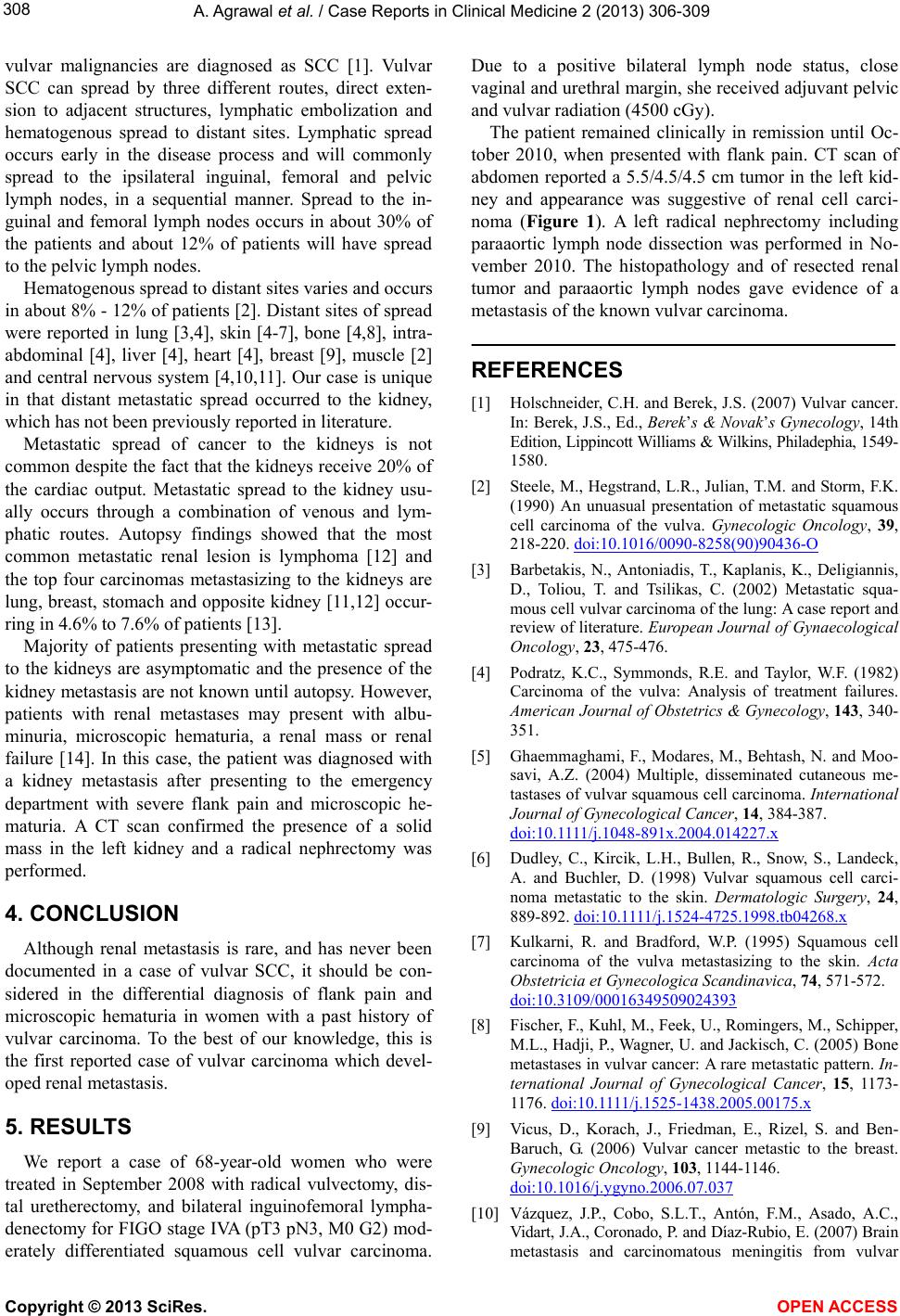 A. Agrawal et al. / Case Reports in Clinical Medicine 2 (2013) 306-309 308 vulvar malignancies are diagnosed as SCC [1]. Vulvar SCC can spread by three different routes, direct exten- sion to adjacent structures, lymphatic embolization and hematogenous spread to distant sites. Lymphatic spread occurs early in the disease process and will commonly spread to the ipsilateral inguinal, femoral and pelvic lymph nodes, in a sequential manner. Spread to the in- guinal and femoral lymph nodes occurs in about 30% of the patients and about 12% of patients will have spread to the pelvic lymph nod es. Hematogenous spread to distant sites varies and occurs in about 8% - 1 2% of patients [2]. Distan t sites of spread were reported in lung [3,4], skin [4-7], bone [4,8], intra- abdominal [4], liver [4], heart [4], breast [9], muscle [2] and central nervous system [4,10,11]. Our case is un ique in that distant metastatic spread occurred to the kidney, which has not been previously reported in literature. Metastatic spread of cancer to the kidneys is not common despite the fact that the kidneys receive 20% of the cardiac output. Metastatic spread to the kidney usu- ally occurs through a combination of venous and lym- phatic routes. Autopsy findings showed that the most common metastatic renal lesion is lymphoma [12] and the top four carcinomas metastasizing to the kidneys are lung, breast, stomach and opposite kidney [11,12] occur- ring in 4.6% to 7.6% of patients [13]. Majority of patients presenting with metastatic spread to the kidneys are asymptomatic and the presence of the kidney metastasis are not known un til autopsy. However, patients with renal metastases may present with albu- minuria, microscopic hematuria, a renal mass or renal failure [14]. In this case, the patient was diagnosed with a kidney metastasis after presenting to the emergency department with severe flank pain and microscopic he- maturia. A CT scan confirmed the presence of a solid mass in the left kidney and a radical nephrectomy was performed. 4. CONCLUSION Although renal metastasis is rare, and has never been documented in a case of vulvar SCC, it should be con- sidered in the differential diagnosis of flank pain and microscopic hematuria in women with a past history of vulvar carcinoma. To the best of our knowledge, this is the first reported case of vulvar carcinoma which devel- oped renal metastasis. 5. RESULTS We report a case of 68-year-old women who were treated in September 2008 with radical vulvectomy, dis- tal uretherectomy, and bilateral inguinofemoral lympha- denectomy for FIGO stage IVA (pT3 pN3, M0 G2) mod- erately differentiated squamous cell vulvar carcinoma. Due to a positive bilateral lymph node status, close vaginal and urethral margin, she received adjuvant pelvic and vulvar radiation (4500 cGy). The patient remained clinically in remission until Oc- tober 2010, when presented with flank pain. CT scan of abdomen reported a 5.5/4.5/4.5 cm tumor in the left kid- ney and appearance was suggestive of renal cell carci- noma (Figure 1). A left radical nephrectomy including paraaortic lymph node dissection was performed in No- vember 2010. The histopathology and of resected renal tumor and paraaortic lymph nodes gave evidence of a metastasis of the known vulvar carcinoma. REFERENCES [1] Holschneider, C.H. and Berek, J.S. (2007) Vulvar cancer. In: Berek, J.S., Ed., Berek’s & Novak’s Gynecology, 14th Edition, Lippincott Williams & Wilkins, Philadephia, 1549- 1580. [2] Steele, M., Hegstrand, L.R., Julian, T.M. and Storm, F.K. (1990) An unuasual presentation of metastatic squamous cell carcinoma of the vulva. Gynecologic Oncology, 39, 218-220. doi:10.1016/0090-8258(90)90436-O [3] Barbetakis, N., Antoniadis, T., Kaplanis, K., Deligiannis, D., Toliou, T. and Tsilikas, C. (2002) Metastatic squa- mous cell vulvar carcinoma of the lung: A case report and review of literature. European Journal of Gynaecological Oncology, 23, 475-476. [4] Podratz, K.C., Symmonds, R.E. and Taylor, W.F. (1982) Carcinoma of the vulva: Analysis of treatment failures. American Journal of Obstetrics & Gynecology, 143, 340- 351. [5] Ghaemmaghami, F., Modares, M., Behtash, N. and Moo- savi, A.Z. (2004) Multiple, disseminated cutaneous me- tastases of vulvar squamous cell carcinoma. International Journal of Gynecological Cancer, 14, 384-387. d oi: 10. 1111 /j .1048-891x.2004.014227.x [6] Dudley, C., Kircik, L.H., Bullen, R., Snow, S., Landeck, A. and Buchler, D. (1998) Vulvar squamous cell carci- noma metastatic to the skin. Dermatologic Surgery, 24, 889-892. d oi: 10.1111/ j .1524-4725.1998.tb04268.x [7] Kulkarni, R. and Bradford, W.P. (1995) Squamous cell carcinoma of the vulva metastasizing to the skin. Acta Obstetricia et Gynecologica Scandinavica, 74, 571-572. doi:10.3109/00016349509024393 [8] Fischer, F., Kuhl, M., Feek, U., Romingers, M., Schipper, M.L., Hadji, P., Wagner, U. and Jackisch, C. (2005) Bone metastases in vulvar cancer: A rare metastatic pattern. In- ternational Journal of Gynecological Cancer, 15, 1173- 1176. doi:10.1111/j.1525-1438.2005.00175.x [9] Vicus, D., Korach, J., Friedman, E., Rizel, S. and Ben- Baruch, G. (2006) Vulvar cancer metastic to the breast. Gynecologic Oncology, 103, 1144-1146. doi:10.1016/j.ygyno.2006.07.037 [10] Vázquez, J.P., Cobo, S.L.T., Antón, F.M., Asado, A.C., Vidart, J.A., Coronado, P. and Díaz-Rubio, E. (20 07) Brain metastasis and carcinomatous meningitis from vulvar Copyright © 2013 SciRes. OPEN ACCESS  A. Agrawal et al. / Case Reports in Clinical Medicine 2 (2013) 306-309 Copyright © 2013 SciRes. OPEN ACCESS 309 squamous cell carcinoma: Case report. European Journal of Gynaecological Oncology, 28, 152-154. [11] Dursun, P., Ayhan, A., Tarhan, N.C., Coban, G. and Kuscu, E. (2009) Cerebellar metastasis in squamous cell vulvar carcinoma. Archives of Gynecology and Obstetrics, 279, 69-71. doi:10.1007/s00404-008-0635-2 [12] Wagle, D.G., Moore, R.H. and Murphy, G.P. (1975) Sec- ondary carcinomas of the kidney. Journal of Urology, 114 , 30-32. [13] Stoughton, W.B. and Milam, D.F. (1975) Metastatic tu- mors to the kidney. West Virginia Medical Journal, 71, 199-201. [14] Bennington, J.L. and Beckwith, J.B. (1975) Tumors of the kidney, renal pelvis, and ureter. In Atlas of Tumor Pathology, 2nd Series, Fascicle 12, Armed Forces Insti- ture of Pathology, Washington DC, 162-163. |