

Paper Menu >>
Journal Menu >>
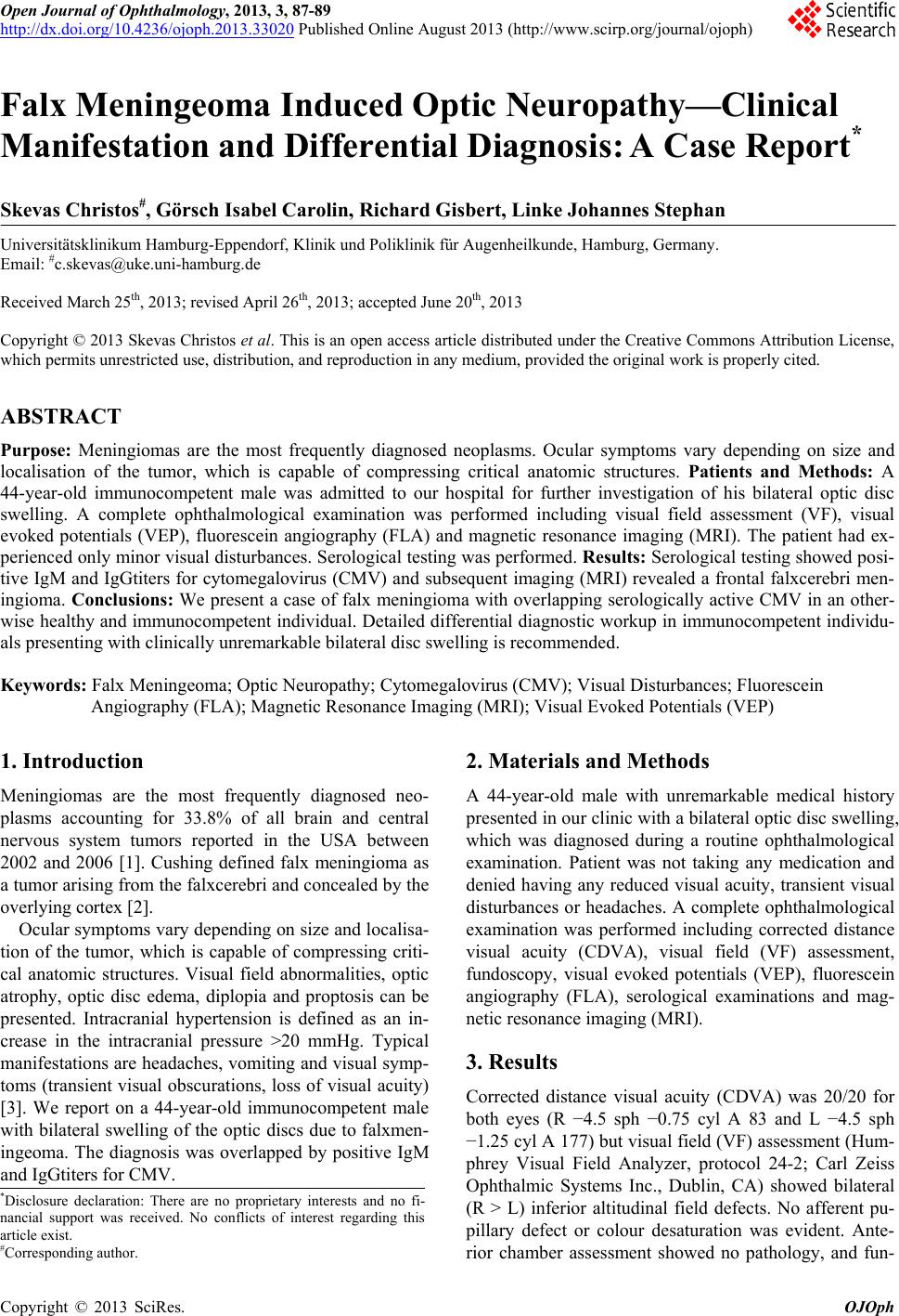 Open Journal of Ophthalmology, 2013, 3, 87-89 http://dx.doi.org/10.4236/ojoph.2013.33020 Published Online August 2013 (http://www.scirp.org/journal/ojoph) 87 Falx Meningeoma Induced Optic Neuropathy—Clinical Manifestation and Differential Diagnosis: A Case Report* Skevas Christos#, Görsch Isabel Carolin, Richard Gisbert, Linke Johannes Stephan Universitätsklinikum Hamburg-Eppendorf, Klinik und Poliklinik für Augenheilkunde, Hamburg, Germany. Email: #[email protected] Received March 25th, 2013; revised April 26th, 2013; accepted June 20th, 2013 Copyright © 2013 Skevas Christos et al. This is an open access article distributed under the Creative Commons Attribution License, which permits unrestricted use, distribution, and reproduction in any medium, provided the original work is properly cited. ABSTRACT Purpose: Meningiomas are the most frequently diagnosed neoplasms. Ocular symptoms vary depending on size and localisation of the tumor, which is capable of compressing critical anatomic structures. Patients and Methods: A 44-year-old immunocompetent male was admitted to our hospital for further investigation of his bilateral optic disc swelling. A complete ophthalmological examination was performed including visual field assessment (VF), visual evoked potentials (VEP), fluorescein angiography (FLA) and magnetic resonance imaging (MRI). The patient had ex- perienced only minor visual disturbances. Serological testing was performed. Results: Serological testing showed posi- tive IgM and IgGtiters for cytomegalovirus (CMV) and subsequent imaging (MRI) revealed a frontal falxcerebri men- ingioma. Conclusions: We present a case of falx meningioma with overlapping serologically active CMV in an other- wise healthy and immunocompetent individual. Detailed differential diagnostic workup in immunocompetent individu- als presenting with clinically unremarkable bilateral disc swelling is recommended. Keywords: Falx Meningeoma; Optic Neuropathy; Cytomegalovirus (CMV); Visual Disturbances; Fluorescein Angiography (FLA); Magnetic Resonance Imaging (MRI); Visual Evoked Potentials (VEP) 1. Introduction Meningiomas are the most frequently diagnosed neo- plasms accounting for 33.8% of all brain and central nervous system tumors reported in the USA between 2002 and 2006 [1]. Cushing defined falx meningioma as a tumor arising from the falxcerebri and concealed by the overlying cortex [2]. Ocular symptoms vary depending on size and localisa- tion of the tumor, which is capable of compressing criti- cal anatomic structures. Visual field abnormalities, optic atrophy, optic disc edema, diplopia and proptosis can be presented. Intracranial hypertension is defined as an in- crease in the intracranial pressure >20 mmHg. Typical manifestations are headaches, vomiting and visual symp- toms (transient visual obscurations, loss of visual acuity) [3]. We report on a 44-year-old immunocompetent male with bilateral swelling of the optic discs due to falxmen- ingeoma. The diagnosis was overlapped by positive IgM and IgGtiters for CMV. 2. Materials and Methods A 44-year-old male with unremarkable medical history presented in our clinic with a bilateral optic disc swelling, which was diagnosed during a routine ophthalmological examination. Patient was not taking any medication and denied having any reduced visual acuity, transient visual disturbances or headaches. A complete ophthalmological examination was performed including corrected distance visual acuity (CDVA), visual field (VF) assessment, fundoscopy, visual evoked potentials (VEP), fluorescein angiography (FLA), serological examinations and mag- netic resonance imaging (MRI). 3. Results Corrected distance visual acuity (CDVA) was 20/20 for both eyes (R −4.5 sph −0.75 cyl A 83 and L −4.5 sph −1.25 cyl A 177) but visual field (VF) assessment (Hum- phrey Visual Field Analyzer, protocol 24-2; Carl Zeiss Ophthalmic Systems Inc., Dublin, CA) showed bilateral (R > L) inferior altitudinal field defects. No afferent pu- pillary defect or colour desaturation was evident. Ante- rior chamber assessment showed no pathology, and fun- *Disclosure declaration: There are no proprietary interests and no fi- nancial support was received. No conflicts of interest regarding this article exist. #Corresponding author. Copyright © 2013 SciRes. OJOph 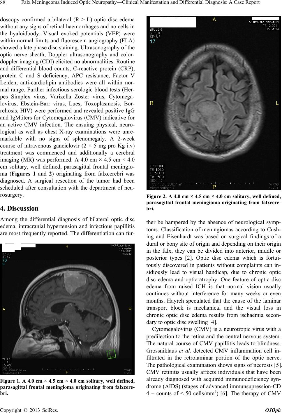 Falx Meningeoma Induced Optic Neuropathy—Clinical Manifestation and Differential Diagnosis: A Case Report 88 doscopy confirmed a bilateral (R > L) optic disc edema without any signs of retinal haemorrhages and no cells in the hyaloidbody. Visual evoked potentials (VEP) were within normal limits and fluorescein angiography (FLA) showed a late phase disc staining. Ultrasonography of the optic nerve sheath, Doppler ultrasonography and color- doppler imaging (CDI) elicited no abnormalities. Routine and differential blood counts, C-reactive protein (CRP), protein C and S deficiency, APC resistance, Factor V Leiden, anti-cardiolipin antibodies were all within nor- mal range. Further infectious serologic blood tests (Her- pes Simplex virus, Varizella Zoster virus, Cytomega- lovirus, Ebstein-Barr virus, Lues, Toxoplasmosis, Bor- reliosis, HIV) were performed and revealed positive IgG and IgMtiters for Cytomegalovirus (CMV) indicative for an active CMV infection. The ensuing physical, neuro- logical as well as chest X-ray examinations were unre- markable with no signs of splenomegaly. A 2-week course of intravenous ganciclovir (2 × 5 mg pro Kg i.v) treatment was commenced and additionally a cerebral imaging (MR) was performed. A 4.0 cm × 4.5 cm × 4.0 cm solitary, well defined, parasagittal frontal meningio- ma (Figures 1 and 2) originating from falxcerebri was diagnosed. A surgical resection of the tumor had been scheduled after consultation with the department of neu- rosurgery. 4. Discussion Among the differential diagnosis of bilateral optic disc edema, intracranial hypertension and infectious papillitis are most frequently reported. The differentiation can fur- Figure 1. A 4.0 cm × 4.5 cm × 4.0 cm solitary, well defined, parasagittal frontal meningioma originating from falxcere- bri. Figure 2. A 4.0 cm × 4.5 cm × 4.0 cm solitary, well defined, parasagittal frontal meningioma originating from falxcere- bri. ther be hampered by the absence of neurological symp- toms. Classification of meningiomas according to Cush- ing and Eisenhardt was based on surgical findings of a dural or bony site of origin and depending on their origin in the falx, they can be divided into anterior, middle or posterior types [2]. Optic disc edema which is fortui- tously discovered in patients without complaints can in- sidiously lead to visual handicap, due to chronic optic disc edema and optic atrophy. One feature of optic disc edema from raised ICH is that normal vision usually continues without interference for many weeks or even months. Hayreh speculated that the cause of the laminar transport block is mechanical and the visual loss in chronic optic disc edema results from ischaemia secon- dary to optic disc swelling [4]. Cytomegalovirus (CMV) is a neurotropic virus with a predilection to the retina and the central nervous system. The natural course of CMV papillitis leads to blindness. Grossniklaus et al. detected CMV inflammation cell in- filtrated in the retrolaminar portion of the optic nerve. The pathological examination shows signs of necrosis [5]. CMV retinitis usually affects individuals that have been already diagnosed with acquired immunodeficiency syn- drome (AIDS) (stages of advanced immunsupression-CD 4 + counts of < 50 cells/mm3) [6]. The therapy of CMV Copyright © 2013 SciRes. OJOph  Falx Meningeoma Induced Optic Neuropathy—Clinical Manifestation and Differential Diagnosis: A Case Report Copyright © 2013 SciRes. OJOph 89 retinitis/optic neuritis consists of intravenous ganciclovir or intravenous foscarnet, but their use in combination is also presented. The combination is more effective in controlling progression of CMV retinitis in relapsed pa- tients than monotherapy alone [7,8]. Besides patients with immune deficiency (AIDS), there are reports of CMV induced optic neuropathy in healthy immunocom- petent individuals [9,10]. We present to the best of our knowledge the first case of falx meningioma with overlapping serologically active CMV in an otherwise healthy and immunocompetent individual. A step by step approach for detailed differen- tial diagnostic workup in immunocompetent individuals presenting with clinically unremarkable bilateral disc swelling is recommended. Treatment should involve a harmonious cooperative effort between the ophthalmolo- gist and physicians of other specialities such as microbi- ology, neurology and neurosurgery. REFERENCES [1] CBTRUS, “Statistical Report: Primary Brain and Central Nervous System Tumors Diagnosed in Eighteen States in 2002-2006,” Central Brain Tumor Registry of the United States, Hinsdale, 2009-2010. [2] H. Cushing and L. Eisenhardt, “Meningiomas. Their Clas- sification, Regional Behavior, Life History and Surgical End Results,” Bulletin of the Medical Library Association, Vol. 27, No. 2, 1938, pp. 224-249. [3] D. I. Friedman and D. M. Jacobson, “Diagnostic Criteria for Idiopathic Intracranial Hypertension,” Neurology, Vol. 59, No. 10, 2002, pp. 1492-1495. [4] S. S. Hayreh, “Optic Disc Edema in Raised Intracranial Pressure. VI. Associated Visual Disturbances and Their Pathogenesis,” Archives of Ophthalmology, Vol. 95, No. 9, 1977, pp. 1566-1579. doi:10.1001/archopht.1977.04450090088007 [5] H. E. Grossniklaus, K. E. Frank and R. L. Tomsak, “Cy- tomegalovirus Retinitis and Optic Neuritis in Acquired Immune Deficiency Syndrome: Report of a Case,” Oph- thalmology, Vol. 94, No. 12, 1987, pp. 1601-1604. [6] J. D. Roarty, E. J. Fisher and J. J. Nussbaum, “Long-Term Visual Morbidity of Cytomegalovirus Retinitis in Patients with Acquired Immune Deficiency Syndrome,” Ophthal- mology, Vol. 100, No. 11, 1993, pp. 1685-1688. [7] D. N. Friedberg, “Cytomegalovirus Retinitis: Diagnosis and Status of Systemic Therapy,” Journal of Acquired Immune Deficiency Syndromes & Human Retrovirology, Vol. 14, No. 1, 1997, pp. S1-S6. doi:10.1097/00042560-199700001-00002 [8] A. M. Mansor and H. K. Li, “Cytomegalovirus Optic Neuritis: Characteristics, Therapy and Survival,” Oph- thalmologica, Vol. 209, No. 5, 1995, pp. 260-266. doi:10.1159/000310627 [9] E. Baglivo, P. M. Leuenberger and K. H. Krause, “Pre- sumed Bilateral Cytomegalovirus-Induced Optic Neu- ropathy in an Immunocompetent Person: A Case Report,” Journal of Neuro-Ophthalmology, Vol. 16, No. 1, 1996, pp. 14-17. doi:10.1097/00041327-199603000-00004 [10] S. R. De Silva, G. Chohan, D. Jones and M. Hu, “Cy- tomegalovirus Papillitis in an Immunocompetent Patient,” Journal of Neuro-Ophthalmology, Vol. 28, No. 2, 2008, pp. 126-127. doi:10.1097/WNO.0b013e3181782fed |