

Paper Menu >>
Journal Menu >>
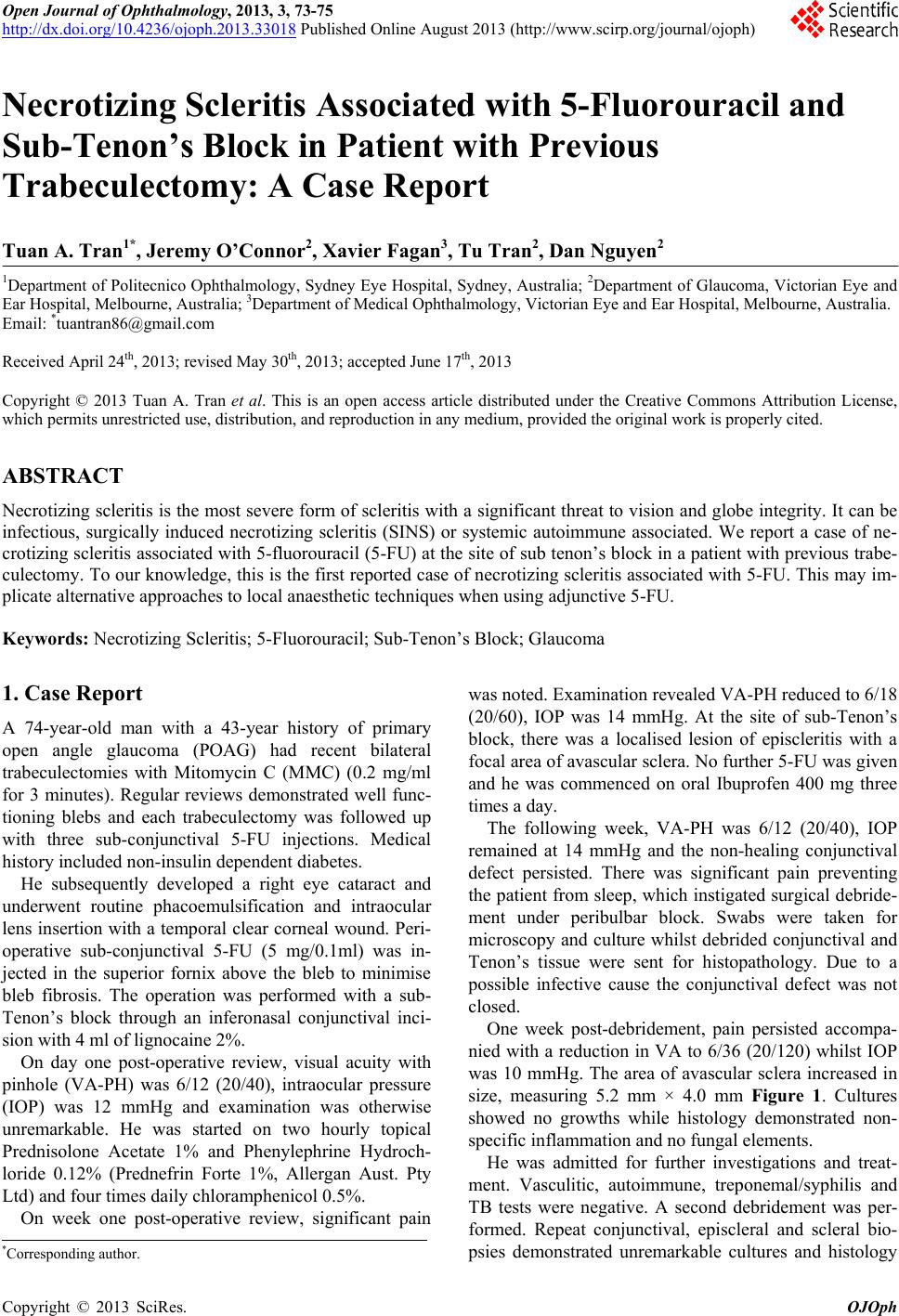 Open Journal of Ophthalmology, 2013, 3, 73-75 http://dx.doi.org/10.4236/ojoph.2013.33018 Published Online August 2013 (http://www.scirp.org/journal/ojoph) 73 Necrotizing Scleritis Associated with 5-Fluorouracil and Sub-Tenon’s Block in Patient with Previous Trabeculectomy: A Case Report Tuan A. Tran1*, Jeremy O’Connor2, Xavier Fagan3, Tu Tran2, Dan Nguyen2 1Department of Politecnico Ophthalmology, Sydney Eye Hospital , Sydney, Austra lia; 2Department of Glaucoma, Victorian Eye and Ear Hospital, Melbourne, Australia; 3Department of Medical Ophthalmology, Victorian Eye and Ear Hospital, Melbourne, Australia. Email: *[email protected] Received April 24th, 2013; revised May 30th, 2013; accepted June 17th, 2013 Copyright © 2013 Tuan A. Tran et al. This is an open access article distributed under the Creative Commons Attribution License, which permits unrestricted use, distribution, and reproduction in any medium, provided the original work is properly cited. ABSTRACT Necrotizing scleritis is the most severe form of scleritis with a significant threat to vision and globe integrity. It can be infectious, surgically induced necrotizing scleritis (SINS) or systemic autoimmune associated. We report a case of ne- crotizing scleritis associated with 5-fluorouracil (5-FU) at the site of sub tenon’s block in a patient with previous trabe- culectomy. To our knowledge, this is the first reported case of necro tizing scleritis associated with 5-FU. This may im- plicate alternative approaches to local anaesthetic techniques when using adjunctive 5-FU. Keywords: Necrotizing Scleritis; 5-Fluorouracil; Sub-Tenon’s Block; Glaucoma 1. Case Report A 74-year-old man with a 43-year history of primary open angle glaucoma (POAG) had recent bilateral trabeculectomies with Mitomycin C (MMC) (0.2 mg/ml for 3 minutes). Regular reviews demonstrated well func- tioning blebs and each trabeculectomy was followed up with three sub-conjunctival 5-FU injections. Medical history included non-insulin dependent diabetes. He subsequently developed a right eye cataract and underwent routine phacoemulsification and intraocular lens insertion with a temporal clear corn eal wound. Peri- operative sub-conjunctival 5-FU (5 mg/0.1ml) was in- jected in the superior fornix above the bleb to minimise bleb fibrosis. The operation was performed with a sub- Tenon’s block through an inferonasal conjunctival inci- sion with 4 ml of lignocaine 2%. On day one post-operative review, visual acuity with pinhole (VA-PH) was 6/12 (20/40), intraocular pressure (IOP) was 12 mmHg and examination was otherwise unremarkable. He was started on two hourly topical Prednisolone Acetate 1% and Phenylephrine Hydroch- loride 0.12% (Prednefrin Forte 1%, Allergan Aust. Pty Ltd) and four times daily chloramphenicol 0.5%. On week one post-operative review, significant pain was noted. Examination revealed VA-PH reduced to 6/18 (20/60), IOP was 14 mmHg. At the site of sub-Tenon’s block, there was a localised lesion of episcleritis with a focal area of avascular sclera. No further 5-FU was given and he was commenced on oral Ibuprofen 400 mg three times a day. The following week, VA-PH was 6/12 (20/40), IOP remained at 14 mmHg and the non-healing conjunctival defect persisted. There was significant pain preventing the patient from sleep, which instigated surg ical debride- ment under peribulbar block. Swabs were taken for microscopy and culture whilst debrided conjunctival and Tenon’s tissue were sent for histopathology. Due to a possible infective cause the conjunctival defect was not closed. One week post-debridement, pain persisted accompa- nied with a reduction in VA to 6/36 (20/120) whilst IOP was 10 mmHg. The area of avascular sclera increased in size, measuring 5.2 mm × 4.0 mm Figure 1. C ultures showed no growths while histology demonstrated non- specific inflammation and no fungal elements. He was admitted for further investigations and treat- ment. Vasculitic, autoimmune, treponemal/syphilis and TB tests were negative. A second debridement was per- formed. Repeat conjunctival, episcleral and scleral bio- psies demonstrated unremarkable cultures and histology *Corresponding a uthor. Copyright © 2013 SciRes. OJOph 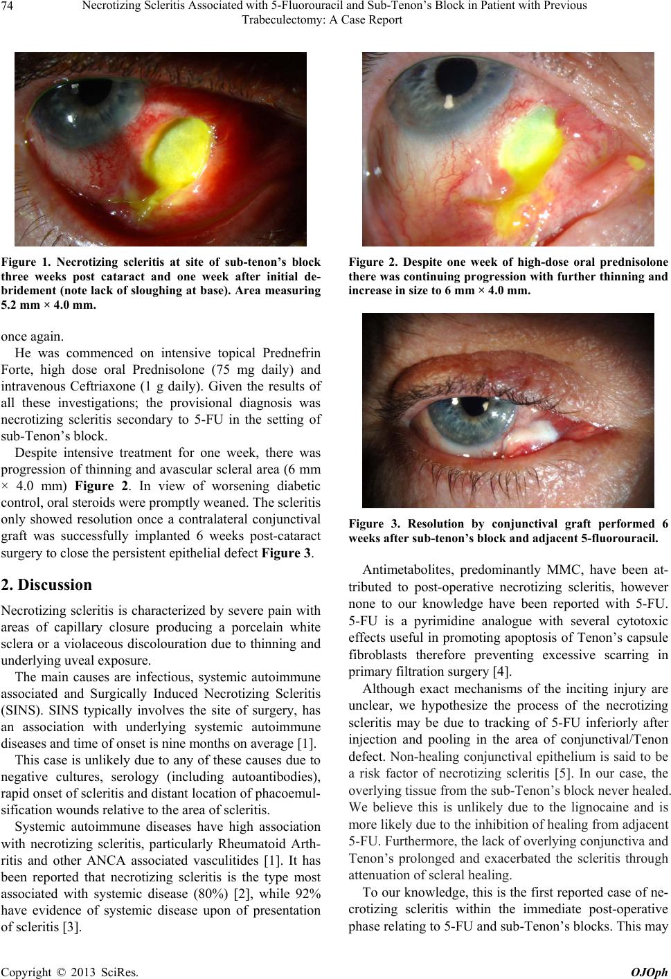 Necrotizing Scleritis Associated with 5-Fluorouracil and Sub-Tenon’s Block in Patient with Previous Trabeculectomy: A Case Report 74 Figure 1. Necrotizing scleritis at site of sub-tenon’s block three weeks post cataract and one week after initial de- bridement (note lack of sloughing at base). Area measuring 5.2 mm × 4.0 mm. once again. He was commenced on intensive topical Prednefrin Forte, high dose oral Prednisolone (75 mg daily) and intravenous Ceftriaxone (1 g daily). Given the results of all these investigations; the provisional diagnosis was necrotizing scleritis secondary to 5-FU in the setting of sub-Tenon’s block. Despite intensive treatment for one week, there was progression of thinning and avascular scleral area (6 mm × 4.0 mm) Figure 2. In view of worsening diabetic control, oral steroids were promptly weaned. The scleritis only showed resolution once a contralateral conjunctival graft was successfully implanted 6 weeks post-cataract surgery to close the persistent epithelial defect Figure 3. 2. Discussion Necrotizing scleritis is characterized by severe pain with areas of capillary closure producing a porcelain white sclera or a violaceous discolouration due to thinning and underlyi ng uveal expos ure . The main causes are infectious, systemic autoimmune associated and Surgically Induced Necrotizing Scleritis (SINS). SINS typically involves the site of surgery, has an association with underlying systemic autoimmune diseases and time of onset is nine months on average [1]. This case is unlikely due to any of these causes due to negative cultures, serology (including autoantibodies), rapid onset of scleritis and distant location of phaco emul- sification wounds relative to the area of scleritis. Systemic autoimmune diseases have high association with necrotizing scleritis, particularly Rheumatoid Arth- ritis and other ANCA associated vasculitides [1]. It has been reported that necrotizing scleritis is the type most associated with systemic disease (80%) [2], while 92% have evidence of systemic disease upon of presentation of scleritis [3]. Figure 2. Despite one week of high-dose oral prednisolone there was continuing progression with further thinning and increase in size to 6 mm × 4.0 mm. Figure 3. Resolution by conjunctival graft performed 6 weeks after sub-tenon’s block and adjace nt 5-fluor our acil. Antimetabolites, predominantly MMC, have been at- tributed to post-operative necrotizing scleritis, however none to our knowledge have been reported with 5-FU. 5-FU is a pyrimidine analogue with several cytotoxic effects useful in promoting apoptosis of Tenon’s capsule fibroblasts therefore preventing excessive scarring in primary filtration surgery [4]. Although exact mechanisms of the inciting injury are unclear, we hypothesize the process of the necrotizing scleritis may be due to tracking of 5-FU inferiorly after injection and pooling in the area of conjunctival/Tenon defect. Non-healing conjunctival epithelium is said to be a risk factor of necrotizing scleritis [5]. In our case, the overlying tissue from the sub-Tenon’s block never healed. We believe this is unlikely due to the lignocaine and is more likely due to the inhibition of healing from adjacent 5-FU. Furthermore, th e lack of overlyin g conjun ctiva and Tenon’s prolonged and exacerbated the scleritis through attenuation of scleral healing. To our knowledge, this is the fir st reported case of ne- crotizing scleritis within the immediate post-operative phase relating to 5-FU and sub-Tenon’s blocks. This may Copyright © 2013 SciRes. OJOph 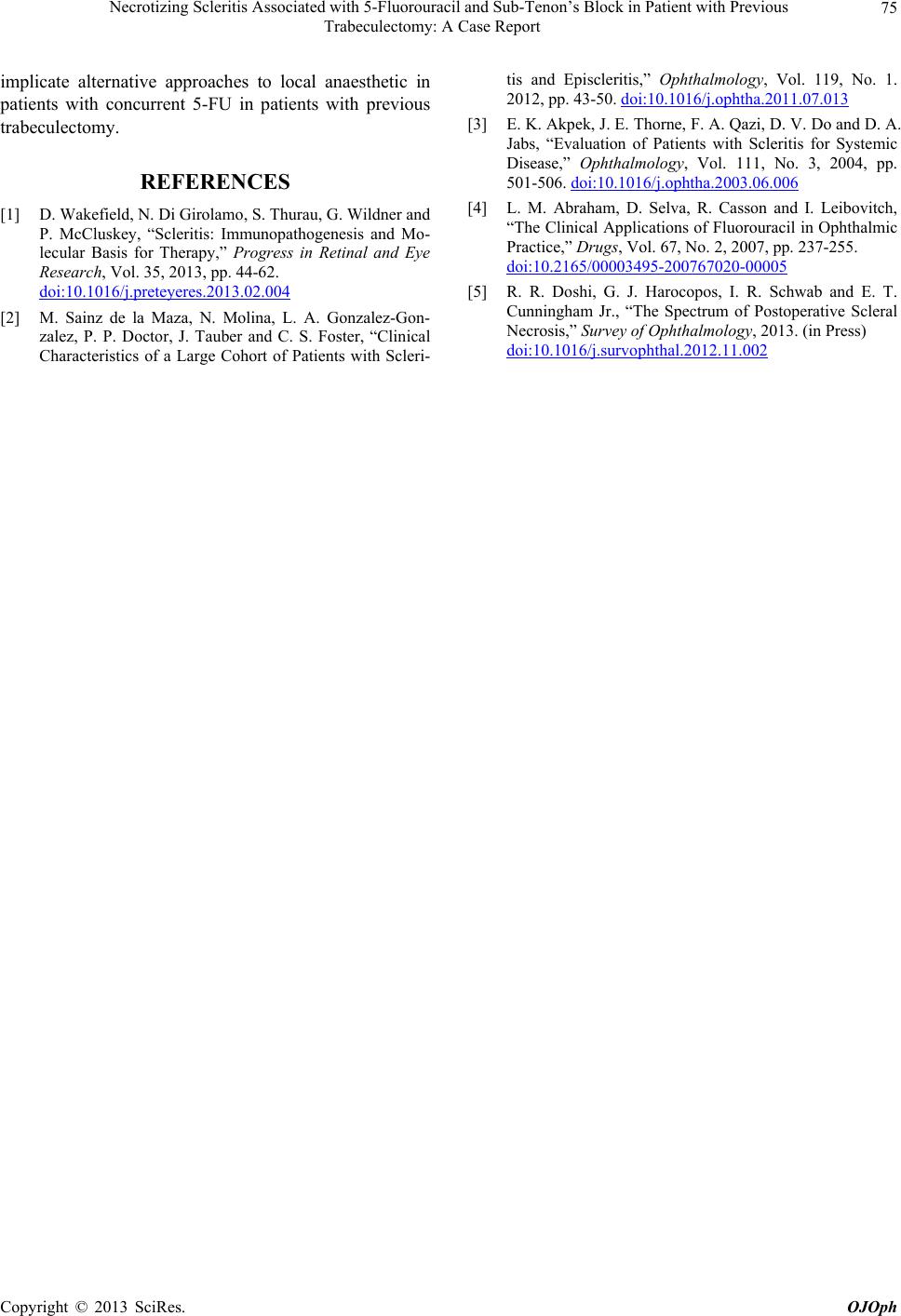 Necrotizing Scleritis Associated with 5-Fluorouracil and Sub-Tenon’s Block in Patient with Previous Trabeculectomy: A Case Report Copyright © 2013 SciRes. OJOph 75 implicate alternative approaches to local anaesthetic in patients with concurrent 5-FU in patients with previous trabeculectomy. REFERENCES [1] D. Wakefield, N. Di Girolamo, S. Thurau, G. Wildner and P. McCluskey, “Scleritis: Immunopathogenesis and Mo- lecular Basis for Therapy,” Progress in Retinal and Eye Research, Vol. 35, 2013, pp. 44-62. doi:10.1016/j.preteyeres.2013.02.004 [2] M. Sainz de la Maza, N. Molina, L. A. Gonzalez-Gon- zalez, P. P. Doctor, J. Tauber and C. S. Foster, “Clinical Characteristics of a Large Cohort of Patients with Scleri- tis and Episcleritis,” Ophthalmology, Vol. 119, No. 1. 2012, pp. 43-50. doi:10.1016/j.ophtha.2011.07.013 [3] E. K. Akpek, J. E. Thorne, F. A. Qazi, D. V. Do and D. A. Jabs, “Evaluation of Patients with Scleritis for Systemic Disease,” Ophthalmology, Vol. 111, No. 3, 2004, pp. 501-506. doi:10.1016/j.ophtha.2003.06.006 [4] L. M. Abraham, D. Selva, R. Casson and I. Leibovitch, “The Clinical Applications of Fluorouracil in Ophthalmic Practice,” Drugs, Vol. 67, No. 2, 2007, pp. 237-255. doi:10.2165/00003495-200767020-00005 [5] R. R. Doshi, G. J. Harocopos, I. R. Schwab and E. T. Cunningham Jr., “The Spectrum of Postoperative Scleral Necrosis,” Survey of Ophthalmology, 2013. (in Press) doi:10.1016/j.survophthal.2012.11.002 |