

Paper Menu >>
Journal Menu >>
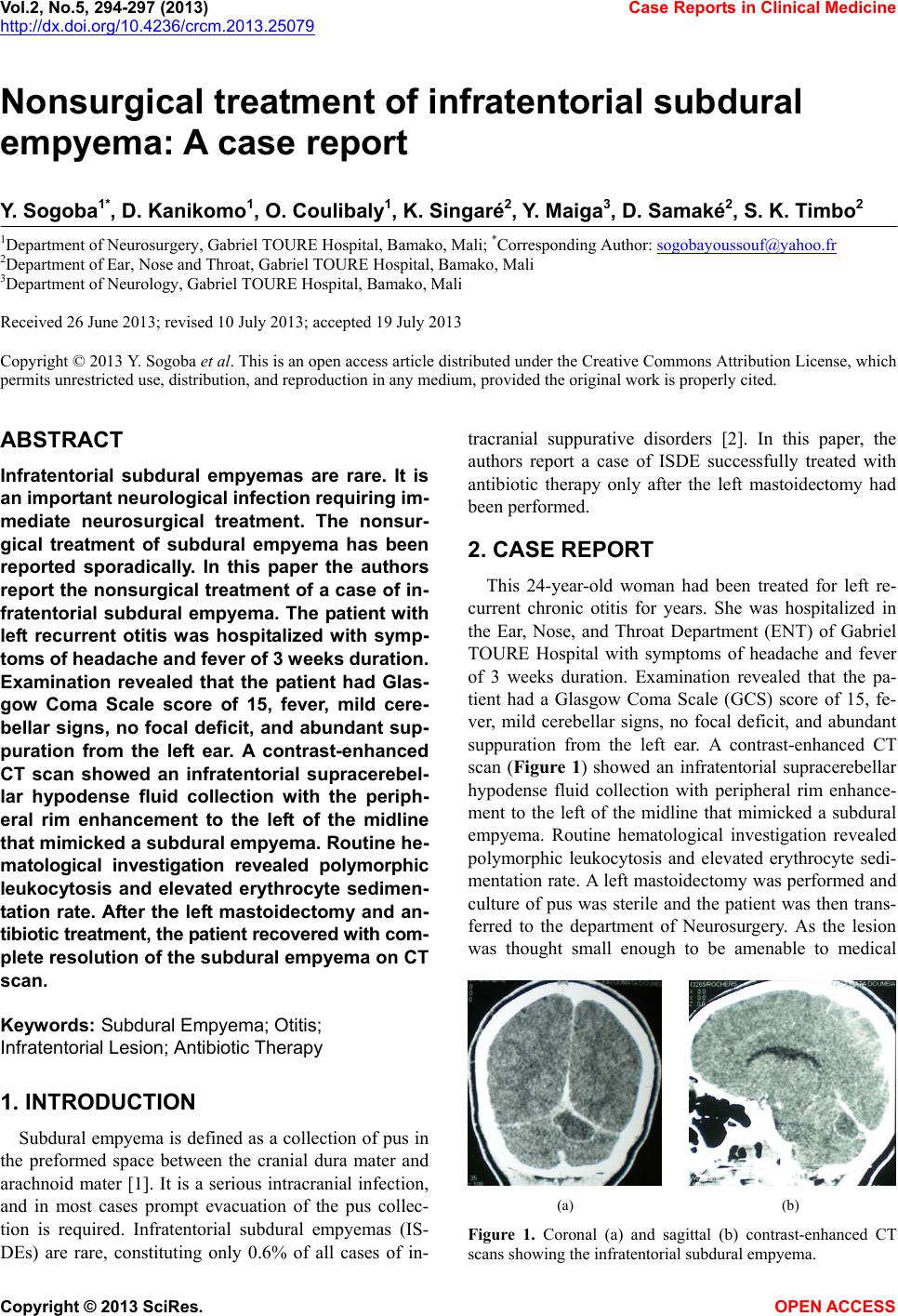 Vol.2, No.5, 294-297 (2013) Case Reports in Clinical Medicine http://dx.doi.org/10.4236/crcm.2013.25079 Nonsurgical treatment of infratentorial subdural empyema: A case report Y. Sogoba1*, D. Kanikomo1, O. Coulibaly1, K. Singaré2, Y. Maiga3, D. Samaké2, S. K. Timbo2 1Department of Neurosurgery, Gabriel TOURE Hospital, Bamako, Mali; *Corresponding Author: [email protected] 2Department of Ear, Nose and Throat, Gabriel TOURE Hospital, Bamako, Mali 3Department of Neurology, Gabriel TOURE Hospital, Bamako, Mali Received 26 June 2013; revised 10 July 2013; accepted 19 July 2013 Copyright © 2013 Y. Sogoba et al. This is an open access article distributed under the Creative Commons Attribution License, which permits unrestricted use, distribution, and reproduction in any medium, provided the original work is properly cited. ABSTRACT Infratentorial subdural empyemas are rare. It is an import ant neurological infection r e quiring im- mediate neurosurgical treatment. The nonsur- gical treatment of subdural empyema has been reported sporadically. In this paper the authors report the nonsurgical treatment of a case of in- fratentorial subdural empyema. The patient with left recurrent otitis was hospitalized with symp- toms of headache and fev er of 3 weeks duration. Examination revealed that the patient had Glas- gow Coma Scale score of 15, fever, mild cere- bellar signs, no focal deficit, and abundant sup- puration from the left ear. A contrast-enhanced CT scan showed an infratentorial supracerebel- lar hypodense fluid collection with the periph- eral rim enhancement to the left of the midline that mimicked a subdural empyema. Routine he- matological investigation revealed polymorphic leukocytosis and elevated erythrocyte sedimen- tation rate. After the left mastoidectomy and an- tibiotic treatment, the patient recovered with com- plete resolution of the subdural empyema on CT scan. Keywords: Subdural Empyema; Otitis; Infratentorial Lesion; Antibiotic Therapy 1. INTRODUCTION Subdural empyema is defined as a collection of pus in the preformed space between the cranial dura mater and arachnoid mater [1]. It is a serious intracranial infection, and in most cases prompt evacuation of the pus collec- tion is required. Infratentorial subdural empyemas (IS- DEs) are rare, constituting only 0.6% of all cases of in- tracranial suppurative disorders [2]. In this paper, the authors report a case of ISDE successfully treated with antibiotic therapy only after the left mastoidectomy had been performed. 2. CASE REPORT This 24-year-old woman had been treated for left re- current chronic otitis for years. She was hospitalized in the Ear, Nose, and Throat Department (ENT) of Gabriel TOURE Hospital with symptoms of headache and fever of 3 weeks duration. Examination revealed that the pa- tient had a Glasgow Coma Scale (GCS) score of 15, fe- ver, mild cerebellar signs, no focal deficit, and abundant suppuration from the left ear. A contrast-enhanced CT scan (Figure 1) showed an infratentorial supracerebellar hypodense fluid collection with peripheral rim enhance- ment to the left of the midline that mimicked a subdural empyema. Routine hematological investigation revealed polymorphic leukocytosis and elevated erythrocyte sedi- mentation rate. A left mastoidectomy was performed and culture of pus was sterile and the patient was then trans- ferred to the department of Neurosurgery. As the lesion was thought small enough to be amenable to medical (a) (b) Figure 1. Coronal (a) and sagittal (b) contrast-enhanced CT scans showing the infratentorial subdural empyema. Copyright © 2013 SciRes. OPEN ACCESS 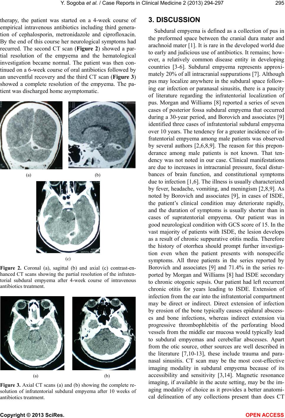 Y. Sogoba et al. / Case Reports in Clinica l Me dicine 2 (2013) 294-297 295 therapy, the patient was started on a 4-week course of empirical intravenous antibiotics including third genera- tion of cephalosporin, metronidazole and ciprofloxacin. By the end of this course he r neurological symptoms h ad recurred. The second CT scan (Figure 2) showed a par- tial resolution of the empyema and the hematological investigation became normal. The patient was then con- tinued on a 6-week course of oral antibiotics followed by an uneventful recovery and the third CT scan (Figure 3) showed a complete resolution of the empyema. The pa- tient was discharged home asymptomati c . (a) (b) (c) Figure 2. Coronal (a), sagittal (b) and axial (c) contrast-en- hanced CT scans showing the partial resolution of the infraten- torial subdural empyema after 4-week course of intravenous antibiotics treatment. (a) (b) Figure 3. Axial CT scans (a) and (b) showing the complete re- solution of infratentorial subdural empyema after 10 weeks of antibiotics treatment. 3. DISCUSSION Subdural empyema is defined as a collection of pus in the preformed space between the cranial dura mater and arachnoid mater [1]. It is rare in the developed world due to early and judicious use o f antibiotics. It remains; how- ever, a relatively common disease entity in developing countries [3-6]. Subdural empyema represents approxi- mately 20% of all intracranial suppurations [7]. Although pus may localize anywhere in the subdural space follow- ing ear infection or paranasal sinusitis, there is a paucity of literature regarding the infratentorial localization of pus. Morgan and Williams [8] reported a series of seven cases of posterior fossa subdural empyema that occurred during a 30-year p eriod, and Borovich and associates [9] identified three cases of infratentorial subdural e mpyema over 10 years. The tendency for a greater incidence of in- fratentorial empyema among male patients was observed by several authors [2,6,8,9]. The reason for this prepon- derance among male patients is not known. That ten- dency was not noted in our case. Clinical manifestations are due to increases in intracranial pressure, focal distur- bances of brain function, and constitutional symptoms due to infection [1,6]. Th e illness is usu ally characterized by fever, headache, vomiting, and meningism [2,8,9]. As noted by Borovich and associates [9], in cases of ISDE, the patient’s clinical condition may deteriorate rapidly, and the duration of symptoms is usually shorter than in cases of supratentorial empyema. Our patient was in good neurological condition with GCS score of 15. In the vast majority of patients with ISDE, the lesion develops as a result of chronic suppurative otitis media. Therefore the history of otorrhea should prompt further investiga- tion even when the patient presents with nonspecific symptoms. All three patients in the series reported by Borovich and associates [9] and 71.4% in the series re- ported by Morgan and Williams [8] had ISDE secondary to chronic otogenic sepsis. Our patient had left recurrent chronic otitis for years leading to ISDE. Extension of infection from the ear into the infratentorial compartment may be direct or indirect. Direct extension of infection by erosion of the bone typically causes epidural abscess- es and bone infections, whereas indirect extension via progressive thrombophlebitis of the perforating blood vessels from the middle ear mucosa would typically lead to subdural empyemas and cerebellar abscesses. Apart from the otic source, other sources are well described in the literature [7,10-13], these include trauma and para- nasal sinusitis. CT scan may be the most cost-effective imaging modality in subdural empyema because of its accessibility and sensitivity [3,14]. Magnetic resonance imaging, if availab le in the acute setting, may be the im- aging modality of choice as it provides a better anatomi- cal delineation of any collections present than does CT Copyright © 2013 SciRes. OPEN ACCESS 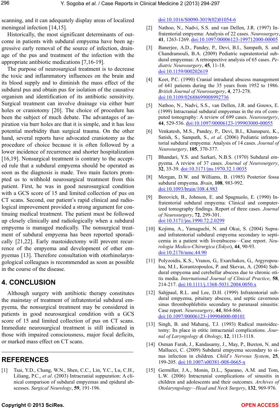 Y. Sogoba et al. / Case Reports in Clinica l Me dicine 2 (2013) 294-297 296 scanning, and it can adequately display areas of localized meningeal infection [14,15]. Historically, the most significant determinants of out- come in patients with subdural empyema have been ag- gressive early removal of the source of infection, drain- age of the pus and treatment of the infection with the appropriate antibiotic medications [7,16-19]. The purpose of neurosurgical treatment is to decrease the toxic and inflammatory influences on the brain and its blood supply and to diminish the mass effect of the subdural pus and ob tain pus for isolation of the causative organism and identification of its antibiotic sensitivity. Surgical treatment can involve drainage via either burr holes or craniotomy [20]. The choice of procedure has been the subject of much debate. The advantages of as- piration via burr holes are that it is simple, and it h as less potential morbidity than surgical trauma. On the other hand, several reports have advocated craniotomy as the procedure of choice because it is often followed by a lower incidence of recurr ence and shorter hospitalization [16,19]. Nonsurgical treatment is contrary to the accept- ed rule that a subdural empyema should be operated as soon as the diagnosis is made. Two main factors prom- pted us to withhold neurosurgical treatment from this patient. First, he was in good neurosurgical condition with a GCS score of 15 and limited collection of pus on CT scans. Second, our patient’s rapid clinical and radio- logical improve ment prov id ed a strong argument for con- tinuing medical treatment. The patient must be followed up closely clinically and radiologically when a subdural empyema is managed medically. The nonsurgical treat- ment of subdural empyema has been reported sporadi- cally [21,22]. Early mastoidectomy will prevent recur- rence of the empyema and development of other em- pyemas [13]. Therefore consultation with otorhinolaryn- gological colleagues is recommended as soon as possible in the course of the disease. 4. CONCLUSION Although surgery with antibiotic therapy constitutes the mainstay of treatment of infratentorial subdural em- pyema, the nonsurgical treatment may be considered in patients in good neurosurgical condition with a GCS score of 15 and limited collection of pus on CT scans. Immediate neurosurgical treatment is still indicated in those with impaired consciousness, major focal deficits, or marked mass effect on CT scans. REFERENCES [1] Tsai, Y.D., Chang, W.N., Shen, C.C., Lin, Y.C., Lu, C.H., Liliang, P.C., et al. (2003) Intracranial suppuration: A cli- nical comparison of subdural empyemas and epidural ab- scesses. Surgical Neurology, 59, 191-196. doi:10.1016/S0090-3019(02)01054-6 [2] Nathoo, N., Nadvi, S.S. and van Dellen, J.R. (1997) In- fratentorial empyema: Analysis of 22 cases. Neurosurgery, 41, 1263-1269. doi:10.1097/00006123-199712000-00005 [3] Banerjee, A.D., Pandey, P., Devi, B.I., Sampath, S. and Chandramouli, B.A. (2009) Pediatric supratentorial sub- dural empyemas: A retrospective analysis of 65 cases. Pe- diatric Neurosurgery, 45, 11-18. doi:10.1159/000202619 [4] Keet, P.C. (1990) Cranial intradural abscess management of 641 patients during the 35 years from 1952 to 1986. British Journal of Neurosurg ery, 4, 273-278. doi:10.3109/02688699008992736 [5] Nathoo, N., Nadvi, S.S., van Dellen, J.R. and Gouws, E. (1999) Intracranial subdural empyemas in the era of com- puted tomography: A review of 699 cases. Neurosurgery, 44, 529-536. doi:10.1097/00006123-199903000-00055 [6] Venkatesh, M.S., Pandey, P., Devi, B.I., Khanapure, K., Satish, S., Sampath, S., et al. (2006) Pediatric infraten- torial subdural empyema: Analysis of 14 cases. Journal of Neurosurgery, 105, 370-377. [7] Bhandari, Y.S. and Sarkari, N.B.S. (1970) Subdural em- pyema. A review of 37 cases. Journal of Neurosurgery, 32, 35-39. doi:10.3171/jns.1970.32.1.0035 [8] Morgan, D.W. and Williams, B. (1985) Posterior fossa subdural empyema. Brain, 108, 983-992. doi:10.1093/brain/108.4.983 [9] Borovich, B., Johnson, E. and Spagnuolo, E. (1990) In- fratentorial subdural empyema: Clinical and computer- ized tomography findings. Report of three cases. Journal of Neurosurgery, 72, 299-301. doi:10.3171/jns.1990.72.2.0299 [10] Kojima, A., Yamaguchi, N. and Okui, S. (2004) Supra- and infratentorial subdural empyema secondary to septi- cemia in a patient with liverabscess—Case report. Neu- rologia Medico-Chirurgica (Tokyo), 44, 90-93. doi:10.2176/nmc.44.90 [11] Polyzoidis, K.S., Vranos, G., Exarchakos, G., Argyropou- lou, M.I., Korantzopoulos, P. and Skevas, A. (2004) Sub- dural empyema and cerebellar abscess due to chronic oti- tis media. International Journal of Clinical Practice, 58, 214-217. d oi :10.1111/j.1368-5031.2004.0050.x [12] Sahjpaul, R.L. and Lee, D.H. (1999) Infratentorial sub- dural empyema, pituitary abscess, and septic cavernous sinus thrombophlebitis secondary to paranasal sinusitis: Case report. Neurosurgery, 44, 864-866. doi:10.1097/00006123-199904000-00101 [13] Singh, B. and Maharaj, T.J. (1993) Radical mastoidec- tomy: Its place in otitic intracranial complications. Jour- nal of Laryngology & Otology, 12, 1113-1118. [14] Osman Farah, J., Kandasamy, J., May, P., Buxton, N. and Mallucci, C. (2009) Subdural empyema secondary to si- nus infection in children. Child’s Nervous System, 25, 199-205. doi:10.1007/s00381-008-0665-x [15] Germiller, J.A., Monin, D.L., Sparano, A.M. and Tom, L.W. (2006) Intracranial complications of sinusitis in children and adolescents and their outcomes. Archives of Otolaryngology—Head and Neck Surgery, 132, 969-976. Copyright © 2013 SciRes. OPEN ACCESS  Y. Sogoba et al. / Case Reports in Clinica l Me dicine 2 (2013) 294-297 Copyright © 2013 SciRes. OPEN ACCESS 297 doi:10.1001/archotol.132.9.969 [16] Bannister, G., Williams, B. and Smith, S. (1981) Treat- ment of subdural empyema. Journal of Neurosurgery, 55, 82-88. doi:10.3171/jns.1981.55.1.0082 [17] Farmer, T.W. and Wise, G.R. (1973) Subdural empyema in infants, children and adults. Neurology, 23, 254-261. doi:10.1212/WNL.23.3.254 [18] Hitchcock, E. and Andreadis, A. (1964) Subdural em- pyema: A review of 29 cases. Journal of Neurology , Neu- ro surg ery & Psyc hi a try, 27, 422-434. doi:10.1136/jnnp.27.5.422 [19] Williams, B. (1983) Subdural empyema. Advances and Technical Standards in Neurosurgery, 9, 133-170. doi:10.1007/978-3-7091-7034-2_6 [20] Bok, A.P. and Peter, J.C. (1993) Subdural empyema: Burr holes or craniotomy? A retrospective computerized to- mography-era analysis of treatment in 90 cases. Journal of Neurosurgery, 78, 574-578. doi:10.3171/jns.1993.78.4.0574 [21] Mauser, H.W., Ravijst, R.A., Elderson, A., van Gijn, J. and Tulleken, C.A. (1985) Nonsurgical treatment of sub- dural empyema. Case report. Journal of Neurosurgery, 63, 128-130. doi:10.3171/jns.1985.63.1.0128 [22] Rosazza, A., de Tribolet, N. and Deonna, T. (1979) Non- surgical treatment of interhemispheric subdural empye- mas. Helvetica Paediatrica Acta, 34, 577-581. |