

Paper Menu >>
Journal Menu >>
 Vol.2, No.5, 291-293 (2013) Case Reports in Clinical Medicine http://dx.doi.org/10.4236/crcm.2013.25078 Non-caseating submental tuberculous lymphadenopathy: A case report M. E. Asuquo1*, V. I. Nwagbara1, S. Akpan1, G. Ebughe2, T. Ugbem2, I. M. Asuquo3 1Department of Surgery, University of Calabar/University of Calabar Teaching Hospital, Calabar, Nigeria; *Corresponding Author: [email protected], [email protected] 2Department of Pathology, University of Calabar/University of Calabar Teaching Hosp ital, Calabar, Nigeria 3Department of Curriculum and Teaching, Faculty of Education, University of Calabar, Calabar, Nigeria Received 10 May 2013; revised 20 June 2013; accepted 10 July 2013 Copyright © 2013 M. E. Asuquo et al. This is an open access article distributed under the Creative Commons Attribution License, which permits unrestricted use, distribution, and reproduction in any medium, provided the original work is properly cited. ABSTRACT Chronic peripheral ly mphadenop athy in adult s is an indication of pathology of which tuberculosis is the commonest in the developing countries. Presented is a 36-year-old with a huge chronic Submental lymphadenopathy in a seronegative patient of 7 years duration. Histology revealed non-caseating tuberculosis. Tuberculosis should be considered in Submental lymphadenopathy. Despite the long duration, the absence of cold abscess, and or sinus formation may be an in- dication of the non-caseating tuberculous lym- phadenopathy. Keywords: Tuberculosis; Submental L ymph Node; Non-Caseating Granuloma 1. INTRODUCTION Chronic peripheral lymph node enlargement in adults is an indication of an underlying disease that may pose a diagnostic dilemma to physicians [1]. The commonest lymph node group affected is reported to be the cervical, the clinical manifestation varies with the challenges of diagnosis not unusual [2,3]. Tuberculosis (TB) has been reported as a common cause of chronic lymphadenopa- thy in the develop ing countries and one of the most com- mon of all extra pulmonary (TB) [4,5]. However, in the developed countries with the rarity of infections, malign- nancies are reported as the predominant cause of periph- eral lymphadenopathy [1]. In developing countries in Asia and Africa where tuberculous infection is common and other granulomatous infections rare, the presence of granulomatous features on histology are suggestive of TB [3]. We present this case of florid non-caseating Su b- mental tuberculous lymphadenopathy in a human im- munodeficiency virus (HIV) seronegative patient to high- light its unusual presentation; diagnostic challenge due to ulceration from topical herbal medication, florid lesion in an unusual group of lymph no de without any identifiab le primary lesion. 2. CASE REPORT A 36 years old applicant (engineer) presented to the surgical out patient department (SOPD) as a referral from the general out patient department with a 7-year history of an anterior neck swelling. The swelling began as a nodule, which he felt but was not visible, however in- creased gradually to the size at presentation. There was no associated fever, cough, and night sweats. There was no history of trauma or dental pain, mass was painless and not associated with pain or difficulty in swallowing. There was no swelling in any other part of the body. He sought treatment in some hospital facilities (private and government) to no avail. Two months prior to presenta- tion, he applied topical herbal preparation that resulted in a wound hence his presentation to the University of Calabar Teaching Hospital (UCTH), Calabar. Examination revealed a young man in relative good health, afebrile, and not pale. There was a firm, painless, lobulated, ulcerated, and immobile mass in the Submen- tal region of the neck. It measured 8 cm × 6 cm. The edge of the ulcer was slopping with the floor that showed granulation tissue with some spots with sloughs and fixed to the underlying structure, Figures 1(a) and (b). Examination of the oral cavity, ear nose, and throat, chest and abdominal examination were normal. A clinical di- agnosis of Submental lymphad enopathy was made. Full blood count (FBC) showed: haemoglobin 12.9 g/dl, white blood cell 6.5 × 199/l (neutrophils 40%, eosi- nophils 2%, and lymphocytes 58%), human immunode- ficiency virus (HIV) serology was negative and urinaly- Copyright © 2013 SciRes. OPEN ACCESS 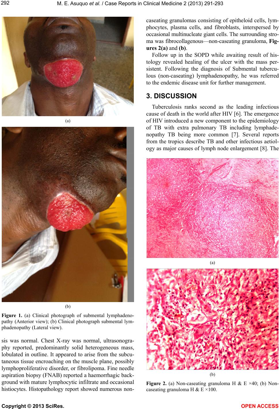 M. E. Asuquo et al. / Case Reports in Clinical Medicine 2 (2013) 2 91-293 292 (a) (b) Figure 1. (a) Clinical photograph of submental lymphadeno- pathy (Anterior view); (b) Clinical photograph submental lym- phadenopathy (Lateral view). sis was normal. Chest X-ray was normal, ultrasonogra- phy reported, predominantly solid heterogeneous mass, lobulated in outline. It appeared to arise from the subcu- taneous tissue encroaching on the muscle plane, possibly lymphoproliferative disorder, or fibrolipoma. Fine needle aspiration biopsy (FNAB) reported a haemorrhagic back- ground with mature lymphocytic infiltrate and occasional histiocytes. Histopathology report showed numerous non- caseating granulomas consisting of epitheloid cells, lym- phocytes, plasma cells, and fibroblasts, interspersed by occasional multinucleate giant cells. The surrounding stro- ma was fibrocollagenous—non-caseating granuloma, Fig- ures 2 (a) and (b). Follow up in the SOPD while awaiting result of his- tology revealed healing of the ulcer with the mass per- sistent. Following the diagnosis of Submental tubercu- lous (non-caseating) lymphadenopathy, he was referred to the endemic disease unit for further management. 3. DISCUSSION Tuberculosis ranks second as the leading infectious cause of death in the world after HIV [6]. The emergence of HIV introduced a new component to the ep idemiology of TB with extra pulmonary TB including lymphade- nopathy TB being more common [7]. Several reports from the tropics describe TB and other infectious aetiol- ogy as major causes of lymph node enlargement [8]. The (a) (b) Figure 2. (a) Non-caseating granuloma H & E ×40; (b) Non- caseating granuloma H & E ×100. Copyright © 2013 SciRes. OPEN ACCESS  M. E. Asuquo et al. / Case Reports in Clinical Medicine 2 (2013) 2 91-293 Copyright © 2013 SciRes. OPEN ACCESS 293 commonest site of tuberculous lymphadenopathy re- ported was cervical. Its involvement of cervical lymph nodes has been known a long time as Scrofula or the Kings Evil [9]. Olu-Eddo and Omoti reported lympha- denopathy as the single commonest cause of cervical lymphadenopathy constituting 35% of cases [4], and in Saudi Arabia, Al-Sohaibani reported 28% [10]. This com- munication describes a huge tuberculous lymphadeno- pathy in an HIV seronegative patient in an uncommon location for tuberculous cervical lymphadenopathy. Tuberculous lymphadenopathy is largely confined to the cervical lymph nodes mostly because tonsils and ade- noids provide an easy portal of entry for inhaled myco- bacteria [5]. It may also result from lymphatic or ha- ematogenous dissemination from an original focus in the lungs [5], our patient had no identifiable dental or oral lesion. However, some lesions may be healed without being detected and may be the case in our patient whose evaluation revealed no primary focus despite the huge Submental lymphadenopathy. Majority of TB is diagnosed on clinical grounds with or without histological appearance of the biopsy [5]. Ac- tive lesions are seen as characteristic granulomatous in- flammatory reaction that forms caseating and non-caseat- ing tubercles [6]. Our patient’s histologic features were consistent with non-caseating TB, Figure 2. It is less common (20%) and described as hypertrophic. The firm consistency was in keeping with the clinical evaluation despite the long duration of the lesion; this variety was unlikely to form cold ab scess and eventually a sinus seen in the caseating form. The non-caseating TB is seen in patients with good immunity [11], consistent with our patient. In the authors’ setting tradition al healers, enjo y a lot of patronage especially with long standing lesions. Topical herbal medication induced chemical inflammation of the skin and ulceration and was capable of misdirecting cli- nical judgement and therapy. Delay in presentation and diagnosis was due to the inability of previous consulta- tions to result in a proper diagnosis while the morbidity associated with topical herbal medication, ulceration, prompted presentation. Health education is pivotal for early presentation. Physicians should subject chronic cer- vical lymphadenopathy to histologic evaluation for di- agnosis and proper treatment as this is crucial for sat- isfactory outcome. Huge Submental chronic lymphadenopathy may be tuberculous. The effect of topical herbal medication on clinical evaluation should not be underestimated like- wise the morbidity. Diagnosis is histologic, long-standing chronic lymphadenopathy without suppuration in a sero- negative patient should arouse the diagnosis of non- caseating TB. REFERENCES [1] Olu-Eddo, A.N. and Ohanaka, C.E. (2006) Peripheral ly m- phadenopathy in Nigerian adults. Journal of Pakistan Medical Association, 56, 405-408. [2] Adeniji, K.A. and Anjorin, A.S. (2000) Peripheral lym- phadenopathy in Nigeria. African Journal of Medicine & Medical Sciences, 29, 233-237. [3] Nwagbara, V.I., Asuquo, M.E., Ebughe, G., Agbor, C., Akpan, S., Ugbem, T. and Asuquo, I.M. (2013) Tubercu- lous lymphadenitis of the neck. Case series. International Journal of Medicine, 1, 4-8. [4] Olu-Eddo, A.N. and Omoti, C.E. (2011) Diagnostic evaluation of primary cervical adenopathies in a devel- oping country. Pan African Medical Journal, 10, 52. [5] Shubha, A.B., Sapna, H. and Dinesh Rao, B. (2010) Tu- berculous lymphadenitis presenting a diagnostic dilemma. A Case Report. International Journal of Dental Clinics, 2, 48-52. [6] Udoh, M.O. (2009) Pathogenesis and Morphology of Tuberculosis. Benin Journal of Postgraduate Medicine, 11, 91-96. [7] Ben, C., Patel, P.S., Bharucha, H., Namaambo, K. and Luo, N. (1996) Importance of human immunodeficiency virus associated lymphadenopathy and tuberculous lym- phadenitis in patients undergoing lymph node biopsy in Zambia. British Journal of Surgery, 83, 75-78. doi:10.1002/bjs.1800830124 [8] Thomas, J.O., Ladikpo, J.K. and Yawe, T. (1995) Histo- pathology of lymphadenopathy in a tropical country. East African Medical Journal, 72, 703-705. [9] Tan, K.K. (1988) Tuberculous lymphadenitis in Singa- pore. Singapore Medical Journal, 29, 441-446. [10] Al-Shohaibani, M.O. (1996) Chronic lymphadenopathy in the eastern province of Saudi Arabia. East African Medical Journal, 73, 533-537. [11] Sriran Bhat, M. (2009) SRB’s mannual of surgery. 3rd Edition, Jaypee, New Delhi, 391-393. |