

Paper Menu >>
Journal Menu >>
 Surgical Science, 2013, 4, 6-14 http://dx.doi.org/10.4236/ss.2013.49A002 Published Online September 2013 (http://www.scirp.org/journal/ss) Surgeon-Operated In-Office Ultrasonography for the Diagnosis of Rotator Cuff Tears: A Comparison with Magnetic Resonance Imaging Tsutomu Kobayashi1*#, Atsushi Yamamoto1*, Hitoshi Shitara1, Tsuyoshi Ichinose1, Eiji Takasawa1, Daisuke Shimoyama1, Toshihisa Osawa2, Kenji Takagishi1 1Department of Orthopaedi c Surgery, Graduate School of Medicine, Gunma University, Maebashi, Japan 2Department of Orthopae d ic Surgery, Takasaki General Medical Center, Takasaki, Japa n Email: #[email protected] Received May 26, 2013; revised June 27, 2013; accepted July 6, 2013 Copyright © 2013 Tsutomu Kobayashi et al. This is an open access article distributed under the Creative Commons Attribution Li- cense, which permits unrestricted use, distribution, and reproduction in any medium, provided the original work is properly cited. ABSTRACT Obje ctive: Few investigators have evaluated whether ultrasonography operated by a surgeon during a patient’s clinic visit is capable of obtaining a similar degree of accuracy as magnetic resonance imaging in regard to the diagnosis of rotator cuff tears and lesions of the biceps tendon. Th e purpose of this study was to clarify the accur acy of in-office ul- trasonography for the diagnosis of rotator cuff tears in comp arison to magnetic resonance imaging. Methods: One hun - dred and three patients (105 shoulders) with a clinical diagnosis of impingement and suspected rotator cuff tear, who subsequently underwent arthroscopic surgery were retrospectively enrolled in this study, including 7 males with 89 shoulders, and 33 females with 33 shoulders, and their mean age was 60.9 years (range, 30 to 83 years). The subjects were examined using ultrasonography and magnetic resonance imaging within three months pre-operatively per normal practice of the outpatient clinic. The two modalities were then compared to the reference standard, arthrosco pic finding s. Results: Intra-operatively, 79 full-thickness and 15 partial-thickness rotator cuff tears were found. The agreement be- tween ultrasonography and magnetic resonance imaging for diagnosis of rotator cuff tears was statistically good; ob- served degree of agreement was 87 % with Kappa coefficient of 0.73. Ultrasonograph y showed a sensitiv ity of 94% an d a specificity of 100% for full-thickness tears, and a sensitivity of 80% and a specificity of 91% for partial-thickness tears. The agreement of the two modalities for diagno sis of lesions of the biceps ten don was also good; observed degree of agreement was 93% with Kappa coefficient of 0.76. In addition, ultrasonography showed comparable accuracy for classifying the size of rotator cuff tears to that of magnetic resonance imaging. Conclusion: Surgeon-operated in-office ultrasonography is an appropriate technique for the assessment of rotator cuff tears with a comparable sensitivity and specificity to that of magnetic resonance imaging. Keywords: Diagnostic Accuracy; Magnetic Resonance Imaging; Rotator Cuff Tear; Ultrasonography; Arthroscopic Surgery 1. Introduction A rotator cuff tear is one of the most common disorders affecting the shoulder and a recent population-based study showed that approximately one-fourth of residents of a rural area over 50 years of age had full-thickness rotator cuff tears [1]. A proper diagnosis based on accu- rate imaging is indispensable for deciding on the appro- priate management. The first study using ultrasonography to detect rotator cuff tears was reported in 1979 by Seltzer et al. [2]. Al- though several studies tried to develop accurate diagnos- tic methods, the early reports of ultrasonography for ro- tator cuff tears were not able to show favorable results, probably due to the immaturity of the technique related to the procedure and the instrument itself [3,4]. Thus, magnetic resonance imaging had been considered the first-choice imaging modality for the detection of rotator cuff tears because of its high accuracy, despite its rela- tively high cost and occasional limited availability [5,6]. However, following the development of new devices, such as high-frequency transducers and improvements in *Both authors contributed equally to this work. #Corresponding author. C opyright © 2013 SciRes. SS  T. KOBAYASHI ET AL. 7 real-time imaging, a number of studies have reported the high accuracy of ultrasonography for detecting rotator cuff tears [7-9]. Ultrasonography of the shoulder has be- come an accepted method for evaluating rotator cuff tears. Although both ultrasonography and magnetic reso- nance imaging are highly accurate, widely-used and non- invasive imaging modalities for the diagnosis of rotator cuff tears [10] ultrasonography is relatively less expen- sive, less time-consuming, and permits a dynamic eval- uation of the shoulder. Furthermore, the portability of ultrasonography means that the integrity of the rotator cuff can be assessed in geographically isolated locations or by a surgeon during a clinic session to allow more ef- ficient planning of treatment [8]. There have been several studies that compared the accuracy of ultrasonography and magnetic resonance imaging [9,11,12], however, few investigators have evaluated whether ultrasonography performed by a surgeon during the patient’s clinic visit is capable of obtaining a similar degree of accuracy as magnetic resonance imaging with regard to the diagnosis of rotator cuff tears and lesions of the biceps tendon. The purpose of this study was to compare the accuracy of surgeon-operated in-office ultrasonography and magnetic resonance imaging for the detection and measurement of the size of rotator cuff tears, with the results of arthro- scopic surgery as the reference standard. 2. Materials and Methods 2.1. Subjects After institutional review board approval was obtained, 122 patients (122 shoulders) with a clinical diagnosis of impingement and a suspected rotator cuff tear, experi- encing pain, decreased function, and/or weakness, who subsequently underwent arthroscopic surgery in our in- stitute from January 2010 to August 2012, were retro- spectively enrolled in th is study. All patients gave informed consent to particip ate in the study. The patients were allocated to one of three ortho- paedic surgeo ns, with a specialist interest in the shoulder for more than 7 years. And these surgeons have more than 5-years experiences of orthopaedic ultrasonography. Each of these surgeons performed a preoperative evalua- tion including ultrasonography and magnetic resonance imaging, and subsequently performed arthroscopic sur- gery, independently. This process occurred as part of the standard outpatient allo cation, but did not involve patient pre-selection by the clinicians. Of these pa tients, the sub- jects who were involved in the study included patients who 1) had undergone a preoperative examination by both ultrasonography and magnetic resonance imaging within three months before surgery, 2) had complete data of all target evaluation items. The exclusion criteria were patients 1) with claustro- phobia, 2) who had metal devices in the field of view, 3) has undergone a previous surgery, 4) had a previous fracture, or 5) had a known inflammatory arthropathy. Based on these criteria, one patient was excluded because of claustrophobia and 16 patients were excluded because they had incomplete data. Thus, this study comprised 103 patients (105 shoul- ders); there were 87 males with 89 shoulders, and 33 females with 33 shoulders, and their mean age was 60.9 years (range, 30 to 83 years). 2.2. Ultrasonography All ultrasonographic examinations were performed be- fore MRI within the normal flow of the outpatient clinic by one of the three aforementioned orthopaedic surgeons, using a LOGIQ e instrument (GE Healthcare, Bucking- hamshire, England) with linear-array probes at 12 MHz. The standard technique was a modification of the technique described by Middleton et al. [13] and Teefey et al. [14]. Parameters such as the scanning frequency, focal zone number and placement, field of view, and gain were not standardized, but left to the discretion of the observer. Both the patient and the observer were seated on backless stools facing each other. First, the biceps tendon was examined in front of the shoulder by transverse and longitudinal scans. Then the patient was asked to hold the arm in external rotation, and the lon gitudinal scans of the subscapularis tendon were examined. Next, the lon- gitudinal scans of the supraspinatus tendon was exam- ined with the shoulder extended, the elbow flexed, and the hand placed on the iliac wing to expose as much of the supraspinatus tendon as possible from under the ac- romion. The transducer was moved anteriorly to posteri- orly in order to provide the best longitudinal scans of the infraspinatus tendon. The transducer was then rotated 90 degrees in order to examine the transverse areas of the supraspinatus and infrasupinatus tendons. A full-thickness rotator cuff tear was diagnosed when there was a focal discontinuity or thinning of the rotator cuff or when the cuff could not be visualized because of complete avulsion and retraction under the acromion [14, 15]. A partial-thickness tear was diagnosed when there was minimal flattening of the bursal side of the rotator cuff or a distinct hypoechoic or mixed hyperechoic and hypoechoic defect was visualized in both the longitudinal and the transverse planes at the deep articular side of the rotator cuff [14]. The size of the tear in centimeters was also measured directly on freeze-frame images with use of the cursor software function. When the torn cuff was retracted un- der the acromion, the size was recorded as a “not lower than” measured length. Additionally, a finding of a rup- Copyright © 2013 SciRes. SS  T. KOBAYASHI ET AL. 8 ture of the biceps tendon was recorded when the tendon was not identified within or medial to the intertubercular sulcus. Dislocation of the biceps tendon was recorded when the tendon was anterior or medial to the lesser tu- berosity [14]. 2.3. Magnetic Resonance Imaging After examining clinical findings and ultrasonography, magnetic resonance imaging was performed with the same equipment at various facilities, but all examinations were performed under the same standardized conditions. Patients were positioned in the supine positio n with their arms in a neutral position. A 1.5-T system with standard coil was used. The slice thickness was 3 - 4 mm, the field of view was small (12 - 16 cm) and the imaging matrix was 256 mm × 192 mm or higher. The sequences per- formed in all patients were T1- and T2-weighted images in the oblique coronal, oblique sagittal and axial planes. All magnetic resonance imaging was also referred blind- ly within the normal flow of the outpatient clinic by an- other orthopaedic surgeon who ultrasonographically ex- amined the patient. A full-thickness rotator cuff tear was diagnosed if there was a fluid-filled gap on the T2-weighted oblique coronal or oblique sagittal plane that extended through the entire thickness of the tendon, or a complete disrupt- tion of all tendon fibers with retraction. A partial-thick- ness tear was defined as an increase in the signal noted on the T1-weighted images with a brighter signal on the T2-weighted images, as well as an identification of a focal defect on either the bursal or the articular surface of the involved tendon [16]. The size of the tear was meas- ured in centimeters with use of the magnetic resonance imaging scale noted on the images. Additionally, a find- ing of a rupture and dislocation of the biceps tendon was recorded. 2.4. Arthroscopic Surgery One of the three aforementioned orthopaedic surgeons who examined the ultrasonography scans and magnetic resonance images of the patient performed the same pa- tient’s surgery independently. There were 92 cases of arthroscopic rotator cuff repair and 13 cases of arthro- scopic subacromial decompression. A capsular release was added in five cases, biceps tenotomy was added in three cases and resection of the distal clavicle was added in one case. All surgeries were performed with completely arthro- scopic techniques, placing the patient in the beach-chair position under general anesthesia. Intraoperatively, the following findings were recorded: the presence or ab- sence of a rotator cuff tear, the type (full- or partial- thickness) of the tear and the size of the tear in centime- ters. A calibrated arthroscopic probe was used to define both the anteroposterior and the medilolateral size of the tear. In addition, the presence or absence of a complete rupture and complete dislocation of the biceps tendon were recorded. Any suspected cases of partial-thickness tears and subluxation of the biceps tendon were consid- ered to be negative findings in this study. 2.5. Data Analysis First, the accuracy of ultrasonography and magnetic reso- nance imaging for the diagnosis of rotator cuff tears was calculated with 95% con fidence intervals (95% CI) using the arthroscopic diagnosis as the reference standard. The two modalities were then compared with regard to the observed degree of agreement, with Cohen’s Kappa co- efficient and McNemar’s test for paired proportions. Subsequently, the diagnostic parameters for a diagnosis of full- and partial-thickness rotator cuff tears, such as the sensitivit y, specif icity, po sitive pr edictive value (PPV), negative predictive value (NPV) and accuracy, were cal- culated with the 95% CI. When counting the full-thick- ness rotator cuff tears, partial-thickness tears were con- sid ered to be no tear, and when counting for partial-thick - ness tears, full-thickness tears were considered to be par- tial-thickness tears, because a tear was identified [14]. Second, the accuracy of ultrasonography and magnetic resonance imaging for the diagnosis of the lesions of the biceps tendon was calculated and data were compared in the same way. When counting for the rupture of the bi- ceps tendon, the dislocation of the biceps tendon was considered to be a negative finding, because the tendon was assumed to be visualized, and when counting for the dislocation of the biceps tendon, a rupture of the biceps tendon was considered to be a positive finding, because the tendon w as assume d to h ave not be en visualized. Finally, the size of the tear was examined. According to the classification of DeOrio and Cofield [17], th e leng- th of the greatest diameter was used to divide the tear int o one of four categories: small (one centimeter or less) , medium (one to three centimeters), large (three to five centimeters), massive (greater than five centimeters). Be- cause ultrasonography could not measure the exact di- ameter of a huge tear due to the interference of the ac- romion, the categories of large and massive wer e counted together. Thus, each case was classified as following four cate- gories in this study; no tear, small (one centimeter or less), medium (one to three centimeters), large/massive (greater than three centimeters). The accuracy of ultra- sonography and magnetic resonance imaging for the classification of the tear size was calculated with the 95% CI. In addition, the agreement of the two modalities was examined with the observed degree of agreement, and Copyright © 2013 SciRes. SS 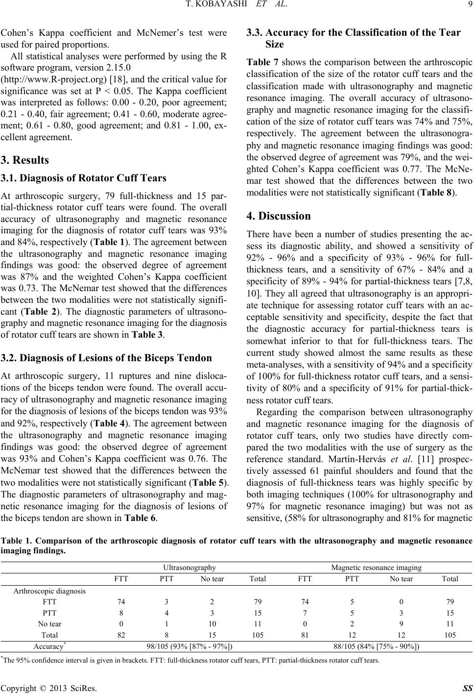 T. KOBAYASHI ET AL. Copyright © 2013 SciRes. SS 9 Cohen’s Kappa coefficient and McNemer’s test were used for paired proportions. All statistical analyses were performed by using the R software program, version 2.15.0 (http://www.R-project.org) [18], and the critical value for significance was set at P < 0.05. The Kappa coefficient was interpreted as follows: 0.00 - 0.20, poor agreement; 0.21 - 0.40, fair agreement; 0.41 - 0.60, moderate agree- ment; 0.61 - 0.80, good agreement; and 0.81 - 1.00, ex- cellent agreement. 3. Results 3.1. Diagnosis of Rotator Cuff Tears At arthroscopic surgery, 79 full-thickness and 15 par- tial-thickness rotator cuff tears were found. The overall accuracy of ultrasonography and magnetic resonance imaging for the diagnosis of rotator cuff tears was 93% and 84%, respectively (Table 1). The agreeme nt b etwe en the ultrasonography and magnetic resonance imaging findings was good: the observed degree of agreement was 87% and the weighted Cohen’s Kappa coefficient was 0.73. The McNemar test showed that the differences between the two modalities were not statistically signifi- cant (Table 2). The diagnostic parameters of ultrasono- graphy and magnetic resonance imaging for the diagnosis of rotator cuff tears are shown in Table 3. 3.2. Diagnosis of Lesions of the Biceps Tendon At arthroscopic surgery, 11 ruptures and nine disloca- tions of the biceps tendon were found. The overall accu- racy of ultrasonography and magnetic resonance imaging for the diagnosis of lesion s of the bicep s tendon was 93% and 92%, respectively (Table 4). The agreeme nt b etwe en the ultrasonography and magnetic resonance imaging findings was good: the observed degree of agreement was 93% and Cohen’s Kappa coefficient was 0.76. The McNemar test showed that the differences between the two modalities were not statistically significan t (Table 5). The diagnostic parameters of ultrasonography and mag- netic resonance imaging for the diagnosis of lesions of the biceps tendon are shown in Table 6. 3.3. Accuracy for the Classification of the Tear Size Table 7 shows the comparison between the arthroscopic classification of the size of the rotator cuff tears and the classification made with ultrasonography and magnetic resonance imaging. The overall accuracy of ultrasono- graphy and magnetic resonance imaging for the classifi- cation of the size of rotator cuff tears was 74% and 75%, respectively. The agreement between the ultrasonogra- phy and magnetic resonance imaging findings was good: the observed degree of agreement was 79%, and the wei- ghted Cohen’s Kappa coefficient was 0.77. The McNe- mar test showed that the differences between the two modalities were not statistically significant (Table 8). 4. Discussion There have been a number of studies presenting the ac- sess its diagnostic ability, and showed a sensitivity of 92% - 96% and a specificity of 93% - 96% for full- thickness tears, and a sensitivity of 67% - 84% and a specificity of 89% - 94% for partial-thickness tears [7,8, 10]. They all agreed that ultrasonography is an appropri- ate technique for assessing rotator cuff tears with an ac- ceptable sensitivity and specificity, despite the fact that the diagnostic accuracy for partial-thickness tears is somewhat inferior to that for full-thickness tears. The current study showed almost the same results as these meta-analyses, with a sensitivity of 94 % and a specificity of 100% for full-thickness ro tator cuff tears, and a sensi- tivity of 80% and a specificity of 91% for partial-thick- ness rotator cuff tears. Regarding the comparison between ultrasonography and magnetic resonance imaging for the diagnosis of rotator cuff tears, only two studies have directly com- pared the two modalities with the use of surgery as the reference standard. Martín-Hervás et al. [11] prospec- tively assessed 61 painful shoulders and found that the diagnosis of full-thickness tears was highly specific by both imaging techniques (100% for ultrasonography and 97% for magnetic resonance imaging) but was not as sensitive, (58% for ultrasonography and 81% for magnetic Table 1. Comparison of the arthroscopic diagnosis of rotator cuff tears with the ultrasonography and magnetic resonance imaging findings. Ultrasonography Magnetic reso nance imaging FTT PTT No tear Total FTT PTT No tear Total Arthroscopic diagnosis FTT 74 3 2 79 74 5 0 79 PTT 8 4 3 15 7 5 3 15 No tear 0 1 10 11 0 2 9 11 Total 82 8 15 105 81 12 12 105 Accuracy* 98/105 (93% [87% - 97%]) 88/105 (84% [75% - 90%]) *The 95% confidence interval is given in brackets. FTT: full-thickness rotator cuff tears, PTT: partial-thickness rotator cuff tears. 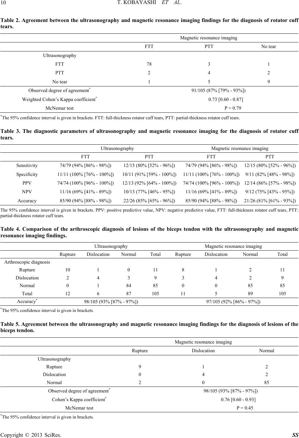 T. KOBAYASHI ET AL. 10 Table 2. Agreement between the ultrasonography and magnetic resonance imaging findings for the diagnosis of rotator cuff tears. Magnetic resona n ce imaging FTT PTT No tear Ultrasonography FTT 78 3 1 PTT 2 4 2 No tear 1 5 9 Observed degree of agreement* 91/105 (87% [79% - 93%]) Weighted Cohen’s Kappa coefficient* 0.73 [0.60 - 0.87] McNemar test P = 0.79 *The 95% confidence interval is given in brackets. FTT: full-thickness rotator cuff tears, PTT: partial-thickness rotator cuff tears. Table 3. The diagnostic parameters of ultrasonography and magnetic resonance imaging for the diagnosis of rotator cuff tears. Ultrasonography Magnetic reso nance imaging FTT PTT FTT PTT Sensitivity 74/79 (94% [86% - 98%]) 12/15 (80% [52% - 96%]) 74/79 (94% [86% - 98%]) 12/15 (80% [52% - 96%]) Specificity 11/11 (100% [76% - 100%]) 10/11 (91% [59% - 100%])11/11 (100% [76% - 100%]) 9/11 (82% [48% - 98%]) PPV 74/74 (100% [96% - 100%]) 12/13 (92% [64% - 100%])74/74 (100% [96% - 1 00 %]) 12/14 (86% [57% - 98 %]) NPV 11/16 (69% [41% - 89%]) 10/13 (77% [46% - 95%]) 11/16 (69% [41% - 89%]) 9/12 (75% [43% - 95 %]) Accuracy 85/90 (94% [88% - 98 %]) 22/26 (85% [65% - 96%]) 85/90 (94% [88% - 98%]) 21/26 (81% [61% - 93%]) The 95% confidence interval is given in brackets. PPV: positive predictive value, NPV: negative predictive value, FTT: full-thickness rotator cuff tears, PTT: partial-thickness rotator cuff tears. Table 4. Comparison of the arthroscopic diagnosis of lesions of the biceps tendon with the ultrasonography and magnetic resonance imaging findings. Ultrasonography Magnetic resonance imaging Rupture Dislocation Normal Total Rupture Dislocation Normal Total Arthroscopic diagnosis Rupture 10 1 0 11 8 1 2 11 Dislocation 2 4 3 9 3 4 2 9 Normal 0 1 84 85 0 0 85 85 Total 12 6 87 105 11 5 89 105 Accuracy* 98/105 (93% [87% - 97%]) 97/105 (92% [86% - 97%]) *The 95% conf i d ence interval is given in brackets. Table 5. Agreement betw een the ultrasonography and magnetic re sonance imaging findings for the diagnosis of lesions of the biceps tendon. Magnetic r esonance imaging Rupture Dislocation Normal Ultrasonography Rupture 9 1 2 Dislocation 0 4 2 Normal 2 0 85 Observed degree of agreement* 98/105 (93% [87% - 97%]) Cohen’s Kappa coefficient* 0.76 [0.60 - 0.93] McNemar test P = 0.45 *The 95% conf i d ence interval is given in brackets. Copyright © 2013 SciRes. SS 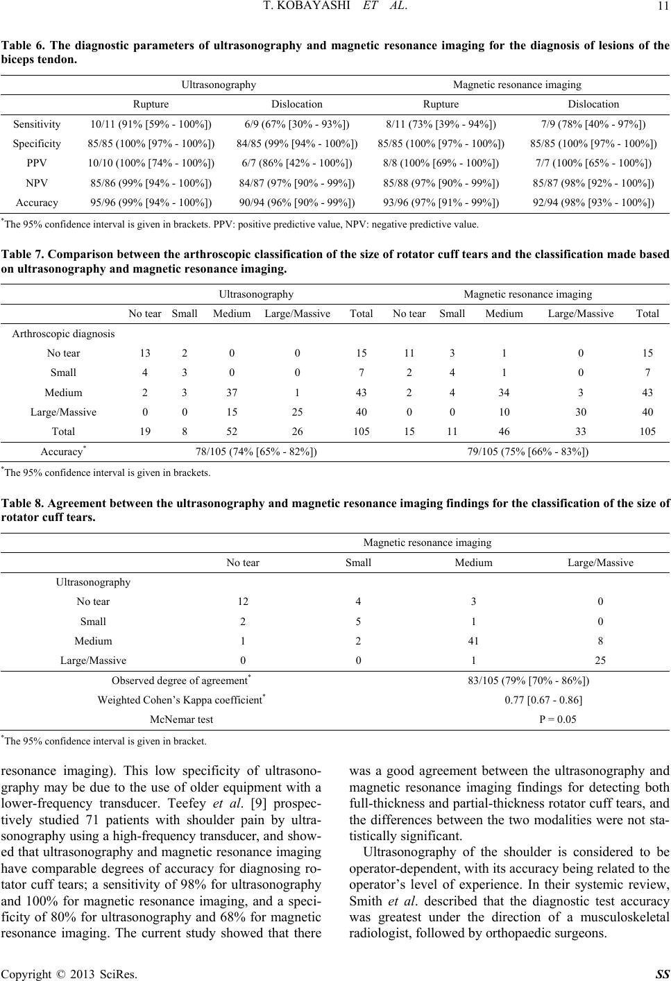 T. KOBAYASHI ET AL. Copyright © 2013 SciRes. SS 11 Table 6. The diagnostic parameters of ultrasonography and magnetic resonance imaging for the diagnosis of lesions of the biceps tendon. Ultrasonography Magnetic reso nance imaging Rupture Dislocation Rupture Dislocation Sensitivity 10/11 (91% [59% - 100%]) 6/9 (67% [30% - 93%]) 8/11 (73% [39% - 94%]) 7/9 (78% [40% - 97%]) Specificity 85/85 (100% [97% - 100%]) 84/85 (99% [94% - 100%])85/85 (100% [97% - 100%]) 85/85 (100% [97% - 100%]) PPV 10/10 (100% [74% - 100%]) 6/7 (86% [42% - 100%]) 8/8 (100% [69% - 100%]) 7/7 (100% [65% - 100%]) NPV 85/86 (99% [94% - 100%]) 84/87 (97% [90% - 99% ])85/88 (97% [90% - 99%]) 85/87 (98% [92% - 10 0%]) Accuracy 95/96 (99% [94% - 10 0% ]) 90/94 (96% [90% - 99%])93/96 (97% [91% - 99% ]) 92/94 (98% [93% - 100%]) *The 95% conf i d ence interval is given in brackets. PPV: positive predictive value, NPV: negative predictive value. Table 7. Comparison between the arthroscopic classification of the size of rotator cuff tears and the classification made based on ultrasonography and magnetic resonance imaging. Ultrasonography Magnetic reso nance imaging No tear Small Medium Large/MassiveTotal No tearSmallMedium Large/Massive Total Arthroscopic diagnosis No tear 13 2 0 0 15 11 3 1 0 15 Small 4 3 0 0 7 2 4 1 0 7 Medium 2 3 37 1 43 2 4 34 3 43 Large/Massive 0 0 15 25 40 0 0 10 30 40 Total 19 8 52 26 105 15 11 46 33 105 Accuracy* 78/105 (74% [65% - 82%]) 79/105 (75% [66% - 83%]) *The 95% conf i d ence interval is given in brackets. Table 8. Agreement between the ultrasonography and magnetic resonance imaging findings for the classification of the size of rotator cuff tears. Magnetic resonance imaging No tear Small Medium Large/Massive Ultrasonography No tear 12 4 3 0 Small 2 5 1 0 Medium 1 2 41 8 Large/Massive 0 0 1 25 Observed degree of agreement* 83/105 (79% [70% - 86%]) Weighted Cohen’s Kappa coefficient* 0.77 [0.67 - 0.86] McNemar test P = 0.05 *The 95% confi dence interval is given in bracket. resonance imaging). This low specificity of ultrasono- graphy may be due to the use of older equipment with a lower-frequency transducer. Teefey et al. [9] prospec- tively studied 71 patients with shoulder pain by ultra- son ogr aphy using a high-frequency transducer, and sho w- ed that ultrasonography and magnetic resonance imaging have comparable degrees of accuracy for diagnosing ro- tator cuff tears; a sensitivity of 98% for ultrasonography and 100% for magnetic resonance imaging, and a speci- ficity of 80% for ultrasonography and 68% for magnetic resonance imaging. The current study showed that there was a good agreement between the ultrasonography and magnetic resonance imaging findings for detecting both full-thickness and partial-thickness rotator cuff tears, and the differences between the two modalities were not sta- tistically significant. Ultrasonography of the shoulder is considered to be operator-dependent, with its accuracy being related to the operator’s level of experience. In their systemic review, Smith et al. described that the diagnostic test accuracy was greatest under the direction of a musculoskeletal radiologist, followed by orthopaedic surgeons. 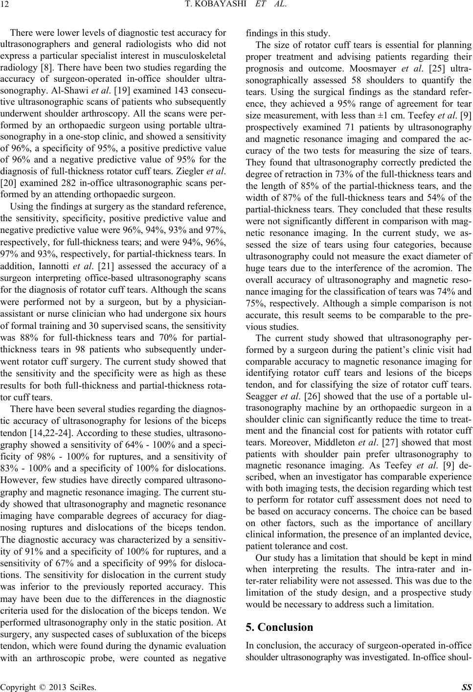 T. KOBAYASHI ET AL. 12 There were lower levels of diagnostic test accuracy for ultrasonographers and general radiologists who did not express a particular specialist interest in musculoskeletal radiology [8]. There have been two studies regarding the accuracy of surgeon-operated in-office shoulder ultra- sonography. Al-Sh awi et al. [19] examined 143 consecu- tive ultrasonographic scans of patients who subsequently underwent shoulder arthroscopy. All the scans were per- formed by an orthopaedic surgeon using portable ultra- sonography in a one-stop clinic, and sho wed a sensitivity of 96%, a specificity of 95%, a positive predictive value of 96% and a negative predictive value of 95% for the dia gno sis of f ull- thic kness rotator cuff tears. Ziegler et al. [20] examined 282 in-office ultrasonographic scans per- formed by an attending orthopaedic surgeon. Using the findings at surgery as the standard reference, the sensitivity, specificity, positive predictive value and negativ e p r ed ictiv e v alu e w ere 96 %, 94 %, 93 % a nd 97 %, re sp ect iv el y, for full-thick ness tears; and wer e 9 4% , 96 %, 97% and 93%, respectively, for partial-thickn ess tears. In addition, Iannotti et al. [21] assessed the accuracy of a surgeon interpreting office-based ultrasonography scans for the diagn osis of rotator cuff tears. Although the scans were performed not by a surgeon, but by a physician- assistant or nurse clinician who had undergone six hours of formal training and 30 supervised scans, the sensitivity was 88% for full-thickness tears and 70% for partial- thickness tears in 98 patients who subsequently under- went rotator cuff surgery. The current study showed that the sensitivity and the specificity were as high as these results for both full-thickness and partial-thickness rota- tor cuff tears. There have been several studies regarding the diagnos- tic accuracy of ultrasonography for lesions of the biceps tendon [14,22-24]. According to these studies, ultrasono- graphy showed a sensitivity of 64% - 100% and a speci- ficity of 98% - 100% for ruptures, and a sensitivity of 83% - 100% and a specificity of 100% for dislocations. However, few studies have directly compared ultrasono- graphy and magnetic resonance imaging. The current stu- dy showed that ultrasonography and magnetic resonance imaging have comparable degrees of accuracy for diag- nosing ruptures and dislocations of the biceps tendon. The diagnostic accuracy was characterized by a sensitiv- ity of 91% and a specificity of 100% for ruptures, and a sensitivity of 67% and a specificity of 99% for disloca- tions. The sensitivity for dislocation in the current study was inferior to the previously reported accuracy. This may have been due to the differences in the diagnostic criteria used for the dislocation of the biceps tendon. We performed ultrasonography only in th e static position. At surgery, any suspected cases of subluxation of the biceps tendon, which were found during the dynamic evaluation with an arthroscopic probe, were counted as negative findings in this study. The size of rotator cuff tears is essential for planning proper treatment and advising patients regarding their prognosis and outcome. Moosmayer et al. [25] ultra- sonographically assessed 58 shoulders to quantify the tears. Using the surgical findings as the standard refer- ence, they achieved a 95% range of agreement for tear size measurement, with less than ±1 cm. Teefey et al. [9] prospectively examined 71 patients by ultrasonography and magnetic resonance imaging and compared the ac- curacy of the two tests for measuring the size of tears. They found that ultrasonography correctly predicted the degree of retraction in 73% of the full-th ickness tears and the length of 85% of the partial-thickness tears, and the width of 87% of the full-thickness tears and 54% of the partial-thickness tears. They concluded that these results were not significantly different in comparison with mag- netic resonance imaging. In the current study, we as- sessed the size of tears using four categories, because ultrasonograp hy could not measure the exact diameter of huge tears due to the interference of the acromion. The overall accuracy of ultrasonography and magnetic reso- nance imaging for the classification of tears was 74% and 75%, respectively. Although a simple comparison is not accurate, this result seems to be comparable to the pre- vious studies. The current study showed that ultrasonography per- formed by a surgeon during the patient’s clinic visit had comparable accuracy to magnetic resonance imaging for identifying rotator cuff tears and lesions of the biceps tendon, and for classifying the size of rotator cuff tears. Seagger et al. [26] showed that the use of a portable ul- trasonography machine by an orthopaedic surgeon in a shoulder clinic can significantly reduce the time to treat- ment and the financial cost for patients with rotator cuff tears. Moreover, Middleton et al. [27] showed that most patients with shoulder pain prefer ultrasonography to magnetic resonance imaging. As Teefey et al. [9] de- scribed, when an investigator has comparable experience with both imaging tests, the decision regarding which test to perform for rotator cuff assessment does not need to be based on accuracy concerns. The choice can be based on other factors, such as the importance of ancillary clinical information , the presence of an implanted device, patient tolerance and co st. Our study has a limitation that should be kept in mind when interpreting the results. The intra-rater and in- ter-rater reliability were not assessed. This was due to the limitation of the study design, and a prospective study would be necessary to address such a limitation. 5. Conclusion In conclusion, the accuracy of surgeon-operated in-office shoulder ultrasonography was investigated. In-office shoul- Copyright © 2013 SciRes. SS  T. KOBAYASHI ET AL. 13 der ultrasonography had comparable accuracy to mag- netic resonance imaging for detecting rotator cuff tears and lesions of the biceps tendon, as well as for classify- ing the size of rotator cuff tears. REFERENCES [1] A. Al-Shawi, R. Badge and T. Bunker, “The Detection of Full Thickness Rotator Cuff Tears Using Ultrasound,” The Journal of Bone and Joint Surgery. British Volume, Vol. 90, No. 7, 2008, pp. 889-892. doi:10.1302/0301-620X.90B7.20481 [2] A. Armstrong, S. A. Teefey, T. Wu, A. M. Clark, W. D. Middleton, K. Yamaguchi and L. M. Galatzl, “The Effi- cacy of Ultrasound in the Diagnosis of Long Head of the Biceps Tendon Pathology,” Journal of Shoulder and El- bow Surgery, Vol. 15, No. 1, 2006, pp. 7-11. doi:10.1016/j.jse.2005.04.008 [3] T. D. Brandt, B. W. Cardone, T. H. Grant, M. Post and C. A. Weiss, “Rotator Cuff Sonography: A Reassessment,” Radiology, Vol. 173, No. 2, 1989, pp. 323-327. [4] D. L. Burk Jr., D. Karasick, A. B. Kurtz, D. G. Mitchell, M. D. Rifkin, C. L. Mille r, D. W. Levy, J. M. Fenlin and A. R. Bartolozzi, “Rotator Cuff Tears: Prospective Com- parison of MR Imaging with Arthrography, Sonography, and Surgery,” American Journal of Roentgenology, Vol. 153, No. 1, 1989, pp. 87-92. doi:10.2214/ajr.153.1.87 [5] J. O. de Jesus, L. Parker, A. J. Frangos and L. N. Nazar- ian, “Accuracy of MRI, MR Arthrography, and Ultra- sound in the Diagnosis of Rotator Cuff Tears: A Meta- Analysis,” American Journal of Roentgenology, Vol. 192, No. 6, 2009, pp. 1701-1707. doi:10.2214/AJR.08.1241 [6] J. K. DeOrio and R. H. Cofield, “Results of a Second Attempt at Surgical Repair of a Failed Initial Rotator-Cuff Repair,” The Journal of Bone and Joint Surgery. Ameri- can Volume, Vol. 66, No. 4, 1984, pp. 563-567. [7] A. N. Fotiadou, M. Vlychou, P. Papadopoulos, D. S. Kar- ataglis, P. Palladas and I. V. Fezoulidis, “ Ultrasonogra- phy of Symptomatic Rotator Cuff Tears Compared with MR Imaging and Surgery,” European Journal of Radiol- ogy, Vol. 68, No. 1, 2008, pp. 174-179. doi:10.1016/j.ejrad.2007.11.002 [8] J. P. Iannotti, J. Ciccone, D. D. Buss, J. L. Visotsky, E. Mascha, K. Cotman an d N. M. Rawool, “Accuracy of Of- fice-Based Ultrasonography of the Shoulder for the Di- agnosis of Rotator Cuff Tears,” The Journal of Bone and Joint Surgery. American Volume, No. 6, Vol. 87, 2005, pp. 1305-1311. [9] J. P. Iannotti, M. B. Zlatkin, J. K. Esterhai, H. Y. Kressel, M. K. Dalinka and K. P. Spindler, “Magnetic Resonance Imaging of the Shoulder. Sensitivity, Specificity, and Pre- dictive Value,” The Journal of Bone and Joint Surgery. American Volume, Vol. 73, No. 1, 1991, pp. 17-29. [10] J. B. Kneeland, W. D. Middleton, G. F. Carrera, R. C. Zeuge, A. Jesmanowicz, W. Froncisz and J. S. Hyde, “MR Imaging of the Shoulder: Diagnosis of Rotator Cuff Tears,” American Journal of Roentgenology, Vol. 149, No. 2, 1987, pp. 333-337. doi:10.2214/ajr.149.2.333 [11] C. Martín-Hervás, J. Romero, A. Navas-Acién, J. J. Re- boiras and L. Munuera, “Ultrasonographic and Magnetic Resonance Images of Rotator Cuff Lesions Compared with Arthroscopy or Open Surgery Findings,” Journal of Shoulder and Elbow Surgery, Vol. 10, No. 5, 2001, pp. 410-415. doi:10.1067/mse.2001.116515 [12] W. D. Middleton, W. T. Payne, S. A. Tee fey, C. F. Hilde - bolt, D. A. Rubin and K. Yamaguchi, “Sonography and MRI of the Shoulder: Comparison of Patient Satisfac- tion,” American Journal of Roentgenology, Vol. 183, No. 5, 2004, pp. 1449-1452. doi:10.2214/ajr.183.5.1831449 [13] W. D. Middleton, W. R. Reinus, W. G. Totty, C. L. Mel- son and W. A. Murphy, “Ultrasonographic Evaluation of the Rotator Cuff and Biceps Tendon,” The Journal of Bone and Joint Surgery. American Volume, Vol. 68, No. 3, 1986, pp. 440-450. [14] S. Moosmayer, S. Heir and H. J. Smith, “Sonography of the Rotator Cuff in Painful Shoulders Performed without Knowledge of Clinical Information: Results from 58 Sonographic Examinations with Surgical Correlation,” Journal of Clinical Ultrasound, Vol. 35, No. 1, 2007, pp. 20-26. doi:10.1002/jcu.20286 [15] M. C. Nelson, G. P. Leather, R. P. Nirschl, F. A. Pettrone and M. T. Freedman, “Evaluation of the Painful Shoulder. A Prospective Comparison of Magnetic Resonance Im- aging, Computerized Tomographic Arthrography, Ultra- sonography, and Operative Findings,” The Journal of Bone and Joint Surgery. American Volume, Vol. 73, No. 5, 1991, pp. 707-716. [16] R. P. Ottenheijm, M. J. Jansen, J. B. Staal, A. van den Bruel, R. E. Weijers, R. A. de Bie and G. J. Dinant, “Ac- curacy of Diagnostic Ultrasound in Patients with Sus- pected Subacromial Disorders: A Systematic Review and Meta-Analysis,” Archives of Physical Medicine and Re- habilitation, Vol. 91, No. 10, 2010, pp. 1616-1625. doi:10.1016/j.apmr.2010.07.017 [17] J. W. Read and M. Perko, “Shoulder Ultrasound: Diag- nostic Accuracy for Impingement Syndrome, Rotator Cuff Tear, and Biceps Tendon Pathology,” Journal of Shoulder and Elbow Surgery, Vol. 7, No. 3, 1998, pp. 264-271. doi:10.1016/S1058-2746(98)90055-6 [18] R. Seagger, T. Bunker and P. Hamer, “Surgeon-Operated Ultrasonography in a One-Stop Shoulder Clinic,” Annals of The Royal College of Surgeons of England, Vol. 93, No. 7, 2011, pp. 528-531. doi:10.1308/147870811X13137608454939 [19] S. E. Seltzer, H. J. Finberg, B. N. Weissman, D. K. Kido and B. D. Collier, “Arthrosonography: Gray-Scale Ultra- sound Evaluation of the Shoulder,” Radiology, Vol. 132, No. 2, 1979, pp. 467-468. [20] J. G. Skendzel, J. A. Jacobson, J. E. Carpenter and B. S. Miller, “Long Head of Biceps Brachii Tendon Evaluation: Accuracy of Preoperative Ultrasound,” American Journal of Roentgenology, Vol. 197, No. 4, 2011, pp. 942-948. doi:10.2214/AJR.10.5012 [21] T. O. Smith, T. Back, A. P. Toms and C. B. Hing, “Di- agnostic Accuracy of Ultrasound for Rotator Cuff Tears in Adults: A Systematic Review and Meta-Analysis,” Cli- nical Radiology, Vol. 66, No. 11, 2011, pp. 1036-1048. doi:10.1016/j.crad.2011.05.007 Copyright © 2013 SciRes. SS  T. KOBAYASHI ET AL. Copyright © 2013 SciRes. SS 14 [22] K. Takagishi, K. Makino, N. Takahira, T. Ikeda, K. Tsu- runo and M. Itoman, “Ultrasonography for Diagnosis of Rotator Cuff Tear,” Skeletal Radiology, Vol. 25, No. 3, 1996, pp. 221-224. doi:10.1007/s002560050068 [23] R Development Core Team, “R: A Language and Envi- ronment for Statistical Computing,” R Foundation for Statistical Computing, Vienna, 2005. http://www.R-project.org [24] S. A. Teefey, S. A. Hasan, W. D. Middleton, M. Patel, R. W. Wright and K. Yamaguchi, “Ultrasonography of the Rotator Cuff. A Comparison of Ultrasonographic and Ar- throscopic Findings in one Hundred Consecutive Cases,” The Journal of Bone and Joint Surgery. American Vol- ume, Vol. 82, No. 4, 2000, pp. 498-504. [25] S. A. Teefey, D. A. Rubin, W. D. Middleton, C. F. Hilde- bolt, R. A. Leibold and K. Yamaguchi, “Detection and Quantification of Rotator Cuff Tears. Comparison of Ul- trasonographic, Magnetic Resonance Imaging, and Ar- throscopic Findings in Seventy-One Consecutive Cases,” The Journal of Bone and Joint Surgery. American Vol- ume, Vol. 86, No. 4, 2004, pp. 708-716. [26] A. Yamamoto, K. Takagishi, T. Osawa, T. Yanagawa, D. Nakajima, H. Shitara and T. Kobayashi, “Prevalence and Risk Factors of a Rotator Cuff Tear in the General Popu- lation,” Journal of Shoulder and Elbow Surgery, Vol. 19, No. 1, 2010, pp. 116-120. doi:10.1016/j.jse.2009.04.006 [27] D. W. Ziegler, “The Use of In-Office, Orthopaedist-Per- formed Ultrasound of the Shoulder to Evaluate and Man- age Rotator Cuff Disorders,” Journal of Shoulder and Elbow Surgery, Vol. 13, No. 3, 2004, pp. 291-297. doi:10.1016/j.jse.2004.01.017 |