

Paper Menu >>
Journal Menu >>
 Open Journal of Gastroenterology, 2013, 3, 223-226 OJGas doi:10.4236/ojgas.2013.34037 Published Online August 2013 (http://www.scirp.org/journal/ojgas/) Open fistulectomy with sphincter fixation for anal fistula Tatsuya Abe1*, Masao Kunimoto1, Yoshikazu Hachiro1, Yoshiaki Ebisawa1, Houhei Hishiyama1, Seishu Abe2 1Department of Proctology, Kunimoto Hospital, Asahikawa, Japan 2Department of Plastic Surgery, Kunimoto Hospital, Asahikawa, Japan Email: *[email protected] Received 1 June 2013; revised 1 July 2013; accepted 20 July 2013 Copyright © 2013 Tatsuya Abe et al. This is an open access article distributed under the Creative Commons Attribution License, which permits unrestricted use, distribution, and reproduction in any medium, provided the original work is properly cited. ABSTRACT Purpose: This study aimed to report clinical data and recurrence rates in patients with anterolateral low fistulas who underwent open fistulectomy with sphinc- ter fixation. Methods: The study group consisted of 133 consecutive patients with anterolateral, low in- tersphincteric, or low trans-sphincteric fistulas who had undergone open fistulectomy with sphincter fixa- tion between January 2006 and December 2010. This procedure involves complete removal of the fistula tract by incision of anal sphincters, followed by fixa- tion of the sphincter muscles. Results: Success was achieved in 127 (95.5%) patients with a median fol- low-up time of 12 months. Anal fistula recurred in 4 cases (3%). Non-healing fistula with persistent anal discharge developed in 2 patients. Maximal resting pressure, but not maximal squeeze pressure, was sig- nificantly decreased after surgery. Five patients (4%) developed temporary anal incontinence after surgery. Conclusions: Open fistulectomy with sphincter fixa- tion was effective for the management of patients with anterolateral low fistula in this study. The high success rate suggests that this procedure is a reason- able option in this gro up of patients. Keywords: Anal Fistula; Fistulectomy; Sphincter Fixation; Fecal Incontinence 1. INTRODUCTION Anal fistula is a devastating problem that most com- monly occurs in healthy subjects, with cryptoglandular infection being the most widely accepted etiological fac- tor [1]. On the basis of their anatomical course relative to the sphincter complex, cryptoglandular fistulas are cate- gorized as intersphincteric, trans-sphincteric, suprasphinc- teric, and extrasphincteric types [2]. The most common type is intersphincteric, followed by transsphincteric [2, 3]. Anal fistulas can also be classified as simple or com- plex. The major type is simple, which includes low in- tersphincteric and low trans-sphincteric fistulas that cross <30% of the external sphincter [4]. Most fistulas have been conventionally treated by fis- tulotomy or fistulectomy, which have both proven to be effective [5]. However, these techniques, even for simple fistulas, result in some form of incontinence in approxi- mately 12% - 39% of patients [3,5,6]. In most patients, the internal opening of the fistula is located along the posterior midline [7]. Fistulas located laterally are sig- nificantly associated with recurrence, and those located along the anterior midline are associated with inconti- nence [3,8]. Anterolateral localization results in greater postoperative anal deformity when fistulotomy or fis- tulectomy is used to excise the fistula [9]. Therefore, more attention is needed for the management of anter- olateral fistulas. Open fistulectomy with sphincter fixation (OFSF) is another option for the management of anterolateral fistu- las. The procedure is based on the premise that removal of the chronic epithelialized tract will allow healing by secondary intention of healthier tissue, and sphincter fixation may result in less deformity and prevention of incontinence compared with leaving the sphincters di- vided. However, this is a more extensive procedure, and there is lack of support for it in the literature. Th e aim of this study was to evaluate clinical data and recurrence rates in patients with anterolateral fistulas who under- went OFSF. 2. PATIENTS AND METHODS The study group consisted of 133 consecutive patients with anterolate ral, low intersphincteric, or low transphinc- teric fistulas who had undergone OFSF between January 2006 and December 2010 at our institution. The median *Corresponding a uthor. Published Online August 2013 in SciRes. http://www.scirp.org/journal/ojgas 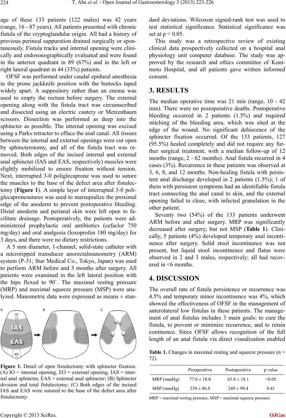 T. Abe et al. / Open Journal of Gastroenterology 3 (2013) 223-226 224 age of these 133 patients (122 males) was 42 years (range, 16 - 87 years). All patients presented with chronic fistula of the cryptoglandular origin. All had a history of previous perineal suppuration drained surgically or spon- taneously. Fistula tracks and internal opening were clini- cally and endosonographically evaluated and were found in the anterior quadrant in 89 (67%) and in the left or right lateral quadrant in 44 (33%) patients. OFSF was performed under caudal epidural anesthesia in the prone jackknife position with the buttocks taped widely apart. A suppository rather than an enema was used to empty the rectum before surgery. The external opening along with the fistula tract was circumscribed and dissected using an electric cautery or Metzenbaum scissors. Dissection was performed as deep into the sphincter as possible. The internal opening was excised using a Parks retracter to efface the anal canal. All tissues between the internal and ex tern al open ing s were cut op en by sphincterotomy, and all of the fistula tract was re- moved. Both edges of the incised internal and external anal sphincter (IAS and EAS, respectively) muscles were slightly mobilized to ensure fixation without tension. Next, interrupted 3-0 poliglecaprone was used to suture the muscles to the base of the defect area after fistulec- tomy (Figure 1). A simple layer of interrupted 3-0 poli- glecapronesutures was used to marsupialize the proximal edge of the anoderm to prevent postoperative bleeding. Distal anoderm and perianal skin were left open to fa- cilitate drainage. Postoperatively, the patients were ad- ministered prophylactic oral antibiotics (cefaclor 750 mg/day) and oral analgesia (loxoprofen 180 mg/day) for 3 days, and the re were n o di etary restricti o ns. A 5 mm diameter, 1-channel, solid-state catheter with a microtipped transducer anorectalmanometry (ARM) system (P-31; Star Medical Co., Tokyo, Japan) was used to perform ARM before and 3 months after surgery. All patients were examined in the left lateral position with the hips flexed to 90˚. The maximal resting pressure (MRP) and maximal squeeze pressure (MSP) were ana- lyzed. Manometric data were expressed as means ± stan- Figure 1. Detail of open fistulectomy with sphincter fixation. (A) IO = internal opening, EO = external opening, IAS = inter- nal anal sphincter, EAS = external anal sphincter; (B) Sphincter division and total fistulectomy; (C) Both edges of the incised IAS and EAS were sutured to the base of the defect area after fistulectomy. dard deviations. Wilcoxon signed-rank test was used to test statistical significance. Statistical significance was set at p < 0.05. This study was a retrospective review of existing clinical data prospectively collected on a hospital anal physiology unit computer database. The study was ap- proved by the research and ethics committee of Kuni- moto Hospital, and all patients gave written informed consent. 3. RESULTS The median operative time was 21 min (range, 10 - 42 min). There were no postoperative deaths. Postoperative bleeding occurred in 2 patients (1.5%) and required stitching of the bleeding area, which was sited at the edge of the wound. No significant dehiscence of the sphincter fixation occurred. Of the 133 patients, 127 (95.5%) healed completely and did not require any fur- ther surgical treatment, with a median follow-up of 12 months (range, 2 - 62 months). Anal fistula recurred in 4 cases (3%). Recurrence in these patients was observed at 3, 6, 8, and 12 months. Non-healing fistula with persis- tent anal discharge developed in 2 patients (1.5%); 1 of them with persistent symptoms had an identifiable fistula tract connecting the anal canal to skin, and the external opening failed to close, with infected granulation in the other pati e nt. Seventy two (54%) of the 133 patients underwent ARM before and after surgery. MRP was significantly decreased after surgery, but not MSP (Table 1). Clini- cally, 5 patients (4%) developed temporary anal inconti- nence after surgery. Solid stool incontinence was not present, but liquid stool incontinence and flatus were observed in 2 and 3 males, respectively; all had recov- ered in <6 months. 4. DISCUSSION The overall rate of fistula persistence or recurrence was 4.5% and temporary minor incontinence was 4%, which showed the effectiveness of OFSF in the management of anterolateral low fistulas in these patients. The manage- ment of anal fistulas includes 3 main goals: to cure the fistula, to prevent or minimize recurrence, and to retain continence. Since OFSF allows recognition of the full length of an anal fistula via direct visualization enabled Table 1. Changes in maximal resting and squeeze pressure (n = 72). Preoperative Postoperative p value MRP (mmHg)77.6 ± 18.0 65.8 ± 18.1 <0.05 MSP (mmHg)239 ± 86.8 249 ± 99.4 0.41 MRP = maximal resting pressure; M SP = maximal squeeze pressure. Copyright © 2013 SciRes. OJGas  T. Abe et al. / Open Journal of Gastroenterology 3 (2013) 223-226 225 by sphincter division, removal can certainly be a primary focus, which eliminates the risk of missing secondary tracts and allows healing by secondary intention of healthier tissue. Moreover, divided sphincters are fixed to prevent reduction in anal resting tone. We have also used this technique successfully to manage complex fis- tulas, but the present study was limited to a more com- mon group, simple fistulas. Both fistulotomy and fistulectomy have long been ac- cepted as the gold standard for simple fistulas. Although fistulotomy has been associated with success rates of 92% - 97%, the procedure will result in some form of incontinence even for simple fistulas in approximately 12% - 39% of patients [3,5,6]. Both fistulotomy and fistulectomy leaves the unepi- thelialized wound opened, which may cause undesired pain and complications, such as bleeding and suppura- tion. Marsupializatio n of the unepithelialized wound was introduced to provide the benefit of shortened healing times and improved continence by minimizing anal de- formity [10]. The addition of marsupialization also has been associated with less postoperative pain and bleeding [11]. However, this procedure leaves the sphincters di- vided, and Pescatori et al. [11] did not observe any supe- riority of marsupialization with respect to incontinence rates. Fistulectomy and immediate sphincter repair for low fistulas, described by Parkash et al. [12], aim to eradicate infection and to anatomically reconstruct the muscular defect. However, very few studies have been published on this technique. Dehiscence of sphincteroplasty is the most fearful complication of this technique and is re- sponsible for its infrequent use [13]. Roig et al. [14] treated 75 patients in whom most of the fistulas were complex (69%) by fistulectomy and end-to-end recon- struction of the disrupted EAS. Five recurrences in total have been described (6.7%), and the postoperative in- continence rate was 21%. Perez et al. [13] included 16 patients with recurrent complex fistulas in whom fistu- lotomy was performed with overlapping repair of the disrupted EAS. Two (25%) of 8 fully continent patients developed incontinence for watery stools and flatus, which gave a recurrence rate of 6.3% (1 patient). By overlapping the muscle, the area in contact is increased and rupture of the sutures may be more unlikely. How- ever, when the width of overlap is too long, the anal ca- nal may narrow and drainage worsen. The strengths of sphincter fixation, our original me- thod, are as follows: 1) since both edges of the sphincters are sutured separately, the tension on the sutures can be reduced by half relative to that on end-to-end sutures; 2) extensive dissection of sphincter muscles is not needed to achieve overlap; 3) the anal canal does not become narrow as can occur with overlap. The edges of the di- vided sphincter muscles will separate rapidly when they are not at all repaired. Even if they are fixed in the origi- nal position, continence will be fully maintainable. In fact, it was found that sphincter fixation was better pre- served for MSP 3 months after surgery. Although MRP was significantly reduced, the extent was only 15%. Cli- nically, 5 patients (4%) developed temporary inconti- nence for liquids and gas, and all of them had recovered in <6 months. 5. CONCLUSION The true advantages of the OFSF procedure may not be clear until larger prosp ective rando mized studies are co n- ducted. However, considering the current reported data, we believe that OFSF can achieve complete removal of fistulas safely and easily with minimal risk of post- operative incontinence and low recurrence rates. REFERENCES [1] Parks, A.G. (1961) Pathogenesis and treatment of fis- tula-in-ano. British Medical Journal, 1, 463-469. doi:10.1136/bmj.1.5224.463 [2] Parks, A.G., Gordon, P.H. and Hardcastle, J.D. (1976) A classification of fistula-in ano. British Journal of Surgery, 63, 1-12. doi:10.1002/bjs.1800630102 [3] Garcia-Aguilar, J., Belmonte, C., Wong, W.D., et al. (1996) Anal fistula surgery. Factors associated with re- currence and incontinence. Disease of the Colon and Rectum, 39, 723-729. doi:10.1007/BF02054434 [4] Parks, A.G. and Stitz, R.W. (1976) The treatment of high fistula-in-ano. Disease of the Colon and Rectum, 19, 487- 499. doi:10.1007/BF02590941 [5] Davies, M., Harris, D. and Lohana, P. (2008) The sur- gical management of fistula-in-ano in a specialist colo- rectal unit. International Journal of Colorectal Disease, 23, 833-838. doi:10.1007/s00384-008-0444-x [6] Roig, J.V., Jordan, J., Garcia-Armengol, J., et al. (2009) Change in anorectal morphologic and functional parame- ters after fistula-in-ano surgery. Disease of the Colon and Rectum, 52, 1462-1469. doi:10.1007/DCR.0b013e3181a80e24 [7] Atkin, G.K., Martins, J., Tozer, P., et al. (2011) For many high anal fistulas, lay open is still a good option. Tech- niques in Coloproctology, 15, 143-150. doi:10.1007/s10151-011-0676-6 [8] Steele, S.R., Kumar, R., Feingold, D.L., et al. (2011) Practice parameters for the management of perianal ab- scess and fistula-in-ano. Disease of the Colon and Rectum, 54, 1465-1474. doi:10.1097/DCR.0b013e31823122b3 [9] Iwadare, J. (2000) Sphincter-preserving techniques for anal fistulas in Japan. Disease of the Colon and Rectum, 43, S69-S77. doi:10.1007/BF02237229 [10] Ho, Y.H., Tan, M., Leong, A.F.P.K., et al. (1998) Marsu- pialization of fistulotomy wounds improves healing: A randomized controlled trial. British Journal of Surgery, Copyright © 2013 SciRes. OJGas  T. Abe et al. / Open Journal of Gastroenterology 3 (2013) 223-226 Copyright © 2013 SciRes. 226 OJGas 85, 105-107. doi:10.1046/j.1365-2168.1998.00529.x [11] Pescatori, M., Ayabaca, S.M., Cafaro, D., et al. (2005) Marsupialization of fistulotomy and fistulectomy wounds improves healing and decreases bleeding: A randomized controlled trial. Colorectal Disease, 8, 11-14. doi:10 .1111/j.1463-1318.2005.00835.x [12] Parkash, S., Lakshmiratan, V. and Gajendran, V. (1985) Fistula-in-ano: Treatment by fistulectomy, primary clo- sure and reconstruction. Australian and New Zealand Journal of Surgery, 55, 23-27. doi:10 .1111/j.1445-2197.1985.tb00849.x [13] Perez, F., Arroyo, A., Serrano, P., et al. (2006) Prospec- tive clinical and manometric study of fistulotomy with primary sphincter reconstruction in the management of recurrent complex fistula-in-ano. International Journal of Colorectal Disease, 21, 522-526. doi:10.1007/s00384-005-0045-x [14] Roig, J.V., Garcia-Armengol, J., Jordan, J.C., et al. (2009) Fistulectomy and sphincteric reconstruction for complex cryptgrandular fistulas. Colorectal Disease, 12, e145- e152. |