

Paper Menu >>
Journal Menu >>
 World Journal of Cardiovascular Diseases, 2013, 3, 1-7 WJCD http://dx.doi.org/10.4236/wjcd.2013.35A001 Published Online August 2013 (http://www.scirp.org/journal/wjcd/) Single intravenous injection of CoQ10 reduces infarct size in a rat model of ischemia and reperfusion injury* Alexander Ivanov1#, Evgenia Gorodetskaya1,2, Elena Kalenikova1,2, Oleg Medvedev1,2 1Department of Pharmacology, Faculty of Basic Medicine, M.V. Lomonosov Moscow State University, Moscow, Russia 2Russian Cardiology Research and Production Complex, Moscow, Russia Email: #[email protected] Received 7 June 2013; revised 7 July 2013; accepted 22 July 2013 Copyright © 2013 Alexander Ivanov et al. This is an open access article distributed under the Creative Commons Attribution License, which permits unrestricted use, distribution, and reproduction in any medium, provided the original work is properly cited. ABSTRACT Maintenance of mitochondrial activity and antioxi- dant features of coenzyme Q10 (CoQ10) could be an effective background for treatment of acute myocar- dial ischemia. Dietary uptake of CoQ10 is limited to only a few percent. In urgent cases, parenteral admi- nistration of CoQ10 could provide fast increase of its plasma and myocardial levels. The aim was to evalu- ate whether a single intravenous (i.v.) injection of so- lubilized CoQ10 before ischemia/reperfusion (IR) could lead to replenishment of its myocardial levels and li- mits subsequent myocardial IR injury. Methods: 30 min prior to coronary artery occlusion rats received i.v. solubilized CoQ10 (30 mg/kg) or saline (1 ml/kg). After 30 min of ischemia and 120 min of reperfusion, infarct zone of left ventricle (LV) and quantity of CoQ10 in LV were determined. Cardiac rhythm was monitored through the whole experiment. Results: At the beginning of reperfusion, arrhythmias were recor- ded in 8 (from 9) in saline and 2 (from 9) in CoQ10- treated rats. Arrhythmias in CoQ10-treated rats arose later (40 ± 8 sec) and had less duration (26 ± 14 sec); 14 ± 13 sec and 52 ± 17 sec in saline treated rats re- spectively. At the end of reperfusion CoQ10 treated rats revealed: 2 fold higher CoQ10 content in LV (p < 0.01), limitation of infarct zone by 35% (p < 0.01). Higher levels of CoQ10 were accompanied by less in- farct size (r = −0.77, p < 0.001). Conclusion: Single i.v. injection of CoQ10 effectively increased its myocardial levels and protected heart against IR injury by dimi- nishing the size of the irreversibly damaged myocar- dium, decreasing frequency and duration of arrhyth- mias. The infarct zone inversely correlated with the quantity of CoQ10 in LV. Keywords: Coenzyme Q10; Intravenous Injection; Myocardial Ischemia; Reperfusion Injury 1. INTRODUCTION Myocardial infarct leads to irreversible loss of cardimyo- cytes accompanied by deterioration of contractile func- tion and arrhythmias. Restoration of coronary blood flow limits necrosis of ischemic myocardium, but from the other hand reperfusion by itself results in myocardium damage [1]. Reperfusion initiates generation of free ra- dicals, intracellular Ca2+-overload, fast pH changes [2]. Excessive formation of free radicals results in cell death. Free radicals trigger inflammatory mediators such nu- clear factor-kB sensitive to reduction/oxidative balance, interleukin-1b, tumor necrosis factor α [3,4]. It is well known that endogenous antioxidants such as glutathione peroxidase, superoxide dismutase and catalase are natu- ral defense attenuating the ischemia/reperfusion (IR) in- jury [5]. Preservation of viable myocardium is possible with help of cardioprotectors. Coenzyme Q10 (CoQ10) is the endogenous compound, essential for mitochondrial function, possesses antioxi- dant and free radical scavenger features [6]. Long per os administration CoQ10 is recommended for prevention and treatment of coronary artery disease, arterial hyperten- sion, heart failure, hyperlipidemia [7]. However, dietary uptake of CoQ10 is limited to only a few percent [8] and elevation of CoQ10 levels for cardioprotection requires long preventive treatment [9]. Replenishment of CoQ10 by cells could be effective during heart surgeries (pre- ventive administration before procedures) or as inhibi- tion of IR injury (restoration of coronary flow after myo- cardial infarction). Fast increase in plasma CoQ10 levels and subsequent uptake by myocardium could be reached with intravenous (i.v.) injection. *The research was supported by grants 11-04-00894-a, 12-04-01246-а (Russian Foundation of Basic Research). #Corresponding author. The aim of the study is to investigate the effects of single i.v. pretreatment of solubilized CoQ10 on its myo- OPEN ACCESS  A. Ivanov et al. / World Journal of Cardiovascular Diseases 3 (2013) 1-7 2 cardial level and IR injury. 2. METHODS 2.1. Animals Animal-handling procedures followed the Guide for the Care and Use of Laboratory Animals [10] and with prior approval by the Bioethics committee of Lomonosov Mo- scow State University. Healthy male Wistar rats were housed separately in cages under a 12:12 hour light/dark cycle at 22˚C with free access to tap water and food. 2.2. Assessment of CoQ10 Levels in Myocardium 30 min after Its Single I.V. Injection Anesthetized rats (sodium pentobarbital, 45 mg/kg, intra- peritoneally) with venous catheters were used. Rats re- ceived i.v. a bolus of CoQ10 (30 mg/kg, solubilized CoQ10 in Kudesan solution, “Akvion”, Russia)— “CoQ10” group (n = 5) or saline (1 ml/kg, 0.9% NaCl)— “Control” group (n = 7). 30 min after injection rats were euthanized with 3 M KCl i.v., left ventricle (LV) samples were collected, frozen and stored at −20˚C for further analysis. Quantitative analysis of myocardial CoQ10 le- vels was performed by reversed-phase HPLC with elec- trochemical detection as described previously [9]. 2.3. Assessment of Cardioprotective Effects of Single I.V. Injection of CoQ10 Surgical preparation. Rats anesthetized as described above were placed on a heated pad (body temperature 37.5˚C ± 0.5˚C). Continuous infusion of pentobarbitallum sodium as maintaining narcosis (20 mg/ml, 200 μl/h) was performed via plastic catheter in femoral vein (“KD Scien- tific 210”, USA). To monitor blood pressure femoral ar- tery catheter was connected to measurement equipment “Macintosh—MacLab” (“ADInstruments”, Australia). Cardiac rhythm was monitored with a standard lead-1 electrocardiogram (ECG) (“MacLab”, “ADInstruments”, Australia). Model of myocardial IR. After intubation (Inspira Advanced Safety Ventilator, Volume Controlled 55 - 7058, Harvard Apparatus) left thoracotomy with fourth rib removing was performed on the rat in supine position and the ligature with an atraumatic needle was placed around the left anterior descending coronary artery. Then a rat was allowed to recover for 30 min. A small plastic snare was threaded through the ligature and placed in contact with the heart. 30 min prior to occlusion rats received i.v. of 1 ml/kg 0.9% NaCl (group “Saline + IR”, n = 11) or 30 mg/kg of solubilized CoQ10 (group “CoQ10 + IR”, n = 10). The artery was occluded by applying tension to the ligature for 30 min and reperfusion was achieved by releasing the tension for 120 min. The sham- operated rats (group “Saline + Sham”, n = 9) received saline bolus after thread placement and underwent the same study procedures except coronary artery occlusion. Through each experiment blood pressure (BP) was mea- sured as BPm = (BPs + 2xBPd)/3, where BPm—mean arterial blood pressure, BPs—systolic blood pressure, BPd—diastolic blood pressure. Arrhythmia analysis was performed accordingly to Lambeth Conventions [11]: 1) number of rats with pre- sence of any ventricle tachyarrhythmia (VTA); 2) num- ber of VTA episodes per one rat; 3) time to development of the first VTA episode; 4) total duration of VRA epi- sodes per one rat; 5) number of rats with lethal VTA. Assessment of myocardial damage. At the end of re- perfusion ligature around coronary artery was tighten again and Evans-Blue stain (5%, 0.5 ml) was infused i.v. to mark the risk zone (the non-stained tissue). Rats were euthanized with 3 M KCl i.v. Heart and liver were quick- ly removed. LV was separated, irrigated with cold water, frozen and stored at −20˚C for further analysis. To dis- tinguish living myocardium within risk zone frozen LV was cut into 2 mm transverse slices, which were incu- bated in 2% triphenyl tetrazolium chloride (TTC) in pH 7.4 buffer at 37˚C for 15 min (Figure 1). On the slices the risk zone (ischemic area) was determined as ratio of not-stained with Evans-Blue myocardium to total myo- cardium area. The volume of infarct zone was calculated as ratio of not stained with TTC myocardium (necrotic tissue) to ischemic area. Coenzyme Q10 assay in rat liver and LV was perform- ed with HPLC [9]. LV myocardial level was estimated after assessment of myocardial damage. 2.4. Data Analysis Values are presented as mean ± SD. Statistical analysis was performed with Statistica 8.0 (Stat Soft, Inc.). The differences in the means between the groups were tested using one-way ANOVA, followed by post hoc analysis for multiple comparisons (Student-Newman-Keuls me- thod) to test for statistical significance (p < 0.05). Cate- gorical values were compared using Fisher test (p < 0.05). 3. RESULTS Single i.v. bolus of solubilized CoQ10 led in 30 min to enhanced myocardial levels by 18.5% (p < 0.05) versus control rats. 30 rats were used in experiments for as- sessment of single i.v. injection of CoQ10 for cardiopro- tection. 2 rats died in “Saline + IR” group and 1 rat in “CoQ10 + IR” group during ischemia. Baseline values of blood pressure were similar in all experimental groups indicating equal initial conditions (Figure 2). Ischemia and reperfusion led to continuous Copyright © 2013 SciRes. OPEN ACCESS 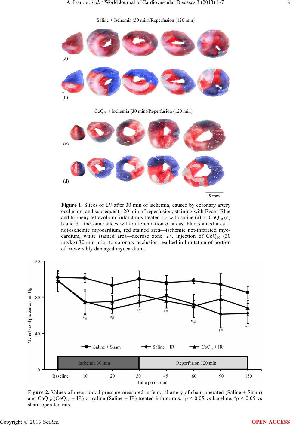 A. Ivanov et al. / World Journal of Cardiovascular Diseases 3 (2013) 1-7 Copyright © 2013 SciRes. 3 5 mm (d) (c) (a) (b) CoQ 10 + Ischemia (30 min)/Reperfusion (120 min) Saline + Ischemia (30 min)/Reperfusion (120 min) Figure 1. Slices of LV after 30 min of ischemia, caused by coronary artery occlusion, and subsequent 120 min of reperfusion, staining with Evans Blue and triphenyltetrazolium: infarct rats treated i.v. with saline (a) or CoQ10 (c). b and d—the same slices with differentiation of areas: blue stained area— not-ischemic myocardium, red stained area—ischemic not-infarcted myo- cardium, white stained area—necrose zone. I.v. injection of CoQ10 (30 mg/kg) 30 min prior to coronary occlusion resulted in limitation of portion of irreversibly damaged myocardium. Figure 2. Values of mean blood pressure measured in femoral artery of sham-operated (Saline + Sham) and CoQ10 (CoQ10 + IR) or saline (Saline + IR) treated infarct rats. *p < 0.05 vs baseline, #p < 0.05 vs sham-operated rats. OPEN ACCESS 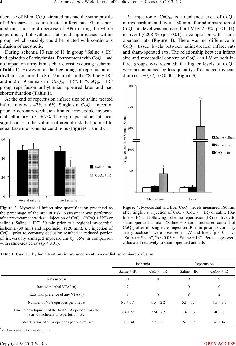 A. Ivanov et al. / World Journal of Cardiovascular Diseases 3 (2013) 1-7 4 decrease of BPm. CoQ10-treated rats had the same profile of BPm curve as saline treated infarct rats. Sham-oper- ated rats had slight decrease of BPm during the whole experiment, but without statistical significance within group, which possibly could be related with continuous infusion of anesthetic. During ischemia 10 rats of 11 in group “Saline + IR” had episodes of arrhythmias. Pretreatment with CoQ10 had no impact on arrhythmias characteristics during ischemia (Table 1). However, at the beginning of reperfusion ar- rhythmias occurred in 8 of 9 animals in the “Saline + IR” and in 2 of 9 animals in “CoQ10 + IR”. In “CoQ10 + IR” group reperfusion arrhythmias appeared later and had shorter duration (Table 1). At the end of reperfusion infarct size of saline treated infarct rats was 47% ± 6%. Single i.v. CoQ10 injection prior to coronary occlusion limited irreversible myocar- dial cell injury to 31 ± 7%. These groups had no statistical significance in the volume of area at risk that pointed to equal baseline ischemia conditions (Figures 1 and 3). Figure 3. Myocardial infarct size quantification presented as the percentage of the area at risk. Assessment was performed after pre-treatment with i.v. injection of CoQ10 (“CoQ + IR”) or saline (“Saline + IR”) 30 min prior to a regional myocardial ischemia (30 min) and reperfusion (120 min). I.v. injection of CoQ10 prior to coronary occlusion resulted in reduced portion of irreversibly damaged myocardium by 35% in comparison with saline-treated rats (p < 0.01). I.v. injection of CoQ10 led to enhance levels of CoQ10 in myocardium and liver: 180 min after administration of CoQ10 its level was increased in LV by 210% (p < 0.01), in liver by 2081% (p < 0.01) in comparison with sham- operated rats (Figure 4). There was no difference in CoQ10 tissue levels between saline-treated infarct rats and sham-operated rats. The relationship between infarct size and myocardial content of CoQ10 in LV of both in- farct groups was revealed: the higher levels of CoQ10 were accompanied by less quantity of damaged myocar- dium (r = −0,77, p < 0.001; Figure 5). Figure 4. Myocardial and liver CoQ10 levels measured 180 min after single i.v. injection of CoQ10 (CoQ10 + IR) or saline (Sa- line + IR) and following ischemia-reperfusion (IR) relatively to sham-operated animals (Saline + Sham). Increased content of CoQ10 after its single i.v. injection 30 min prior to coronary artery occlusion were observed in LV and liver. *p < 0.05 vs “Saline + Sham”, #p < 0.05 vs “Saline + IR”. Percentages were calculated relatively to sham-operated animals. Table 1. Cardiac rhythm alterations in rats underwent myocardial ischemia/reperfusion. Ischemia Reperfusion Saline + IR CoQ10 + IR Saline + IR CoQ10 + IR Rats used, n 11 10 9 9 Rats with lethal VTA* (n) 2 1 0 0 Rats with presence of any VTA (n) 8 8 8 2 Number of VTA episodes per one rat 6.7 ± 1.4 6.5 ± 2.2 5.1 ± 1.7 6.5 ± 3.5 Time to development of the first VTA episode from the start of ischemia or reperfusion, sec 364 ± 55 374 ± 62 14 ± 13 40 ± 8 Total duration of VTA episodes per one rat, sec 103 ± 41 92 ± 58 52 ± 17 26 ± 14 *VTA—ventricle tachyarrhythmia. Copyright © 2013 SciRes. OPEN ACCESS 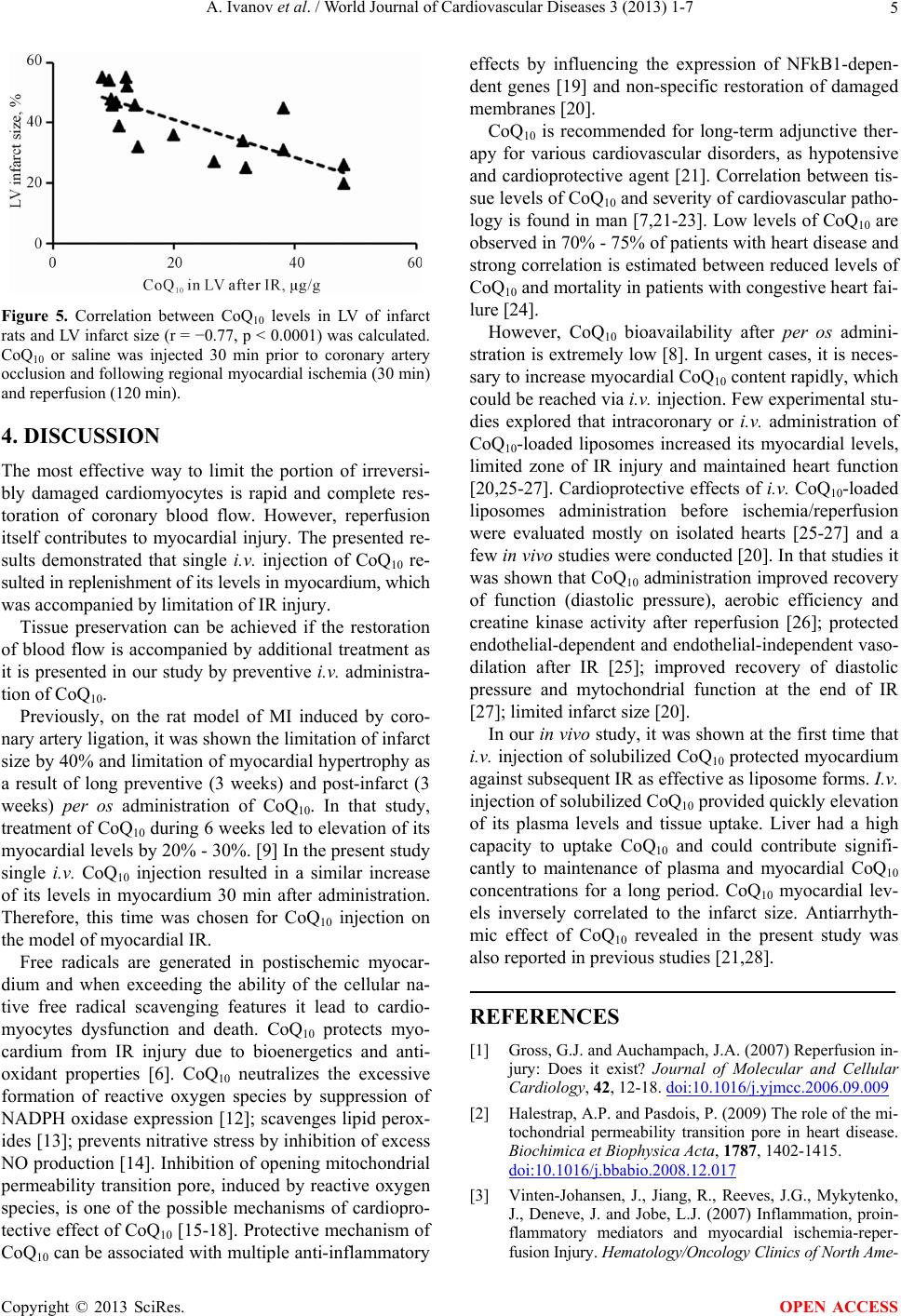 A. Ivanov et al. / World Journal of Cardiovascular Diseases 3 (2013) 1-7 5 Figure 5. Correlation between CoQ10 levels in LV of infarct rats and LV infarct size (r = −0.77, p < 0.0001) was calculated. CoQ10 or saline was injected 30 min prior to coronary artery occlusion and following regional myocardial ischemia (30 min) and reperfusion (120 min). 4. DISCUSSION The most effective way to limit the portion of irreversi- bly damaged cardiomyocytes is rapid and complete res- toration of coronary blood flow. However, reperfusion itself contributes to myocardial injury. The presented re- sults demonstrated that single i.v. injection of CoQ10 re- sulted in replenishment of its levels in myocardium, which was accompanied by limitation of IR injury. Tissue preservation can be achieved if the restoration of blood flow is accompanied by additional treatment as it is presented in our study by preventive i.v. administra- tion of CoQ10. Previously, on the rat model of MI induced by coro- nary artery ligation, it was shown the limitation of infarct size by 40% and limitation of myocardial hypertrophy as a result of long preventive (3 weeks) and post-infarct (3 weeks) per os administration of CoQ10. In that study, treatment of CoQ10 during 6 weeks led to elevation of its myocardial levels by 20% - 30%. [9] In the present study single i.v. CoQ10 injection resulted in a similar increase of its levels in myocardium 30 min after administration. Therefore, this time was chosen for CoQ10 injection on the model of myocardial IR. Free radicals are generated in postischemic myocar- dium and when exceeding the ability of the cellular na- tive free radical scavenging features it lead to cardio- myocytes dysfunction and death. CoQ10 protects myo- cardium from IR injury due to bioenergetics and anti- oxidant properties [6]. CoQ10 neutralizes the excessive formation of reactive oxygen species by suppression of NADPH oxidase expression [12]; scavenges lipid perox- ides [13]; prevents nitrative stress by inhibition of excess NO production [14]. Inhibition of opening mitochondrial permeability transition pore, induced by reactive oxygen species, is one of the possible mechanisms of cardiopro- tective effect of CoQ10 [15-18]. Protective mechanism of CoQ10 can be associated with multiple anti-inflammatory effects by influencing the expression of NFkB1-depen- dent genes [19] and non-specific restoration of damaged membranes [20]. CoQ10 is recommended for long-term adjunctive ther- apy for various cardiovascular disorders, as hypotensive and cardioprotective agent [21]. Correlation between tis- sue levels of CoQ10 and severity of cardiovascular patho- logy is found in man [7,21-23]. Low levels of CoQ10 are observed in 70% - 75% of patients with heart disease and strong correlation is estimated between reduced levels of CoQ10 and mortality in patients with congestive heart fai- lure [24]. However, CoQ10 bioavailability after per os admini- stration is extremely low [8]. In urgent cases, it is neces- sary to increase myocardial CoQ10 content rapidly, which could be reached via i.v. injection. Few experimental stu- dies explored that intracoronary or i.v. administration of CoQ10-loaded liposomes increased its myocardial levels, limited zone of IR injury and maintained heart function [20,25-27]. Cardioprotective effects of i.v. CoQ10-loaded liposomes administration before ischemia/reperfusion were evaluated mostly on isolated hearts [25-27] and a few in vivo studies were conducted [20]. In that studies it was shown that CoQ10 administration improved recovery of function (diastolic pressure), aerobic efficiency and creatine kinase activity after reperfusion [26]; protected endothelial-dependent and endothelial-independent vaso- dilation after IR [25]; improved recovery of diastolic pressure and mytochondrial function at the end of IR [27]; limited infarct size [20]. In our in vivo study, it was shown at the first time that i.v. injection of solubilized CoQ10 protected myocardium against subsequent IR as effective as liposome forms. I.v. injection of solubilized CoQ10 provided quickly elevation of its plasma levels and tissue uptake. Liver had a high capacity to uptake CoQ10 and could contribute signifi- cantly to maintenance of plasma and myocardial CoQ10 concentrations for a long period. CoQ10 myocardial lev- els inversely correlated to the infarct size. Antiarrhyth- mic effect of CoQ10 revealed in the present study was also reported in previous studies [21,28]. REFERENCES [1] Gross, G.J. and Auchampach, J.A. (2007) Reperfusion in- jury: Does it exist? Journal of Molecular and Cellular Cardiology, 42, 12-18. doi:10.1016/j.yjmcc.2006.09.009 [2] Halestrap, A.P. and Pasdois, P. (2009) The role of the mi- tochondrial permeability transition pore in heart disease. Biochimica et Biophysica Acta, 1787, 1402-1415. doi:10.1016/j.bbabio.2008.12.017 [3] Vinten-Johansen, J., Jiang, R., Reeves, J.G., Mykytenko, J., Deneve, J. and Jobe, L.J. (2007) Inflammation, proin- flammatory mediators and myocardial ischemia-reper- fusion Injury. Hematology/Oncology Clinics of North Ame- Copyright © 2013 SciRes. OPEN ACCESS 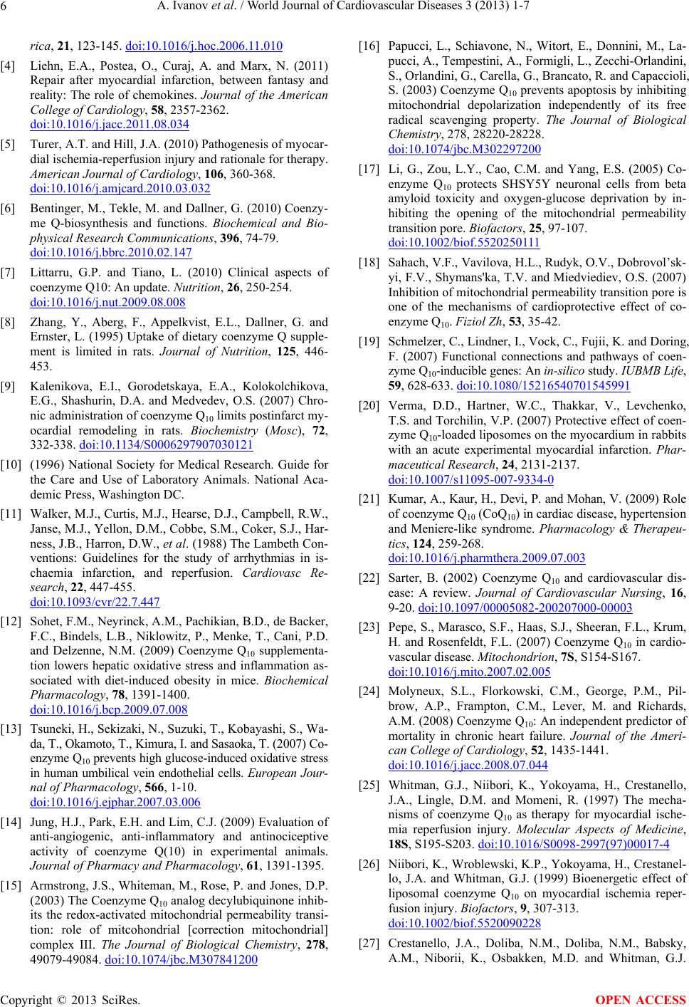 A. Ivanov et al. / World Journal of Cardiovascular Diseases 3 (2013) 1-7 6 rica, 21, 123-145. doi:10.1016/j.hoc.2006.11.010 [4] Liehn, E.A., Postea, O., Curaj, A. and Marx, N. (2011) Repair after myocardial infarction, between fantasy and reality: The role of chemokines. Journal of the American College of Cardiology, 58, 2357-2362. doi:10.1016/j.jacc.2011.08.034 [5] Turer, A.T. and Hill, J.A. (2010) Pathogenesis of myocar- dial ischemia-reperfusion injury and rationale for therapy. American Journal of Cardiology, 106, 360-368. doi:10.1016/j.amjcard.2010.03.032 [6] Bentinger, M., Tekle, M. and Dallner, G. (2010) Coenzy- me Q-biosynthesis and functions. Biochemical and Bio- physical Research Communications, 396, 74-79. doi:10.1016/j.bbrc.2010.02.147 [7] Littarru, G.P. and Tiano, L. (2010) Clinical aspects of coenzyme Q10: An update. Nutrition, 26, 250-254. doi:10.1016/j.nut.2009.08.008 [8] Zhang, Y., Aberg, F., Appelkvist, E.L., Dallner, G. and Ernster, L. (1995) Uptake of dietary coenzyme Q supple- ment is limited in rats. Journal of Nutrition, 125, 446- 453. [9] Kalenikova, E.I., Gorodetskaya, E.A., Kolokolchikova, E.G., Shashurin, D.A. and Medvedev, O.S. (2007) Chro- nic administration of coenzyme Q10 limits postinfarct my- ocardial remodeling in rats. Biochemistry (Mosc), 72, 332-338. doi:10.1134/S0006297907030121 [10] (1996) National Society for Medical Research. Guide for the Care and Use of Laboratory Animals. National Aca- demic Press, Washington DC. [11] Walker, M.J., Curtis, M.J., Hearse, D.J., Campbell, R.W., Janse, M.J., Yellon, D.M., Cobbe, S.M., Coker, S.J., Har- ness, J.B., Harron, D.W., et al. (1988) The Lambeth Con- ventions: Guidelines for the study of arrhythmias in is- chaemia infarction, and reperfusion. Cardiovasc Re- search, 22, 447-455. doi:10.1093/cvr/22.7.447 [12] Sohet, F.M., Neyrinck, A.M., Pachikian, B.D., de Backer, F.C., Bindels, L.B., Niklowitz, P., Menke, T., Cani, P.D. and Delzenne, N.M. (2009) Coenzyme Q10 supplementa- tion lowers hepatic oxidative stress and inflammation as- sociated with diet-induced obesity in mice. Biochemical Pharmacology, 78, 1391-1400. doi:10.1016/j.bcp.2009.07.008 [13] Tsuneki, H., Sekizaki, N., Suzuki, T., Kobayashi, S., Wa- da, T., Okamoto, T., Kimura, I. and Sasaoka, T. (2007) Co- enzyme Q10 prevents high glucose-induced oxidative stress in human umbilical vein endothelial cells. European Jour- nal of Pharmacology, 566, 1-10. doi:10.1016/j.ejphar.2007.03.006 [14] Jung, H.J., Park, E.H. and Lim, C.J. (2009) Evaluation of anti-angiogenic, anti-inflammatory and antinociceptive activity of coenzyme Q(10) in experimental animals. Journal of Pharmacy and Pharmacology, 61, 1391-1395. [15] Armstrong, J.S., Whiteman, M., Rose, P. and Jones, D.P. (2003) The Coenzyme Q10 analog decylubiquinone inhib- its the redox-activated mitochondrial permeability transi- tion: role of mitcohondrial [correction mitochondrial] complex III. The Journal of Biological Chemistry, 278, 49079-49084. doi:10.1074/jbc.M307841200 [16] Papucci, L., Schiavone, N., Witort, E., Donnini, M., La- pucci, A., Tempestini, A., Formigli, L., Zecchi-Orlandini, S., Orlandini, G., Carella, G., Brancato, R. and Capaccioli, S. (2003) Coenzyme Q10 prevents apoptosis by inhibiting mitochondrial depolarization independently of its free radical scavenging property. The Journal of Biological Chemistry, 278, 28220-28228. doi:10.1074/jbc.M302297200 [17] Li, G., Zou, L.Y., Cao, C.M. and Yang, E.S. (2005) Co- enzyme Q10 protects SHSY5Y neuronal cells from beta amyloid toxicity and oxygen-glucose deprivation by in- hibiting the opening of the mitochondrial permeability transition pore. Biofactors, 25, 97-107. doi:10.1002/biof.5520250111 [18] Sahach, V.F., Vavilova, H.L., Rudyk, O.V., Dobrovol’sk- yi, F.V., Shymans'ka, T.V. and Miedviediev, O.S. (2007) Inhibition of mitochondrial permeability transition pore is one of the mechanisms of cardioprotective effect of co- enzyme Q10. Fiziol Zh, 53, 35-42. [19] Schmelzer, C., Lindner, I., Vock, C., Fujii, K. and Doring, F. (2007) Functional connections and pathways of coen- zyme Q10 -inducible genes: An in-silico study. IUBMB Life, 59, 628-633. doi:10.1080/15216540701545991 [20] Verma, D.D., Hartner, W.C., Thakkar, V., Levchenko, T.S. and Torchilin, V.P. (2007) Protective effect of coen- zyme Q10-loaded liposomes on the myocardium in rabbits with an acute experimental myocardial infarction. Phar- maceutical Research, 24, 2131-2137. doi:10.1007/s11095-007-9334-0 [21] Kumar, A., Kaur, H., Devi, P. and Mohan, V. (2009) Role of coenzyme Q10 (CoQ10) in cardiac disease, hypertension and Meniere-like syndrome. Pharmacology & Therapeu- tics, 124, 259-268. doi:10.1016/j.pharmthera.2009.07.003 [22] Sarter, B. (2002) Coenzyme Q10 and cardiovascular dis- ease: A review. Journal of Cardiovascular Nursing, 16, 9-20. doi:10.1097/00005082-200207000-00003 [23] Pepe, S., Marasco, S.F., Haas, S.J., Sheeran, F.L., Krum, H. and Rosenfeldt, F.L. (2007) Coenzyme Q10 in cardio- vascular disease. Mitochondrion, 7S, S154-S167. doi:10.1016/j.mito.2007.02.005 [24] Molyneux, S.L., Florkowski, C.M., George, P.M., Pil- brow, A.P., Frampton, C.M., Lever, M. and Richards, A.M. (2008) Coenzyme Q10: An independent predictor of mortality in chronic heart failure. Journal of the Ameri- can College of Cardiology, 52, 1435-1441. doi:10.1016/j.jacc.2008.07.044 [25] Whitman, G.J., Niibori, K., Yokoyama, H., Crestanello, J.A., Lingle, D.M. and Momeni, R. (1997) The mecha- nisms of coenzyme Q10 as therapy for myocardial ische- mia reperfusion injury. Molecular Aspects of Medicine, 18S, S195-S203. doi:10.1016/S0098-2997(97)00017-4 [26] Niibori, K., Wroblewski, K.P., Yokoyama, H., Crestanel- lo, J.A. and Whitman, G.J. (1999) Bioenergetic effect of liposomal coenzyme Q10 on myocardial ischemia reper- fusion injury. Biofactors, 9, 307-313. doi:10.1002/biof.5520090228 [27] Crestanello, J.A., Doliba, N.M., Doliba, N.M., Babsky, A.M., Niborii, K., Osbakken, M.D. and Whitman, G.J. Copyright © 2013 SciRes. OPEN ACCESS  A. Ivanov et al. / World Journal of Cardiovascular Diseases 3 (2013) 1-7 Copyright © 2013 SciRes. 7 OPEN ACCESS (2002) Effect of coenzyme Q10 supplementation on mi- tochondrial function after myocardial ischemia reperfu- sion. Journal of Surgical Research, 102, 221-228. doi:10.1006/jsre.2001.6324 [28] Makhija, N., Sendasgupta, C., Kiran, U., Lakshmy, R., Hote, M.P., Choudhary, S.K., Airan, B. and Abraham, R. (2008) The role of oral coenzyme Q10 in patients under- going coronary artery bypass graft surgery. Journal of Cardiothoracic and Vascular Anesthesia, 22, 832-839. doi:10.1053/j.jvca.2008.03.007 |