

Paper Menu >>
Journal Menu >>
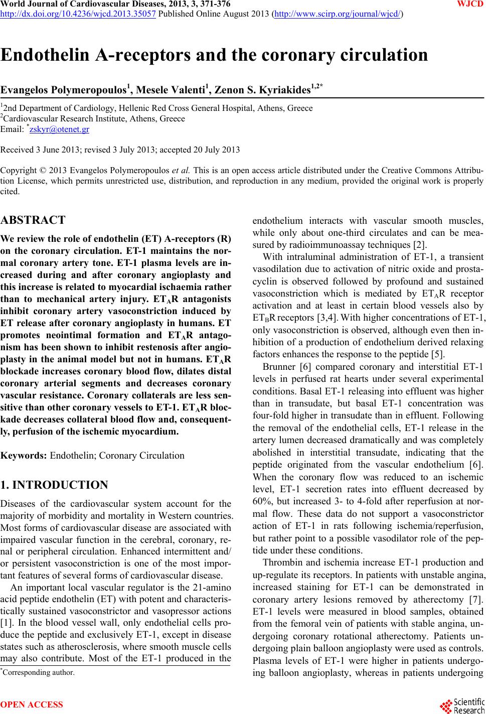 World Journal of Cardiovascular Diseases, 2013, 3, 371-376 WJCD http://dx.doi.org/10.4236/wjcd.2013.35057 Published Online August 2013 (http://www.scirp.org/journal/wjcd/) Endothelin A-receptors and the coronary circulation Evangelos Polymeropoulos1, Mesele Valenti1, Zenon S. Kyriakides1,2* 12nd Department of Cardiology, Hellenic Red Cross General Hospital, Athens, Greece 2Cardiovascular Research Institute, Athens, Greece Email: *[email protected] Received 3 June 2013; revised 3 July 2013; accepted 20 July 2013 Copyright © 2013 Evangelos Polymeropoulos et al. This is an open access article distributed under the Creative Commons Attribu- tion License, which permits unrestricted use, distribution, and reproduction in any medium, provided the original work is properly cited. ABSTRACT We review the role of endothelin (ET) A-receptors (R) on the coronary circulation. ET-1 maintains the nor- mal coronary artery tone. ET-1 plasma levels are in- creased during and after coronary angioplasty and this increase is related to myocardial ischaemia rather than to mechanical artery injury. ETAR antagonists inhibit coronary artery vasoconstriction induced by ET release after coronary angioplasty in humans. ET promotes neointimal formation and ETAR antago- nism has been shown to inhibit restenosis after angio- plasty in the animal model but not in humans. ETAR blockade increases coronary blood flow, dilates distal coronary arterial segments and decreases coronary vascular resistance. Coronary collaterals are less sen- sitive than other coronary vessels to ET-1. ETAR bloc- kade decreases collateral blood flow and, consequent- ly, perfusion of the ischemic myocardium. Keywords: Endothelin; Coronary Circulation 1. INTRODUCTION Diseases of the cardiovascular system account for the majority of morbidity and mortality in Western countries. Most forms of cardiovascular disease are associated with impaired vascular function in the cerebral, coronary, re- nal or peripheral circulation. Enhanced intermittent and/ or persistent vasoconstriction is one of the most impor- tant features of several forms of cardiovascular disease. An important local vascular regulator is the 21-amino acid peptide endothelin (ET) with potent and characteris- tically sustained vasoconstrictor and vasopressor actions [1]. In the blood vessel wall, only endothelial cells pro- duce the peptide and exclusively ET-1, except in disease states such as atherosclerosis, where smooth muscle cells may also contribute. Most of the ET-1 produced in the endothelium interacts with vascular smooth muscles, while only about one-third circulates and can be mea- sured by ra dioimm un oas say techniques [2]. With intraluminal administration of ET-1, a transient vasodilation due to activation of nitric oxide and prosta- cyclin is observed followed by profound and sustained vasoconstriction which is mediated by ETAR receptor activation and at least in certain blood vessels also by ETBR receptors [3,4]. With higher concentrations of ET-1, only vasoconstriction is observed, although even then in- hibition of a production of endothelium derived relaxing factors enhances the response to the peptide [5]. Brunner [6] compared coronary and interstitial ET-1 levels in perfused rat hearts under several experimental conditions. Basal ET-1 releasing into effluent was higher than in transudate, but basal ET-1 concentration was four-fold higher in transudate than in effluent. Following the removal of the endothelial cells, ET-1 release in the artery lumen decreased dramatically and was completely abolished in interstitial transudate, indicating that the peptide originated from the vascular endothelium [6]. When the coronary flow was reduced to an ischemic level, ET-1 secretion rates into effluent decreased by 60%, but increased 3- to 4-fold after reperfusion at nor- mal flow. These data do not support a vasoconstrictor action of ET-1 in rats following ischemia/reperfusion, but rather point to a possible vasodilator role of the pep- tide under these conditions. Thrombin and ischemia increase ET-1 production and up-regulate its receptors. In patients with unstable angina, increased staining for ET-1 can be demonstrated in coronary artery lesions removed by atherectomy [7]. ET-1 levels were measured in blood samples, obtained from the femoral vein of patients with stable angina, un- dergoing coronary rotational atherectomy. Patients un- dergoing plain balloon angioplasty were used as controls. Plasma levels of ET-1 were higher in patients undergo- ing balloon angioplasty, whereas in patients undergoing *Corresponding a uthor. OPEN ACCESS 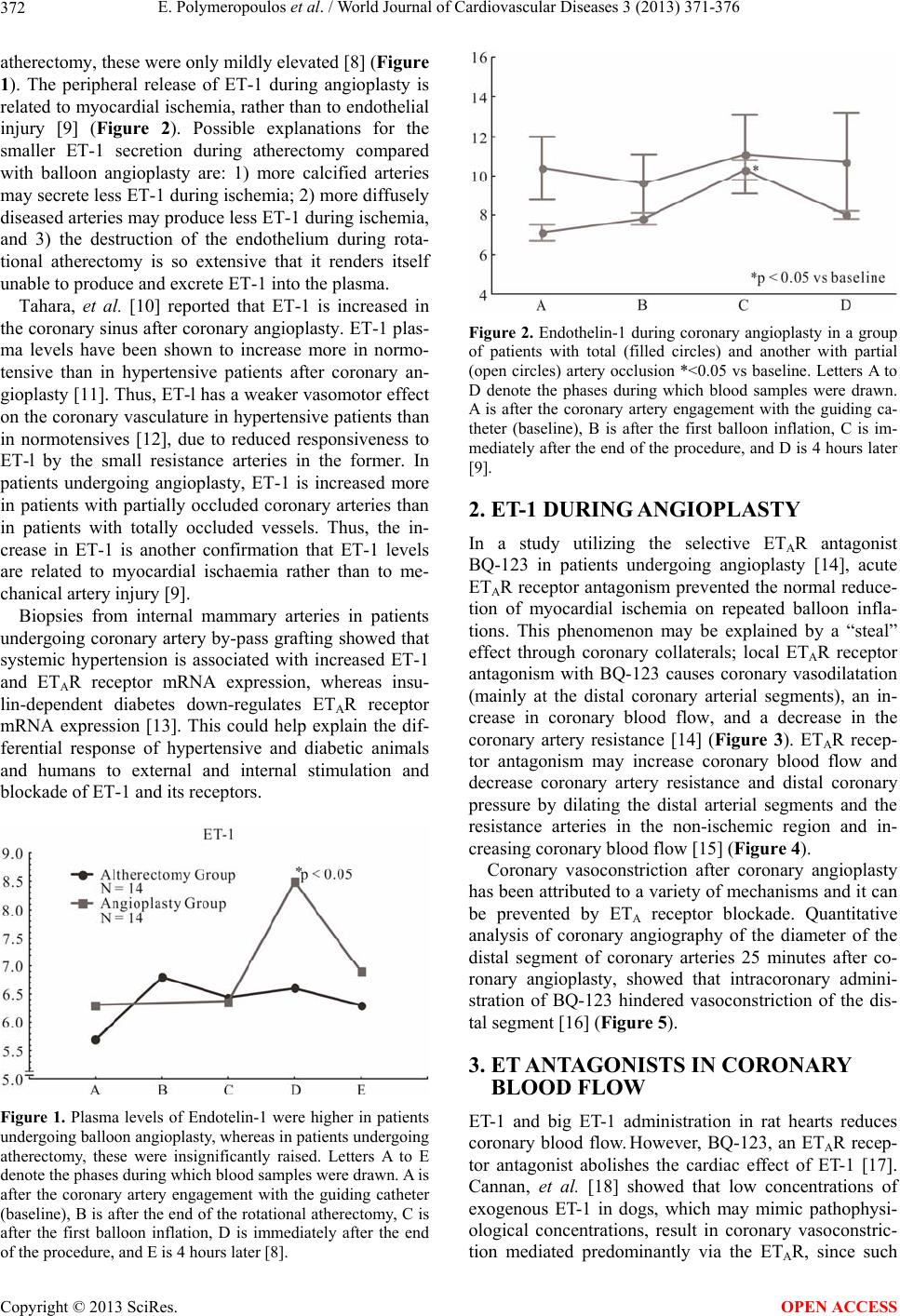 E. Polymeropoulos et al. / World Journal of Cardiovascular Diseases 3 (2013) 371-376 372 atherectomy, these were only mildly elevated [8] (Figure 1). The peripheral release of ET-1 during angioplasty is related to myocardial ischemia, rather than to endoth elial injury [9] (Figure 2). Possible explanations for the smaller ET-1 secretion during atherectomy compared with balloon angioplasty are: 1) more calcified arteries may secrete less ET-1 during ischemia; 2) more diffusely diseased arteries may produce less ET-1 during ischemia, and 3) the destruction of the endothelium during rota- tional atherectomy is so extensive that it renders itself unable to produce and excrete ET-1 into the plasma. Tahara, et al. [10] reported that ET-1 is increased in the coronary sinus after coronary angioplasty. ET-1 plas- ma levels have been shown to increase more in normo- tensive than in hypertensive patients after coronary an- gioplasty [11]. Thus, ET-l has a weaker vasomotor effect on the coronary vasculature in hypertensive patients than in normotensives [12], due to reduced responsiveness to ET-l by the small resistance arteries in the former. In patients undergoing angioplasty, ET-1 is increased more in patients with partially occluded coronary arteries than in patients with totally occluded vessels. Thus, the in- crease in ET-1 is another confirmation that ET-1 levels are related to myocardial ischaemia rather than to me- chanical artery injury [9]. Biopsies from internal mammary arteries in patients undergoing coronary artery by-pass grafting showed that systemic hypertension is associated with increased ET-1 and ETAR receptor mRNA expression, whereas insu- lin-dependent diabetes down-regulates ETAR receptor mRNA expression [13]. This could help explain the dif- ferential response of hypertensive and diabetic animals and humans to external and internal stimulation and blockade of ET-1 and its receptors. Figure 1. Plasma levels of Endotelin-1 were higher in patients undergoing balloon angioplasty, whereas in patients undergoing atherectomy, these were insignificantly raised. Letters A to E denote the phases during which blood samples were drawn. A is after the coronary artery engagement with the guiding catheter (baseline), B is after the end of the rotational atherectomy, C is after the first balloon inflation, D is immediately after the end of the procedure, and E is 4 hours later [8]. Figure 2. Endothelin-1 during coronary angioplasty in a group of patients with total (filled circles) and another with partial (open circles) artery occlusion *<0.05 vs baseline. Letters A to D denote the phases during which blood samples were drawn. A is after the coronary artery engagement with the guiding ca- theter (baseline), B is after the first balloon inflation, C is im- mediately after the end of the procedure, and D is 4 hours later [9]. 2. ET-1 DURING ANGIOPLASTY In a study utilizing the selective ETAR antagonist BQ-123 in patients undergoing angioplasty [14], acute ETAR receptor antagonism prevented the normal reduce- tion of myocardial ischemia on repeated balloon infla- tions. This phenomenon may be explained by a “steal” effect through coronary collaterals; local ETAR receptor antagonism with BQ-123 causes coronary vasodilatation (mainly at the distal coronary arterial segments), an in- crease in coronary blood flow, and a decrease in the coronary artery resistance [14] (Figure 3). ETAR recep- tor antagonism may increase coronary blood flow and decrease coronary artery resistance and distal coronary pressure by dilating the distal arterial segments and the resistance arteries in the non-ischemic region and in- creasing coronary blood flow [15] (Figure 4). Coronary vasoconstriction after coronary angioplasty has been attributed to a variety of mechanisms and it can be prevented by ETA receptor blockade. Quantitative analysis of coronary angiography of the diameter of the distal segment of coronary arteries 25 minutes after co- ronary angioplasty, showed that intracoronary admini- stration of BQ-123 hindered vasoconstriction of the dis- tal segment [16] (Figure 5). 3. ET ANTAGONISTS IN CORONARY BLOOD FLOW ET-1 and big ET-1 administration in rat hearts reduces coronary blood flow. However, BQ-123, an ETAR recep- tor antagonist abolishes the cardiac effect of ET-1 [17]. Cannan, et al. [18] showed that low concentrations of exogenous ET-1 in dogs, which may mimic pathophysi- ological concentrations, result in coronary vasoconstric- tion mediated predominantly via the ETAR, since such Copyright © 2013 SciRes. OPEN ACCESS 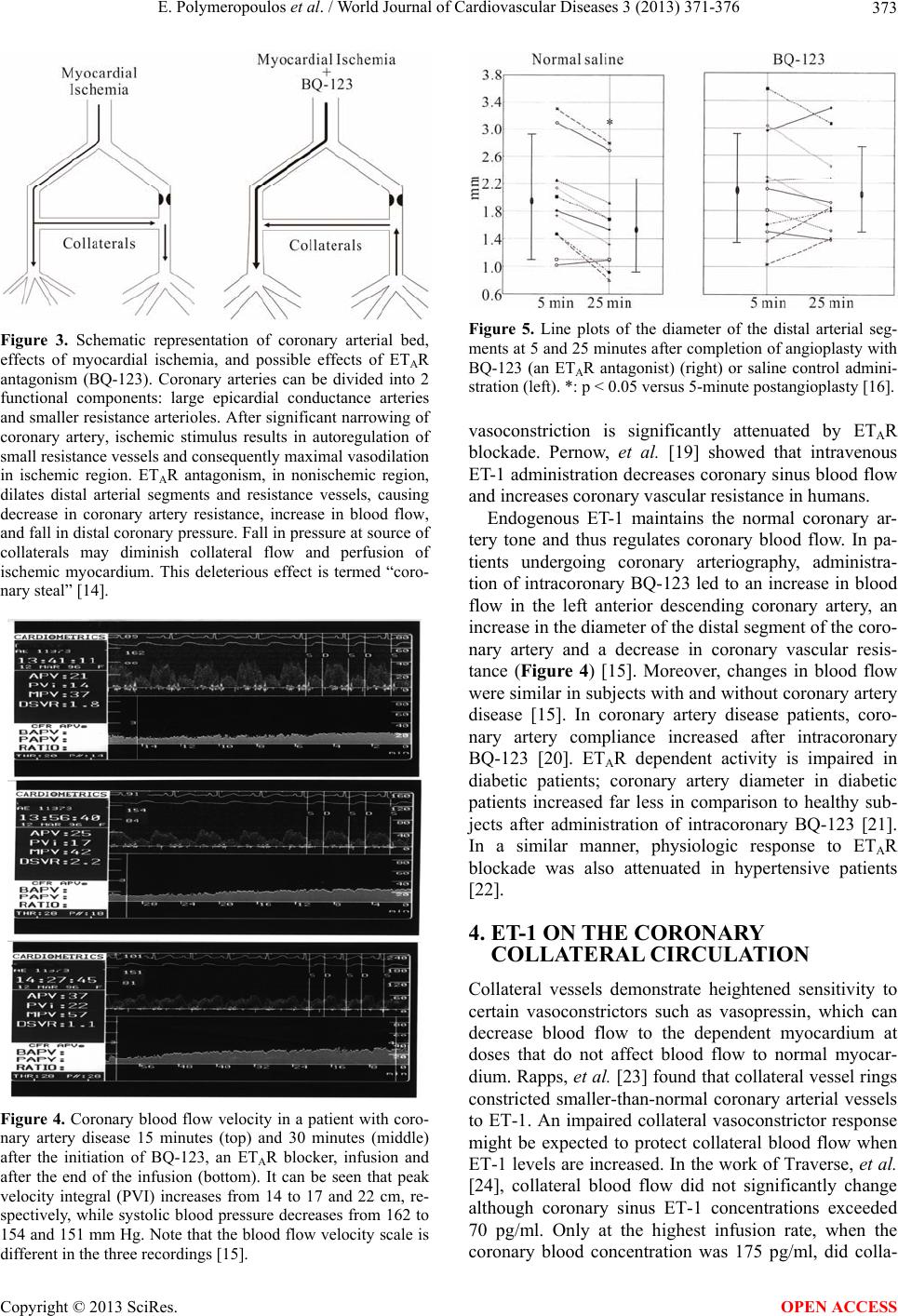 E. Polymeropoulos et al. / World Journal of Cardiovascular Diseases 3 (2013) 371-376 373 Figure 3. Schematic representation of coronary arterial bed, effects of myocardial ischemia, and possible effects of ETAR antagonism (BQ-123). Coronary arteries can be divided into 2 functional components: large epicardial conductance arteries and smaller resistance arterioles. After significant narrowing of coronary artery, ischemic stimulus results in autoregulation of small resistance vessels and consequently maximal vasodilation in ischemic region. ETAR antagonism, in nonischemic region, dilates distal arterial segments and resistance vessels, causing decrease in coronary artery resistance, increase in blood flow, and fall in distal coronary pressure. Fall in pressure at source of collaterals may diminish collateral flow and perfusion of ischemic myocardium. This deleterious effect is termed “coro- nary steal” [14]. Figure 4. Coronary blood flow velocity in a patient with coro- nary artery disease 15 minutes (top) and 30 minutes (middle) after the initiation of BQ-123, an ETAR blocker, infusion and after the end of the infusion (bottom). It can be seen that peak velocity integral (PVI) increases from 14 to 17 and 22 cm, re- spectively, while systolic blood pressure decreases from 162 to 154 and 151 mm Hg. Note that the blood flow velocity scale is different in the three recordings [15]. Figure 5. Line plots of the diameter of the distal arterial seg- ments at 5 and 25 minutes after completion of angioplasty with BQ-123 (an ETAR antagonist) (right) or saline control admini- stration (left). *: p < 0.05 versus 5-minute postangioplasty [16]. vasoconstriction is significantly attenuated by ETAR blockade. Pernow, et al. [19] showed that intravenous ET-1 administration decreases coronary sinus blood flow and increases coronary vascular resistance in humans. Endogenous ET-1 maintains the normal coronary ar- tery tone and thus regulates coronary blood flow. In pa- tients undergoing coronary arteriography, administra- tion of intracoronary BQ-123 led to an increase in blood flow in the left anterior descending coronary artery, an increase in the diameter of the distal segment of the coro- nary artery and a decrease in coronary vascular resis- tance (Figure 4) [15]. Moreover, changes in blood flow were similar in subjects with and without coronary artery disease [15]. In coronary artery disease patients, coro- nary artery compliance increased after intracoronary BQ-123 [20]. ETAR dependent activity is impaired in diabetic patients; coronary artery diameter in diabetic patients increased far less in comparison to healthy sub- jects after administration of intracoronary BQ-123 [21]. In a similar manner, physiologic response to ETAR blockade was also attenuated in hypertensive patients [22]. 4. ET-1 ON THE CORONARY COLLATERAL CIRCULATION Collateral vessels demonstrate heightened sensitivity to certain vasoconstrictors such as vasopressin, which can decrease blood flow to the dependent myocardium at doses that do not affect blood flow to normal myocar- dium. Rapps, et al. [23] found that collateral vessel rings constricted smaller-than-normal coronary arterial vessels to ET-1. An impaired collateral vasoconstrictor response might be expected to protect collateral blood flow when ET-1 levels are increased. In the work of Traverse, et al. [24], collateral blood flow did not significantly change although coronary sinus ET-1 concentrations exceeded 70 pg/ml. Only at the highest infusion rate, when the coronary blood concentration was 175 pg/ml, did colla- Copyright © 2013 SciRes. OPEN ACCESS 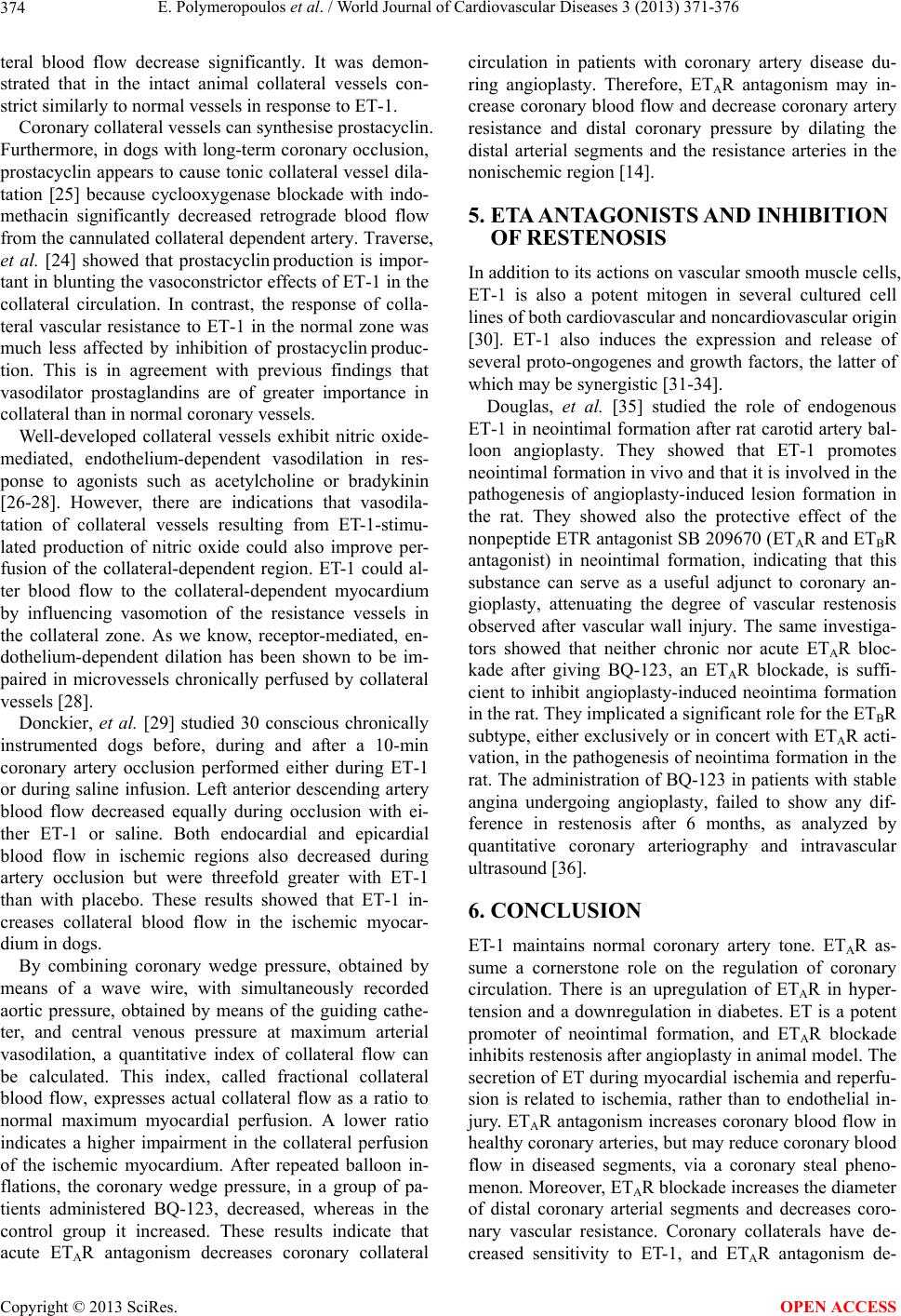 E. Polymeropoulos et al. / World Journal of Cardiovascular Diseases 3 (2013) 371-376 374 teral blood flow decrease significantly. It was demon- strated that in the intact animal collateral vessels con- strict similarly to normal vessels in response to ET-1. Coronary collateral vessels can synthesise prostacyclin. Furthermore, in dogs with long-term coronary occlusion, prostacyclin appears to cause tonic collateral vessel dila- tation [25] because cyclooxygenase blockade with indo- methacin significantly decreased retrograde blood flow from the cannulated collateral dependent artery. Traverse, et al. [24] showed that prostacyclin production is impor- tant in blunting th e vasoconstrictor effects of ET-1 in th e collateral circulation. In contrast, the response of colla- teral vascular resistance to ET-1 in the normal zone was much less affected by inhibition of prostacyclin produc- tion. This is in agreement with previous findings that vasodilator prostaglandins are of greater importance in collateral than in normal coronary vessels. Well-developed collateral vessels exhibit nitric oxide- mediated, endothelium-dependent vasodilation in res- ponse to agonists such as acetylcholine or bradykinin [26-28]. However, there are indications that vasodila- tation of collateral vessels resulting from ET-1-stimu- lated production of nitric oxide could also improve per- fusion of the collateral-dependent region. ET-1 could al- ter blood flow to the collateral-dependent myocardium by influencing vasomotion of the resistance vessels in the collateral zone. As we know, receptor-mediated, en- dothelium-dependent dilation has been shown to be im- paired in microvessels chronically perfused by collateral vessels [28 ]. Donckier, et al. [29] studied 30 conscious chronically instrumented dogs before, during and after a 10-min coronary artery occlusion performed either during ET-1 or during saline infusion. Left anterior descending artery blood flow decreased equally during occlusion with ei- ther ET-1 or saline. Both endocardial and epicardial blood flow in ischemic regions also decreased during artery occlusion but were threefold greater with ET-1 than with placebo. These results showed that ET-1 in- creases collateral blood flow in the ischemic myocar- dium in dogs. By combining coronary wedge pressure, obtained by means of a wave wire, with simultaneously recorded aortic pressure, obtained by means of the guiding cathe- ter, and central venous pressure at maximum arterial vasodilation, a quantitative index of collateral flow can be calculated. This index, called fractional collateral blood flow, expresses actual collateral flow as a ratio to normal maximum myocardial perfusion. A lower ratio indicates a higher impairment in the collateral perfusion of the ischemic myocardium. After repeated balloon in- flations, the coronary wedge pressure, in a group of pa- tients administered BQ-123, decreased, whereas in the control group it increased. These results indicate that acute ETAR antagonism decreases coronary collateral circulation in patients with coronary artery disease du- ring angioplasty. Therefore, ETAR antagonism may in- crease coronary blood flow and decrease coronary artery resistance and distal coronary pressure by dilating the distal arterial segments and the resistance arteries in the nonischemic region [14]. 5. ETA ANTAGONISTS AND INHIBITION OF RESTENOSIS In addition to its actions on vascular smooth muscle cells, ET-1 is also a potent mitogen in several cultured cell lines of both cardiovascular and noncardiovascular origin [30]. ET-1 also induces the expression and release of several proto-ongogenes and growth factors, the latter of which may be synergistic [31-34]. Douglas, et al. [35] studied the role of endogenous ET-1 in neointimal formation after rat carotid artery bal- loon angioplasty. They showed that ET-1 promotes neointimal formation in vivo and that it is involved in the pathogenesis of angioplasty-induced lesion formation in the rat. They showed also the protective effect of the nonpeptide ETR an tagonist SB 209670 ( ETAR and ETBR antagonist) in neointimal formation, indicating that this substance can serve as a useful adjunct to coronary an- gioplasty, attenuating the degree of vascular restenosis observed after vascular wall injury. The same investiga- tors showed that neither chronic nor acute ETAR bloc- kade after giving BQ-123, an ETAR blockade, is suffi- cient to inhibit angioplasty-induced neointima formation in the rat. They implicated a significant role for the ETBR subtype, either exclusively or in concert with ETAR acti- vation, in the pathogenesis of neo intima formation in the rat. The administration of BQ-123 in patients with stab le angina undergoing angioplasty, failed to show any dif- ference in restenosis after 6 months, as analyzed by quantitative coronary arteriography and intravascular ultrasound [36]. 6. CONCLUSION ET-1 maintains normal coronary artery tone. ETAR as- sume a cornerstone role on the regulation of coronary circulation. There is an upregulation of ETAR in hyper- tension and a downregulation in diabetes. ET is a potent promoter of neointimal formation, and ETAR blockade inhibits restenosis after ang ioplasty in an imal model. The secretion of ET during myocardial ischemia and reperfu- sion is related to ischemia, rather than to endothelial in- jury. ETAR antagonism increases coronary blood flow in healthy coronary arteries, but may reduce coronary blood flow in diseased segments, via a coronary steal pheno- menon. Moreover, ETAR blockade increases the diameter of distal coronary arterial segments and decreases coro- nary vascular resistance. Coronary collaterals have de- creased sensitivity to ET-1, and ETAR antagonism de- Copyright © 2013 SciRes. OPEN ACCESS  E. Polymeropoulos et al. / World Journal of Cardiovascular Diseases 3 (2013) 371-376 375 creases collateral blood flow and perfusion of the ische- mic myocardium. REFERENCES [1] Yanagisawa, M., Kurihara, H., Kimura, S., Tomobe, Y., Kobayashi, M., Mitsui, Y., et al. (1988) A novel potent vasoconstrictor peptide produced by vascular endothelial cells. Nature, 332, 411-415. doi:10.1038/332411a0 [2] Wagner, O.F., Christ, G., Wojta, J., Vierhapper, H., Par- zer, S., Nowotny, P.J., et al. (1992) Polar secretion of en- dothelin-1 by cultured endothelial cells. The Journal of Biological Chemistry, 267, 16066-16068. [3] Kiowski, W., Luscher, T.F., Linder, L. and Buhler, F.R. (1991) Endothelin-1-induced vasoconstriction in humans. Reversal by calcium channel blockade but not by nitro- vasodilators or endothelium-derived relaxing factor. Cir- culation, 83, 469-475. doi:10.1161/01.CIR.83.2.469 [4] Seo, B., Oemar, B.S., Siebenmann, R., Von, S.L. and Luscher, T.F. (1994) Both ETA and ETB receptors medi- ate contraction to endothelin-1 in human blood vessels. Circulation, 89, 1203-1208 . doi:10.1161/01.CIR.89.3.1203 [5] Luscher, T.F., Yang, Z., Tschudi, M., Von, S.L., Stulz, P., Boulanger, C., et al. (1990) Interaction between endo- thelin-1 and endothelium-derived relaxing factor in hu- man arteries and veins. Circulation Research, 66, 1088- 1094. doi:10.1161/01.RES.66.4.1088 [6] Brunner, F. (1995) Tissue endothelin-1 levels in perfused rat heart following stimulation with agonists and in is- chaemia and reperfusion. Journal of Molecular and Cel- lular Cardiology, 27, 1953-1963. doi:10.1016/0022-2828(95)90017-9 [7] Zeiher, A.M., Goebel, H., Schachinger, V. and Ihling, C. (1995) Tissue endothelin-1 immunoreactivity in the ac- tive coronary atherosclerotic plaque. A clue to the mecha- nism of increased vasoreactivity of the culprit lesion in unstable angina. Circulation, 91, 941-947. doi:10.1161/01.CIR.91.4.941 [8] Kyriakides, Z.S., Markianos, M., Antoniadis, A., Sba- rouni, E., Nikolaou, N., Zarvalis, E., et al. (1998) Effect of rotational coronary atherectomy on peripheral endo- thelin-1, atrial natriuretic peptide, and cyclic adenosine monophosphate plasma levels. Cardiovascular Drugs and Therapy, 12, 245-250. doi:10.1023/A:1007709614835 [9] Kyriakides, Z.S., Markianos, M., Iliodromitis, E.K. and Kremastinos, D.T. (1995) Vein plasma endothelin-1 and cyclic GMP increase during coronary angioplasty is re- lated to myocardial ischaemia. European Heart Journal, 16, 894-898. [10] Tahara, A., Kohno, M., Yanagi, S., Itagane, H., Toda, I., Akioka, K., et al. (1991) Circulating immunoreactive en- dothelin in patients undergoing percutaneous transluminal coronary angioplasty. Metabolism, 40, 1235-1237. doi:10.1016/0026-0495(91)90021-N [11] Kyriakides, Z.S., Markianos, M., Paraskevaidis, I.A., Tousoulis, D., Fragakis, N.K. and Kremastinos, D.T. (1996) Decreased vasomotor effect of endothelin on the coronary arteries during angioplasty in hypertensive pa- tients. International Journal of Cardiology, 55, 41-48. doi:10.1016/0167-5273(96)02653-8 [12] Schiffrin, E.L., Deng, L.Y. and Larochelle, P. (1992) Blunted effects of endothelin upon small subcutaneous resistance arteries of mild essential hypertensive patients. Journal of Hypertension, 10, 437-444. doi:10.1097/00004872-199205000-00006 [13] Zygalaki, E., Kaklamanis, L., Lolaka, M., Nikolaou, N., Koutouzis, M., Lianidou, E.S., et al. (2009) Systemic hypertension augments, whereas insulin-dependent dia- betes down-regulates, endothelin A-receptor expression in the mammary artery in coronary artery disease patients. Cardiology Journal, 16, 348-354. [14] Kyriakides, Z.S., Kremastinos, D.T., Kolettis, T.M., Ta- souli, A., Antoniadis, A. and Webb, D.J. (2000) Acute endothelin A-receptor antagonism prevents normal re- duction of myocardial ischemia on repeated balloon infla- tions during angioplasty. Circulation, 102, 1937-1943. doi:10.1161/01.CIR.102.16.1937 [15] Kyriakides, Z.S., Kremastinos, D.T., Bofilis, E., Tou- soulis, D., Antoniadis, A. and Webb, D.J. (2000) En- dogenous endothelin maintains coronary artery tone by endothelin type A-receptor stimulation in patients under- going coronary arteriography. Heart, 84, 176-182. doi:10.1136/heart.84.2.176 [16] Kyriakides, Z.S., Kremastinos, D.T., Psychari, S.N., Kolettis, T., Sbarouni, E. and Webb, D.J. (2001) Coro- nary vasoconstriction after coronary angioplasty is at- tenuated by endothelin a receptor antagonism. American Journal of Cardiology, 87, 1011-1013. doi:10.1016/S0002-9149(01)01441-2 [17] Grover, G.J., Sleph, P.G., Fox, M. and Trippodo, N.C. (1992) Role of endothelin-1 and big endothelin-1 in mo- dulating coronary vascular tone, contractile function and severity of ischemia in rat hearts. Journal of Pharmacol- ogy and Experimental Therapeutics, 263, 1074-1082. [18] Cannan, C.R., Burnett, Jr. J.C., , Brandt, R.R. and Ler- man, A. (1995) Endothelin at pathophysiological concen- trations mediates coronary vasoconstriction via the endo- thelin-A receptor. Circulation, 92, 3312-3317. doi:10.1161/01.CIR.92.11.3312 [19] Pernow, J., Kaijser, L., Lundberg, J.M. and Ahlborg, G. (1996) Comparable potent coronary constrictor effects of endothelin-1 and big endothelin-1 in humans. Circulation, 94, 2077-2082. doi:10.1161/01.CIR.94.9.2077 [20] Kyriakides, Z.S., Kremastinos, D.T., Kolokathis, F., Kos- topoulou, A., Georgiadis, M. and Webb, D.J. (2002) Acute endothelin (A) receptor antagonism improves coronary artery compliance in coronary artery disease pa- tients. Clinical Science (London), 103, S179S-S183S. [21] Kyriakides, Z.S., Kremastinos, D.T., Raptis, A.E., Johns- ton, N., Raptis, S.A., Webb, D.J., et al. (2006) Impaired effect of endothelin-1 on coronary artery stiffness in type 2 diabetes. International Journal of Cardiology, 112, 207-212. doi:10.1016/j.ijcard.2005.09.018 [22] Kyriakides, Z., Kyrzopoulos, S., Paraskevaidis, I., Kolo- kathis, F., Tsopotos, I., Lyras, T., et al. (2004) Endothe- Copyright © 2013 SciRes. OPEN ACCESS  E. Polymeropoulos et al. / World Journal of Cardiovascular Diseases 3 (2013) 371-376 Copyright © 2013 SciRes. 376 OPEN ACCESS lin-A receptor antagonism promotes decreased vasodila- tion but has no differential effect on coronary artery com- pliance in hypertensive patients. Journal of Cardiovas- cular Pharmacology, 44, S85-S88. doi:10.1097/01.fjc.0000166209.19921.fc [23] Rapps, J.A., Jones, A.W., Sturek, M., Magliola, L. and Parker, J.L. (1997) Mechanisms of altered contractile re- sponses to vasopressin and endothelin in canine coronary collateral arteries. Circulation, 95, 231-239. doi:10.1161/01.CIR.95.1.231 [24] Traverse, J.H., Judd, D. and Bache, R.J. (1996) Dose-de- pendent effect of endothelin-1 on blood flow to normal and collateral-dependent myocardium. Circulation, 93, 558-566. doi:10.1161/01.CIR.93.3.558 [25] Altman, J., Dulas, D. and Bache, R.J. (1992) Effect of cyclooxygenase blockade on blood flow through well- developed coronary collateral vessels. Circulation Re- search, 70, 1091-1098. doi:10.1161/01.RES.70.6.1091 [26] Angus, J.A., Ward, J.E., Smolich, J.J. and McPherson, G.A. (1991) Reactivity of canine isolated epicardial col- lateral coronary arteries. Relation to vessel structure. Cir- culation Research, 69, 1340-1352. doi:10.1161/01.RES.69.5.1340 [27] Flynn, N.M., Kenny, D., Pelc, L.R., Warltier, D.C., Bosnjak, Z.J. and Kampine, J.P. (1991) Endothelium-de- pendent vasodilation of canine coronary collateral vessels. American Journal of Physiology, 261, H1797-H1801. [28] Sellke, F.W., Quillen, J.E., Brooks, L.A. and Harrison, D.G. (1990) Endothelial modulation of the coronary vas- culature in vessels perfused via mature collaterals. Cir- culation, 81, 1938-1947. doi:10.1161/01.CIR.81.6.1938 [29] Donckier, J., Hanet, C., Stoleru, L., Van, M.H., Galanti, L., Hayashida, W., et al. (1994) Effects of endothelin-1 at pathophysiologic concentrations on coronary perfusion and mechanical function of normal and postischemic myocardium. Journal of Cardiovascular Pharmacology, 23, 212-219. doi:10.1097/00005344-199402000-00006 [30] Ohlstein, E.H., Arleth, A., Bryan, H., Elliott, J.D. and Sung, C.P. (1992) The selective endothelin ETA receptor antagonist BQ123 antagonizes endothelin-1-mediated mitogenesis. European Journal of Pharmacology: Mole- cular Pharmacology, 225, 347-350. doi:10.1016/0922-4106(92)90109-9 [31] Bobik, A., Grooms, A., Millar, J.A., Mitchell, A. and Grinpukel, S. (1990) Growth factor activity of endothelin on vascular smooth muscle. American Journal of Physi- ology, 258, C408-C415. [32] Hirata, Y., Takagi, Y., Fukuda, Y. and Marumo, F. (1989) Endothelin is a potent mitogen for rat vascular smooth muscle cells. Atherosclerosis, 78, 225-228. doi:10.1016/0021-9150(89)90227-X [33] Jaffer, F.E., Knauss, T.C., Poptic, E. and Abboud, H.E. (1990) Endothelin stimulates PDGF secretion in cultured human mesangial cells. Kidney International, 38, 1193- 1198. doi:10.1038/ki.1990.333 [34] Komuro, I., Kurihara, H., Sugiyama, T., Yoshizumi, M., Takaku, F. and Yazaki, Y. (1988) Endothelin stimulates c-fos and c-myc expression and proliferation of vascular smooth muscle cells. FEBS Letters, 238, 249-252. doi:10.1016/0014-5793(88)80489-7 [35] Douglas, S.A., Vickery-Clark, L.M., Louden, C., Elliott, J.D. and Ohlstein, E.H. (1995) Endothelin receptor sub- types in the pathogenesis of angioplasty-induced neoin- tima formation in the rat: A comparison of selective ETA receptor antagonism and dual ETA/ETB receptor antago- nism using BQ-123 and SB 209670. Journal of Cardio- vascular Pharmacology, 26, S186-S189. [36] Kyriakides, Z.S., Psychari, S.N., Kolettis, T.M., Geor- giadis, M., Antoniadis, A. and Kremastinos, D.T. (2003) Usefulness of endothelin (A) receptor antagonists for the prevention of in-stent restenosis in patients with stable angina pectoris or silent myocardial ischemia. American Journal of Cardiology, 91, 476-479. doi:10.1016/S0002-9149(02)03253-8 |