

Paper Menu >>
Journal Menu >>
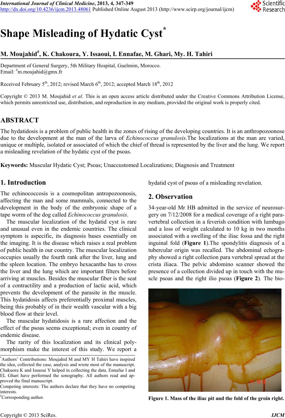 International Journal of Clinical Medicine, 2013, 4, 347-349 http://dx.doi.org/10.4236/ijcm.2013.48061 Published Online August 2013 (http://www.scirp.org/journal/ijcm) 347 Shape Misleading of Hydatic Cyst* M. Moujahid#, K. Chakoura, Y. Issaoui, I. Ennafae, M. Ghari, My. H. Tahiri Department of General Surgery, 5th Military Hospital, Guelmim, Morocco. Email: #[email protected] Received February 5th, 2012; revised March 6th, 2012; accepted March 18th, 2012 Copyright © 2013 M. Moujahid et al. This is an open access article distributed under the Creative Commons Attribution License, which permits unrestricted use, distribution, and reproduction in any medium, provided the original work is properly cited. ABSTRACT The hydatidosis is a problem of public health in the zones of rising of the developing countries. It is an anthropozoonose due to the development at the man of the larva of Echinococcus granulosis.The localizations at the man are varied, unique or multiple, isolated or associated of which th e ch ief of thread is represented b y the liver and the lung . We report a misleading revelation of the hydatic cyst of the psoas. Keywords: Muscular Hydatic Cyst; Psoas; Unaccustomed Localizations; Diagnosis and Treatment 1. Introduction The echinococcosis is a cosmopolitan antropozoonosis, affecting the man and some mammals, connected to the development in the body of the embryonic shape of a tape worm of the dog called Echinococcus granulosis. The muscular localization of the hydatid cyst is rare and unusual even in the endemic countries. The clinical symptom is aspecific, its diagnosis bases essentially on the imaging. It is the disease which raises a real problem of public health in our country. The muscular localization occupies usually the fourth rank after the liver, lung and the spleen location. The embryo hexacanthe has to cross the liver and the lung which are important filters before arriving at muscles. Besides the muscular fiber is the seat of a contractility and a production of lactic acid, which prevents the development of the parasite in the muscle. This hydatidosis affects preferentially proximal muscles, being this probably of in their wealth vascular with a big blood flow at their level. The muscular hydatidosis is a rare affection and the effect of the psoas seems exceptional; even in coun try of endemic disease. The rarity of this localization and its clinical poly- morphism make the interest of this study. We report a hydatid cyst of psoas of a misleading revelation. 2. Observation 34-year-old Mr HB admitted in the service of neurosur- gery on 7/12/2008 for a medical coverage of a right para- vertebral collection in a feverish condition with lumbago and a loss of weight calculated to 10 kg in two months associated with a swelling of the iliac fossa and the right inguinal fold (Figure 1).The spondylitis diagnosis of a tubercular origin was recalled. The abdominal echogra- phy showed a right co llection para verteb ral spread at the crista iliaca. The pelvic abdomino scanner showed the presence of a collection divided up in touch with the mu- scle psoas and the right ilio psoas (Figure 2). The bio- *Authors’ Contributions: Moujahid M and MY H Tahiri have inspired the idea, collected the case, analysis and wrote most of the manuscript. Chakuora K and Issaoui Y helped in collecting the data. Ennafae I and EL Ghari have performed the sonography. All authors read and ap- p roved the final manuscript. Competing interests: The authors declare that they have no competing interests. #Corresponding author. Figure 1. Mass of the iliac pit and the fold of the groin right. Copyright © 2013 SciRes. IJCM 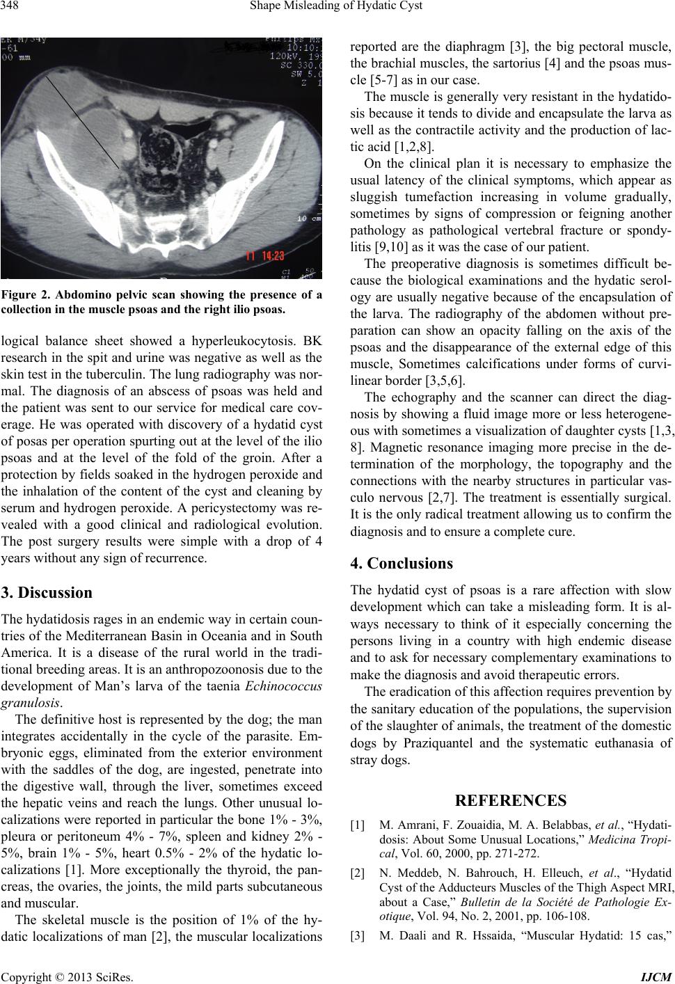 Shape Misleading of Hydatic Cyst 348 Figure 2. Abdomino pelvic scan showing the presence of a collection in the muscle psoas and the right ilio psoas. logical balance sheet showed a hyperleukocytosis. BK research in the spit and urine was negative as well as the skin test in the tuberculin. The lung rad iography was nor- mal. The diagnosis of an abscess of psoas was held and the patient was sent to our service for medical care cov- erage. He was operated with discovery of a hydatid cyst of posas per operation spu rting out at the level of the ilio psoas and at the level of the fold of the groin. After a protection by fields soaked in the hydrogen peroxide and the inhalation of the content of the cyst and cleaning by serum and hydrogen peroxide. A pericystectomy was re- vealed with a good clinical and radiological evolution. The post surgery results were simple with a drop of 4 years without any sign of recurrence. 3. Discussion The hydatidosis rag es in an en d emic way in certain coun- tries of the Mediterranean Basin in Oceania and in South America. It is a disease of the rural world in the tradi- tional breeding ar eas. It is an anthr opozoon osis du e to the development of Man’s larva of the taenia Echinococcus granulosis. The definitive host is represented by the dog; the man integrates accidentally in the cycle of the parasite. Em- bryonic eggs, eliminated from the exterior environment with the saddles of the dog, are ingested, penetrate into the digestive wall, through the liver, sometimes exceed the hepatic veins and reach the lungs. Other unusual lo- calizations were reported in particular the bone 1% - 3%, pleura or peritoneum 4% - 7%, spleen and kidney 2% - 5%, brain 1% - 5%, heart 0.5% - 2% of the hydatic lo- calizations [1]. More exceptionally the thyroid, the pan- creas, the ovaries, the join ts, the mild parts subcutaneous and muscular. The skeletal muscle is the position of 1% of the hy- datic localizations of man [2], the muscular localizations reported are the diaphragm [3], the big pectoral muscle, the brachial muscles, the sartorius [4] and the psoas mus- cle [5-7] as in our case. The muscle is generally very resistant in the hydatido- sis because it tends to divide and encapsulate the larva as well as the contractile activity and the production of lac- tic acid [1,2,8]. On the clinical plan it is necessary to emphasize the usual latency of the clinical symptoms, which appear as sluggish tumefaction increasing in volume gradually, sometimes by signs of compression or feigning another pathology as pathological vertebral fracture or spondy- litis [9,10] as it was the case of our patient. The preoperative diagnosis is sometimes difficult be- cause the biological examinations and the hydatic serol- ogy are usually negative because of the encapsulation of the larva. The radiography of the abdomen without pre- paration can show an opacity falling on the axis of the psoas and the disappearance of the external edge of this muscle, Sometimes calcifications under forms of curvi- linear border [3,5,6]. The echography and the scanner can direct the diag- nosis by showing a fluid image more or less heterogene- ous with sometimes a visualization of daughter cysts [1,3, 8]. Magnetic resonance imaging more precise in the de- termination of the morphology, the topography and the connections with the nearby structures in particular vas- culo nervous [2,7]. The treatment is essentially surgical. It is the only radical treatment allowing us to confirm the diagnosis and to ensure a complete cure. 4. Conclusions The hydatid cyst of psoas is a rare affection with slow development which can take a misleading form. It is al- ways necessary to think of it especially concerning the persons living in a country with high endemic disease and to ask for necessary complementary examinations to make the diagnosis and avoid therapeutic errors. The eradication of this affection requires prevention by the sanitary education of the populations, the supervision of the slaughter of animals, the treatment of the domestic dogs by Praziquantel and the systematic euthanasia of stray dogs. REFERENCES [1] M. Amrani, F. Zouaidia, M. A. Belabbas, et al., “Hydati- dosis: About Some Unusual Locations,” Medicina Tropi- cal, Vol. 60, 2000, pp. 271-272. [2] N. Meddeb, N. Bahrouch, H. Elleuch, et al., “Hydatid Cyst of the Adducteurs Muscles of the Thigh Aspect MRI, about a Case,” Bulletin de la Société de Pathologie Ex- otique, Vol. 94, No. 2, 2001, pp. 106-108. [3] M. Daali and R. Hssaida, “Muscular Hydatid: 15 cas,” Copyright © 2013 SciRes. IJCM  Shape Misleading of Hydatic Cyst Copyright © 2013 SciRes. IJCM 349 Presse Medicale, Paris, Vol. 28, No. 5, 2000, pp. 1166- 1169 [4] T. Hammami, F. Noomane, M. Ketata, et al., “Muscular Hydatid Cyst of the Thigh: A Comment of Three Observations,” Revue de Chirurgie Orthopédique, Vol. 88, 2002, pp. 193-196. [5] A. EL Moussaoui, R. Rabii, H. Rais, et al., “Hydatid Cyst of the Psoas Muscle. A Case Report,” Annales d’Urologie, Vol. 31, No. 6-7, 1997, pp. 357-360. [6] A. Benchekroun, H. Jira, O. J. Cheikhani, et al., “Hy datid Cyst of the Psoas: A Case Report,” Annals of Urology, Vol. 35, No. 2, 2001, pp. 108-110. doi:10.1016/S0003-4401(01)00012-2 [7] M. Melis, L. Marongiu, F. Scintu, Pisano and M. Giu- seppe, “Primary Hydatid Cysts of Psoas Muscle,” ANZ Journal of Surgery, Vol. 72, No. 6, 2002, pp. 443-445. doi:10.1046/j.1445-2197.2002.02420.x [8] M. Daali, R. Bennani-Lahmam and A. Hajji, “Hydatid Cyst of the Psoas about 3 New Observations,” Lyon Chi- ropractic, Vol. 2, No. 93, 1997, pp. 113-115. [9] S. A. Sakka, “Primitif Hydatid Cyst of the Thigh with Painful Demonstration at the Level of the Hip,” Reviews of Orthopaedic Surgery, Vol. 79, 1993, pp. 226-228. [10] S. Karray, M. Karray, M. Zlitni and M. Douik, “Radical Cure of Vertebral Hydatidosis. A Case Report,” Acta Or- thopaedica Belgica, Vol. 70, No. 1, 2004, pp. 80-83. |