

Paper Menu >>
Journal Menu >>
 Open Journal of Fluid Dynamics, 2013, 3, 9-13 http://dx.doi.org/10.4236/ojfd.2013.32A002 Published Online July 2013 (http://www.scirp.org/journal/ojfd) Behavior of Motile Sperm in Taylor-Couette Flow: Effect of Shear Stress on the Behavior of Motile Sperm Yasutaka Hayamizu1, Toru Hyakutake2, Koji Matsuura3, Shinichiro Yanase4, Shinichi Morita1, Shigeru Ohtsuka1, Takeshi Gonda1 1Department of Mechanical Engineering, Yonago National College of Technology, Tottori, Japan 2Graduate School of Engineering, Yokohama National University, Yokohama, Japan 3Research Core for Interdisciplinary Sciences, Okayama University, Okayama, Japan 4Graduate School of Natural Science and Technology, Okayama University, Okayama, Japan Email: [email protected] Received May 27, 2013; revised June 4, 2013; accepted June 11, 2013 Copyright © 2013 Yasutaka Hayamizu et al. This is an open access article distributed under the Creative Commons Attribution Li- cense, which permits unrestricted use, distribution, and reproduction in any medium, provided the original work is properly cited. ABSTRACT Infertility is often cited as one of the causes of a declining birthrate, which has become a serious social problem in re- cent years. Processes by which motile sperm can be safely and easily sorted are therefore important for infertility treat- ment. Therefore, as a new sorting method, microfluidic sperm sorter using the microfluidic system has been developed. To improve more separation efficiency of this device, it is necessary to know the behaviors of motile sperm in the mi- crochannel where the sperm undergo shear flow. The previous study implied the necessity of the modeling of motile sperm in the shear flow. In the present study, therefore, we experimentally investigated the behavior of the motile sperm in the Taylor-Couette flow using PTV (Particle Tracking Velocimetry) method. The experimental results showed that the ascent of the shear stress led to the increase in the sperm velocity, and the direction of the sperm velocity was oppo- site to that of the flow. Keywords: Motile Sperm; Taylor-Couette Flow; Shear Stress; PTV 1. Introduction Infertility is often cited as one of the causes of a declin- ing birthrate, which has become a serious social problem in recent years. Approximately 10% of couples were infertility, and almost 50% of all cases of infertility are associated with a lack of sperm or sperm abnormalities. Processes by which motile sperm can be safely and eas- ily sorted are therefore important for infertility treatment. Currently, the swim up method or the Percoll method is employed for sperm sorting. However, these methods need much time and sort sperm in a non-physiological environment, so damage such as DNA fragmentation is a major problem [1], and it might result in the sorting of sperm that is unsuitable for fertilization [2]. Therefore, as a new sorting method, microfluidic sperm sorter using the microfluidic system has been developed (refer to Fig- ure 1) [3-5]. To improve more separation efficiency of this device, it is necessary to know the behaviors of mo- tile sperm in the microchannel where the sperm undergo shear flow. The previous study implied the necessity of the modeling of motile sperm in the shear flow. In the present study, therefore, we experimentally in- vestigated the behavior of the motile sperm in the Tay- lor-Couette flow using PTV (Particle Tracking Veloci- metry) method. In this paper, we report the effect of shear stress on the behavior of motile sperm. Figure 1. Microfluidic sperm sorter (Life Science Depart- ment, Menicon Co., Ltd.) . C opyright © 2013 SciRes. OJFD 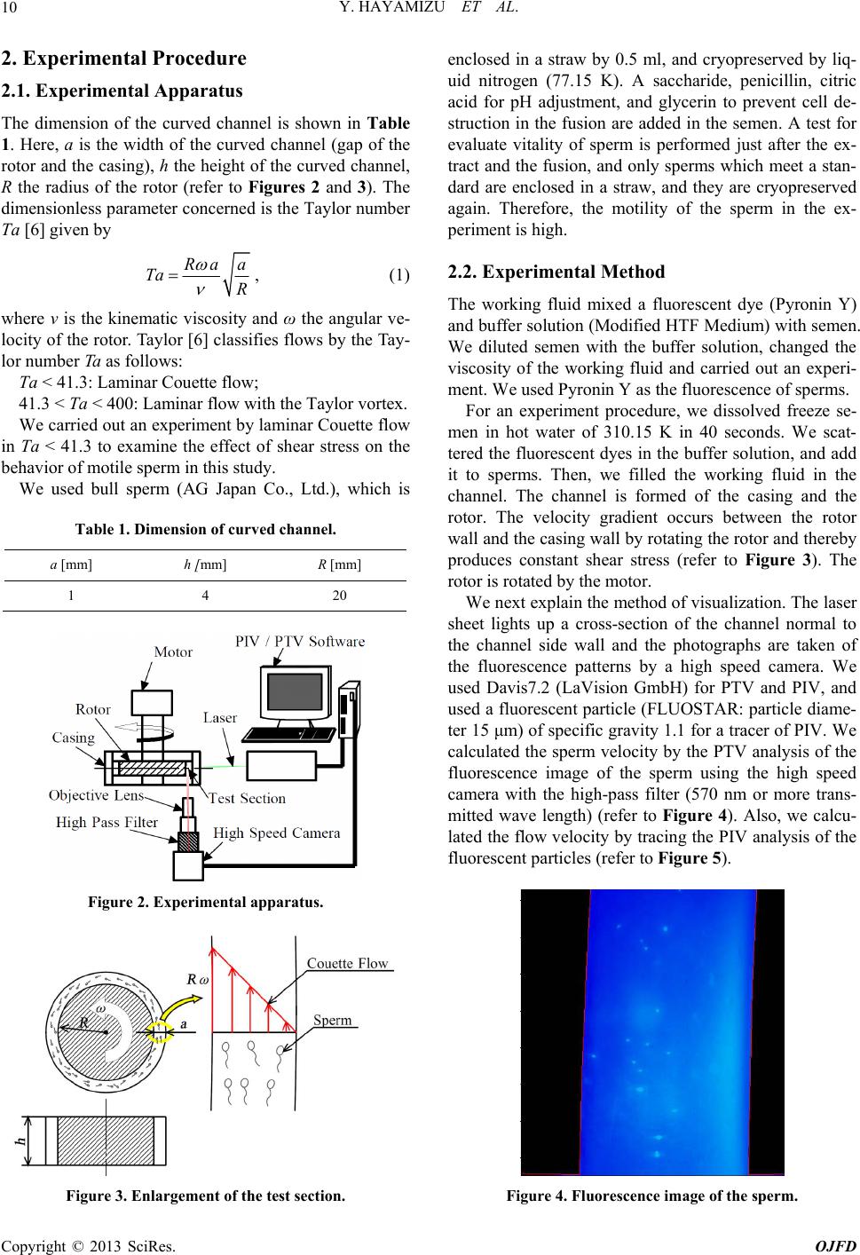 Y. HAYAMIZU ET AL. 10 2. Experimental Procedure 2.1. Experimental Apparatus The dimension of the curved channel is shown in Table 1. Here, a is the width of the curved channel (gap of the rotor and the casing), h the height of the curved channel, R the radius of the rotor (refer to Figures 2 and 3). The dimensionless parameter concerned is the Taylor number Ta [6] given by Ra a Ta R , (1) where ν is the kinematic viscosity and ω the angular ve- locity of the rotor. Taylor [6] classifies flows by the Tay- lor number Ta as follows: Ta < 41.3: Laminar Couette flow; 41.3 < Ta < 400: Laminar flow with the Taylor vortex. We carried out an experiment by laminar Couette flow in Ta < 41.3 to examine the effect of shear stress on the behavior of motile sperm in this study. We used bull sperm (AG Japan Co., Ltd.), which is Table 1. Dimension of curved channel. a [mm] h [mm] R [mm] 1 4 20 Figure 2. Experimental apparatus. Figure 3. Enlargement of the test section. enclosed in a straw by 0.5 ml, and cryopreserved by liq- uid nitrogen (77.15 K). A saccharide, penicillin, citric acid for pH adjustment, and glycerin to prevent cell de- struction in the fusion are added in the semen. A test for evaluate vitality of sperm is performed just after the ex- tract and the fusion, and only sperms which meet a stan- dard are enclosed in a straw, and they are cryopreserved again. Therefore, the motility of the sperm in the ex- periment is high. 2.2. Experimental Method The working fluid mixed a fluorescent dye (Pyronin Y) and buffer solution (Modified HTF Medium) with semen. We diluted semen with the buffer solution, changed the viscosity of the working fluid and carried out an experi- ment. We used Pyronin Y as the fluorescence of sperms. For an experiment procedure, we dissolved freeze se- men in hot water of 310.15 K in 40 seconds. We scat- tered the fluorescent dyes in the buffer solution, and add it to sperms. Then, we filled the working fluid in the channel. The channel is formed of the casing and the rotor. The velocity gradient occurs between the rotor wall and the casing wall by rotating the rotor and thereby produces constant shear stress (refer to Figure 3). The rotor is rotated by the motor. We next explain the method of visualization. The laser sheet lights up a cross-section of the channel normal to the channel side wall and the photographs are taken of the fluorescence patterns by a high speed camera. We used Davis7.2 (LaVision GmbH) for PTV and PIV, and used a fluorescent particle (FLUOSTAR: particle diame- ter 15 μm) of specific gravity 1.1 for a tracer of PIV. We calculated the sperm velocity by the PTV analysis of the fluorescence image of the sperm using the high speed camera with the high-pass filter (570 nm or more trans- mitted wave length) (refer to Figure 4). Also, we calcu- lated the flow velocity by tracing the PIV analysis of the fluorescent particles (refer to Figure 5). Figure 4. Fluorescence image of the sperm. Copyright © 2013 SciRes. OJFD 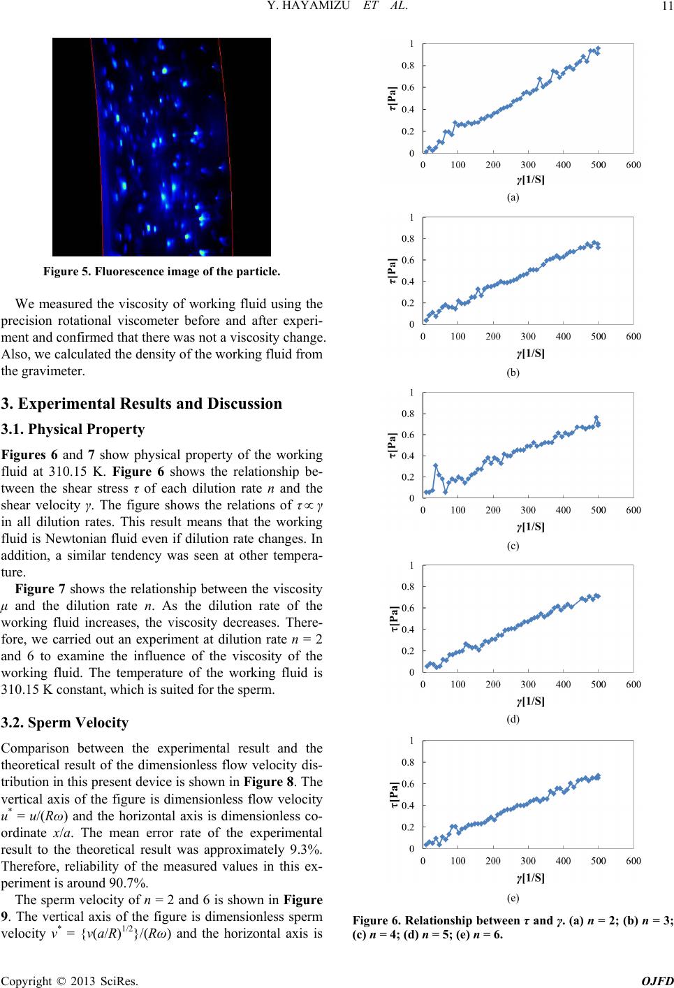 Y. HAYAMIZU ET AL. 11 Figure 5. Fluorescence image of the particle. We measured the viscosity of working fluid using the precision rotational viscometer before and after experi- ment and confirmed that there was not a viscosity change. Also, we calculated the density of the working fluid from the gravimeter. 3. Experimental Results and Discussion 3.1. Physical Property Figures 6 and 7 show physical property of the working fluid at 310.15 K. Figure 6 shows the relationship be- tween the shear stress τ of each dilution rate n and the shear velocity γ. The figure shows the relations of τ γ in all dilution rates. This result means that the working fluid is Newtonian fluid even if dilution rate changes. In addition, a similar tendency was seen at other tempera- tu fluid is 310.15 K constant, which is suited for the sperm. measured values in this ex- pe velocity v* = {v(a/R)1/2}/(Rω) and the horizontal axis is re. Figure 7 shows the relationship between the viscosity μ and the dilution rate n. As the dilution rate of the working fluid increases, the viscosity decreases. There- fore, we carried out an experiment at dilution rate n = 2 and 6 to examine the influence of the viscosity of the working fluid. The temperature of the working 3.2. Sperm Velocity Comparison between the experimental result and the theoretical result of the dimensionless flow velocity dis- tribution in this present device is shown in Figure 8. The vertical axis of the figure is dimensionless flow velocity u* = u/(Rω) and the horizontal axis is dimensionless co- ordinate x/a. The mean error rate of the experimental result to the theoretical result was approximately 9.3%. Therefore, reliability of the riment is around 90.7%. The sperm velocity of n = 2 and 6 is shown in Figure 9. The vertical axis of the figure is dimensionless sperm (a) (b) (c) (d) (e) Figure 6. Relationship between τ and γ. (a) n = 2; (b) n = 3; (c) n = 4; (d) n = 5; (e) n = 6. Copyright © 2013 SciRes. OJFD 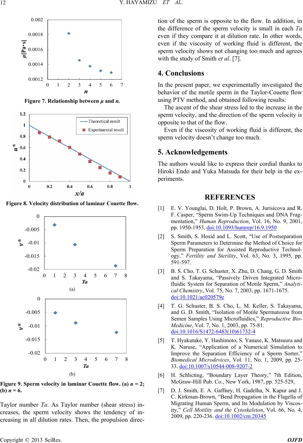 Y. HAYAMIZU ET AL. 12 0.0012 0.0014 0.0016 0.0018 0.002 01234567 μ[Pa・s] n Figure 7. Relationship between μ and n. Figure 8. Velocity distribution of laminar Couette flow. -0.02 -0.015 -0.01 -0.005 0 012345678 v* Ta (a) -0.02 -0.015 -0.01 -0.005 0 012345678 v* Ta (b) Figure 9. Sperm velocity in laminar Couette flow. (a) n = 2; (b) n = 6. Taylor number Ta. As Taylor number (shear stress) in- creases, the sperm velocity shows the tendency of in- creasing in all dilution rates. Then, the propulsion direc- tion of the sperm is opposite to the flow. In addition, in the difference of the sperm velocity is small in each Ta even if they compare it at dilution rate. In other words, even if the viscosity of working fluid is different, the sperm velocity shows not changing too much and agrees with the study of Smith et al. [7]. 4. Conclusions In the present paper, we experimentally investigated the behavior of the motile sperm in the Taylor-Couette flow using PTV method, and obtained following results: The ascent of the shear stress led to the increase in the sperm velocity, and the direction of the sperm velocity is opposite to that of the flow. Even if the viscosity of working fluid is different, the sperm velocity doesn’t change too much. 5. Acknowledgements The authors would like to express their cordial thanks to Hiroki Endo and Yuka Matsuda for their help in the ex- periments. REFERENCES [1] E. V. Younglai, D. Holt, P. Brown, A. Jurisicova and R. F. Casper, “Sperm Swim-Up Techniques and DNA Frag- mentation,” Human Reproduction, Vol. 16, No. 9, 2001, pp. 1950-1953. doi:10.1093/humrep/16.9.1950 [2] S. Smith, S. Hosid and L. Scott, “Use of Postseparation Sperm Parameters to Determine the Method of Choice for Sperm Preparation for Assisted Reproductive Technol- ogy,” Fertility and Sterility, Vol. 63, No. 3, 1995, pp. 591-597. [3] B. S. Cho, T. G. Schuster, X. Zhu, D. Chang, G. D. Smith and S. Takayama, “Passively Driven Integrated Micro- fluidic System for Separation of Motile Sperm,” Analyti- cal Chemistry, Vol. 75, No. 7, 2003, pp. 1671-1675. doi:10.1021/ac020579e [4] T. G. Schuster, B. S. Cho, L. M. Keller, S. Takayama, and G. D. Smith, “Isolation of Motile Spermatozoa from Semen Samples Using Microfluidics,” Reproductive Bio- Medicine, Vol. 7, No. 1, 2003, pp. 75-81. doi:10.1016/S1472-6483(10)61732-4 [5] T. Hyakutake, Y. Hashimoto, S. Yanase, K. Matsuura and K. Naruse, “Application of a Numerical Simulation to Improve the Separation Efficiency of a Sperm Sorter,” Biomedical Microdevices, Vol. 11, No. 1, 2009, pp. 25- 33. doi:10.1007/s10544-008-9207-2 [6] H. Schlicting, “Boundary Layer Theory,” 7th Edition, McGraw-Hill Pub. Co., New York, 1987, pp. 525-529, [7] D. J. Smith, E. A. Gaffney, H. Gadelha, N. Kapur and J. C. Kirkman-Brown, “Bend Propagation in the Flagella of Migrating Human Sperm, and Its Modulation by Viscos- ity,” Cell Motility and the Cytoskeleton, Vol. 66, No. 4, 2009, pp. 220-236. doi:10.1002/cm.20345 Copyright © 2013 SciRes. OJFD 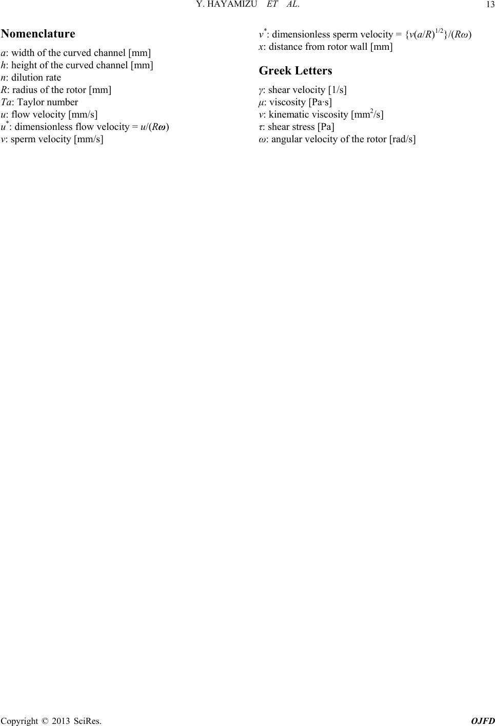 Y. HAYAMIZU ET AL. Copyright © 2013 SciRes. OJFD 13 Nomenclature a: width of the curved channel [mm] h: height of the curved channel [mm] n: dilution rate R: radius of the rotor [mm] Ta: Taylor number u: flow velocity [mm/s] u*: dimensionless flow velocity = u/(Rω) v: sperm velocity [mm/s] v*: dimensionless sperm velocity = {v(a/R)1/2}/(Rω) x: distance from rotor wall [mm] Greek Letters γ: shear velocity [1/s] μ: viscosity [Pa·s] ν: kinematic viscosity [mm2/s] τ: shear stress [Pa] ω: angular velocity of the rotor [rad/s] |