

Paper Menu >>
Journal Menu >>
 Journal of Cancer Therapy, 2013, 4, 1091-1094 http://dx.doi.org/10.4236/jct.2013.46125 Published Online August 2013 (http://www.scirp.org/journal/jct) 1091 Photodynamic Therapy as Palliative Therapy for Invasive Syringoid Eccrine Carcinoma of the Auricle: A Case Report Claudia Gutiérrez Gómez1*, AnailAlvarez2, Ana Lilia Ruelas2, Ixchel Landgrave2, Alfonso Vallarta3, Laura Andrade3 1Department of Plastic and Reconstructive Surgery, Medica Sur Hospital, México City, México; 2Dermatology Department, Medica Sur Hospital, México City, México; 3Plastic Surgery Department, Medica Sur Hospital, México City, México. Email: *[email protected] Received May 9th, 2013; revised June 12th, 2013; accepted June 20th, 2013 Copyright © 2013 Claudia Gutiérrez Gómez et al. This is an open access article distributed under the Creative Commons Attribution License, which permits unrestricted use, distribution, and reproduction in any medium, provided the original work is properly cited. ABSTRACT Recurrent eccrine syringomatous carcinoma is an extremely rare adnexal tumor predominantly found in the head and neck region. We present a case of a 75-year-old woman with a 7-year history of recurrent syringoid eccrine carcinoma in the scalp and auricle. She had undergone five previous oncologic resections and declined further oncologic ablation of the auricle, including temporal bone and external ear canal resection. We performed five sessions of palliative photo- dynamic therapy using topical aminolevulinic acid HCL 20% (ALA) and between 25 and 33 pulses of Intense Pulsed Light (Quantum) with an intensity of 30 Joules. The ALA incubation time was 3 hours, while the time between each session was 4 weeks. This treatment helped to control ulceration, pain, and swellin g. We saw the patient 1 month after the last treatment, by that time she moved and we lost contact. Keywords: Syringoid Eccrine Carcinoma; Photodynamic Therapy 1. Introduction Syringoid Eccrine carcinoma (SEC) is a very rare tumor. It is very locally invasive, destructive, and is often recur- rent. It was originally described as a basal cell tumor with eccrine differentiation in 1969 [1]. SEC differs from basal cell carcinoma in its cytology and enzymatic patterns. Histologically, SEC resembles syringoma in its ductal, cystic, and comma-like ep ithelial components. It differs from syringoma in its cellularity, anaplasia, and deep invasiveness [2]. According to the Cru z Modified Classification System, SEC is also known as a basal cell tumor with eccrine dif- ferentiation, adenocarcinoma of the eccrine sweat gland, syringealhidradenoma, atypical syringoma, sweat gland carcinoma with syringomatous features, eccrinebasalio- ma and eccrine syringomatous carcinoma. With a predi- lection for the scalp, it clinically resembles basal cell carcinoma and tends to histologically invade beyond its gross margins [3]. The tumor presents itself in patients ranging from 1 to 86 years of age, with most cases occurring in the fifth to seventh decades of life. Overall, these tumors occur equally in both sexes, althou gh certain subtypes appear to have a predilection for males or females. Most sweat gland carcinomas grow slowly, with patients frequently having a delayed presentation of 5 years or more. Lym- phatic metastasis typically occurs at a rate of 20% to 24%. Regional cutaneous metastasis and hematogenous spread have also been noted. Local recurrence occurs in 14% to 20% of cases [4]. Most patients survive beyond 10 years and usually die of another cause, although nota- ble exceptions of death within 6 months of diagnosis due to metastasis to the lymph nodes and lungs have been reported. Wide local excision and evaluation of lymph node bi- opsy or regional lymphadenectomy samples are recom- mended. Radiotherapy is reserved for recurrence or me- tastatic lymph node involvement [5]. Chemotherapy is not commonly used, although administration of trastu- zumab has been reported in a metastatic tumor with Her- 2/neu gene amplifi ca t ion [6]. 2. Case Report A 75-year-old female patient developed a small (<1 cm) tumor in the right temporoparietal scalp that had been *Corresponding a uthor. Copyright © 2013 SciRes. JCT 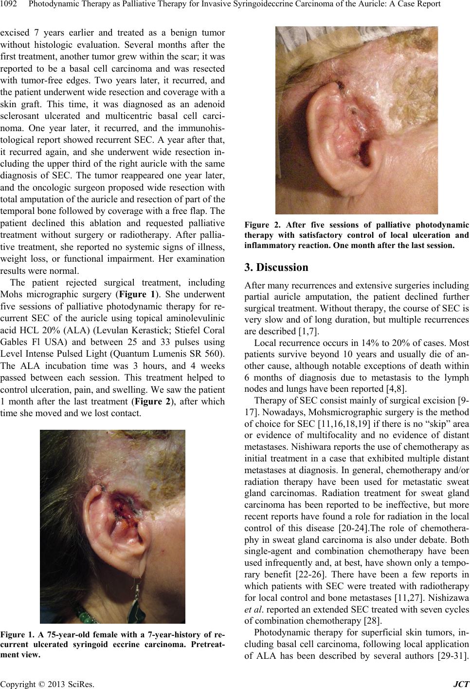 Photodynamic Therapy as Palliative Therapy for Invasive Syringoideccrine Carcinoma of the Auricle: A Case Report 1092 excised 7 years earlier and treated as a benign tumor without histologic evaluation. Several months after the first treatment, another tumor grew within the scar; it was reported to be a basal cell carcinoma and was resected with tumor-free edges. Two years later, it recurred, and the patient underwent wide resection and coverage with a skin graft. This time, it was diagnosed as an adenoid sclerosant ulcerated and multicentric basal cell carci- noma. One year later, it recurred, and the immunohis- tological report showed recurrent SEC. A year after that, it recurred again, and she underwent wide resection in- cluding the upper third of the right auricle with the same diagnosis of SEC. The tumor reappeared one year later, and the oncologic surgeon proposed wide resection with total amputation of the auricle and resection of part of the temporal bone followed by coverage with a free flap. The patient declined this ablation and requested palliative treatment without surgery or radiotherapy. After pallia- tive treatment, she reported no systemic signs of illness, weight loss, or functional impairment. Her examination results were normal. The patient rejected surgical treatment, including Mohs micrographic surgery (Figure 1). She underwent five sessions of palliative photodynamic therapy for re- current SEC of the auricle using topical aminolevulinic acid HCL 20% (ALA) (Levulan Kerastick; Stiefel Coral Gables Fl USA) and between 25 and 33 pulses using Level Intense Pulsed Light (Quantum Lumenis SR 560). The ALA incubation time was 3 hours, and 4 weeks passed between each session. This treatment helped to control ulceration, pain, and swelling. We saw the patient 1 month after the last treatment (Figure 2), after which time she moved and we lost contact. Figure 1. A 75-year-old female with a 7-year-history of re- current ulcerated syringoid eccrine carcinoma. Pretreat- ment view. Figure 2. After five sessions of palliative photodynamic therapy with satisfactory control of local ulceration and inflammatory reaction. One month after the last session. 3. Discussion After many recurrences and extensive surgeries including partial auricle amputation, the patient declined further surgical treatment. Without therapy, the course of SEC is very slow and of long duration, but multiple recurrences are described [1,7]. Local recurrence occurs in 14% to 20% of cases. Most patients survive beyond 10 years and usually die of an- other cause, although notable exceptions of death within 6 months of diagnosis due to metastasis to the lymph nodes and lungs have been reported [4,8]. Therapy of SEC consist mainly of surgical excision [9- 17]. Nowadays, Mohsmicrographic surgery is the method of choice for SEC [11,16,18,19] if there is no “skip” area or evidence of multifocality and no evidence of distant metastases. Nishiwara reports the use of chemotherapy as initial treatment in a case that exhibited multiple distant metastases at diagnosis. In general, chemotherapy and/or radiation therapy have been used for metastatic sweat gland carcinomas. Radiation treatment for sweat gland carcinoma has been reported to be ineffective, but more recent reports have found a role for radiation in the local control of this disease [20-24].The role of chemothera- phy in sweat gland carcinoma is also under debate. Both single-agent and combination chemotherapy have been used infrequently and, at best, have shown only a tempo- rary benefit [22-26]. There have been a few reports in which patients with SEC were treated with radiotherapy for local control and bone metastases [11,27]. Nishizawa et al. reported an extended SEC treated with seven cycles of combination chemotherapy [28]. Photodynamic therapy for superficial skin tumors, in- cluding basal cell carcinoma, following local application of ALA has been described by several authors [29-31]. Copyright © 2013 SciRes. JCT 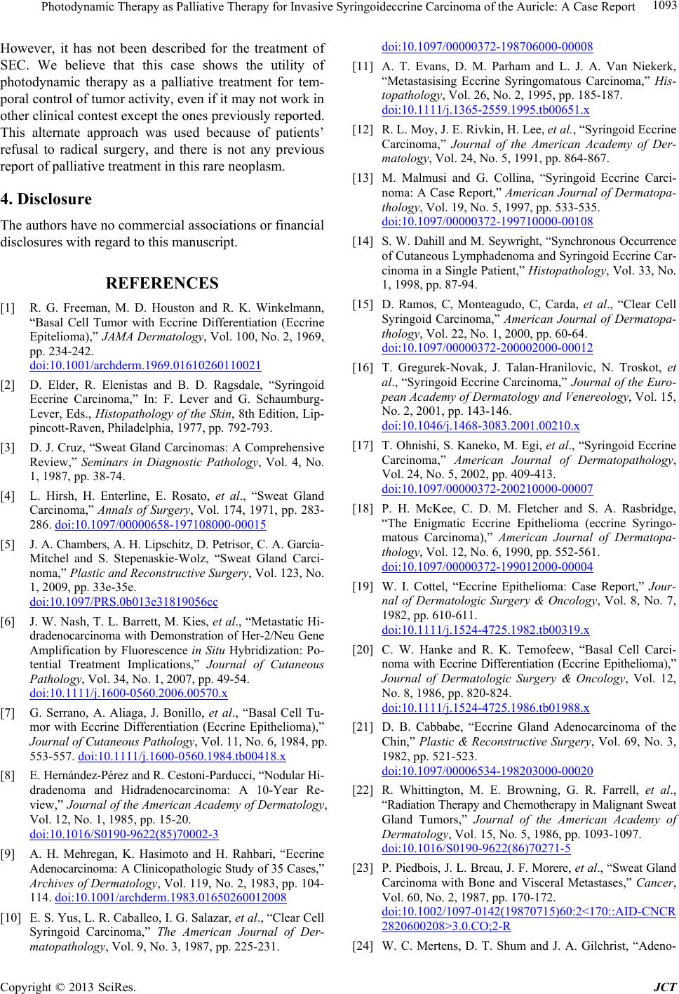 Photodynamic Therapy as Palliative Therapy for Invasive Syringoideccrine Carcinoma of the Auricle: A Case Report 1093 However, it has not been described for the treatment of SEC. We believe that this case shows the utility of photodynamic therapy as a palliative treatment for tem- poral control of tumor activity, even if it may not work in other clinical contest except the ones previously reported. This alternate approach was used because of patients’ refusal to radical surgery, and there is not any previous report of palliative treatment in this rare neoplasm. 4. Disclosure The authors have no commercial associations or financial disclosures with regard to this manuscript. REFERENCES [1] R. G. Freeman, M. D. Houston and R. K. Winkelmann, “Basal Cell Tumor with Eccrine Differentiation (Eccrine Epitelioma),” JAMA Dermatology, Vol. 100, No. 2, 1969, pp. 234-242. doi:10.1001/archderm.1969.01610260110021 [2] D. Elder, R. Elenistas and B. D. Ragsdale, “Syringoid Eccrine Carcinoma,” In: F. Lever and G. Schaumburg- Lever, Eds., Histopathology of the Skin, 8th Edition, Lip- pincott-Raven, Philadelphia, 1977, pp. 792-793. [3] D. J. Cruz, “Sweat Gland Carcinomas: A Comprehensive Review,” Seminars in Diagnostic Pathology, Vol. 4, No. 1, 1987, pp. 38-74. [4] L. Hirsh, H. Enterline, E. Rosato, et al., “Sweat Gland Carcinoma,” Annals of Surgery, Vol. 174, 1971, pp. 283- 286. doi:10.1097/00000658-197108000-00015 [5] J. A. Chambers, A. H. Lipschitz, D. Petrisor, C. A. García- Mitchel and S. Stepenaskie-Wolz, “Sweat Gland Carci- noma,” Plastic and Reconstructive Surgery, Vol. 123, No. 1, 2009, pp. 33e-35e. doi:10.1097/PRS.0b013e31819056cc [6] J. W. Nash, T. L. Barret t, M. Kies, et al., “Metastatic Hi- dradenocarcinoma with De monstration of Her-2/Neu Gene Amplification by Fluorescence in Situ Hybridization: Po- tential Treatment Implications,” Journal of Cutaneous Pathology, Vol. 34, No. 1, 2007, pp. 49-54. doi:10.1111/j.1600-0560.2006.00570.x [7] G. Serrano, A. Aliaga, J. Bonillo, et al., “Basal Cell Tu- mor with Eccrine Differentiation (Eccrine Epithelioma),” Journal of Cutaneous Pathology, Vol. 11, No. 6, 1984, pp. 553-557. doi:10.1111/j.1600-0560.1984.tb00418.x [8] E. Hernández-Pérez and R. Cestoni-Parducci, “Nodular Hi- dradenoma and Hidradenocarcinoma: A 10-Year Re- view,” Journal of the American Academy of Dermatology, Vol. 12, No. 1, 1985, pp. 15-20. doi:10.1016/S0190-9622(85)70002-3 [9] A. H. Mehregan, K. Hasimoto and H. Rahbari, “Eccrine Adenocarcin oma: A Clinicopathologic Study of 35 Cases,” Archives of Dermatology, Vol. 119, No. 2, 1983, pp. 104- 114. doi:10.1001/archderm.1983.01650260012008 [10] E. S. Yus, L. R. Caballeo, I. G. Salazar, et al., “Clear Cell Syringoid Carcinoma,” The American Journal of Der- matopathology, Vol. 9, No. 3, 1987, pp. 225-231. doi:10.1097/00000372-198706000-00008 [11] A. T. Evans, D. M. Parham and L. J. A. Van Niekerk, “Metastasising Eccrine Syringomatous Carcinoma,” His- topathology, Vol. 26, No. 2, 1995, pp. 185-187. doi:10.1111/j.1365-2559.1995.tb00651.x [12] R. L. Moy, J. E. Rivkin, H. Lee, et al., “Syringoid Eccrine Carcinoma,” Journal of the American Academy of Der- matology, Vol. 24, No. 5, 1991, pp. 864-867. [13] M. Malmusi and G. Collina, “Syringoid Eccrine Carci- noma: A Case Report,” American Journal of Dermatopa- thology, Vol. 19, No. 5, 1997, pp. 533-535. doi:10.1097/00000372-199710000-00108 [14] S. W. Dahill and M. Seywright, “Synchronous Occurrence of Cutaneous Lymphadenoma and Syringoid Eccrine Car- cinoma in a Single Patient,” Histopathology, Vol. 33, No. 1, 1998, pp. 87-94. [15] D. Ramos, C, Monteagudo, C, Carda, et al., “Clear Cell Syringoid Carcinoma,” American Journal of Dermatopa- thology, Vol. 22, No. 1, 2000, pp. 60-64. doi:10.1097/00000372-200002000-00012 [16] T. Gregurek-Novak, J. Talan-Hranilovic, N. Troskot, et al., “Syringoid Eccrine Carcinoma,” Journal of the Euro- pean Academy of Dermatology and Venereology, Vol. 15, No. 2, 2001, pp. 143-146. doi:10.1046/j.1468-3083.2001.00210.x [17] T. Ohnishi, S. Kaneko, M. Egi, et al., “Syringoid Eccrine Carcinoma,” American Journal of Dermatopathology, Vol. 24, No. 5, 2002, pp. 409-413. doi:10.1097/00000372-200210000-00007 [18] P. H. McKee, C. D. M. Fletcher and S. A. Rasbridge, “The Enigmatic Eccrine Epithelioma (eccrine Syringo- matous Carcinoma),” American Journal of Dermatopa- thology, Vol. 12, No. 6, 1990, pp. 552-561. doi:10.1097/00000372-199012000-00004 [19] W. I. Cottel, “Eccrine Epithelioma: Case Report,” Jour- nal of Dermatologic Surgery & Oncology, Vol. 8, No. 7, 1982, pp. 610-611. doi:10.1111/j.1524-4725.1982.tb00319.x [20] C. W. Hanke and R. K. Temofeew, “Basal Cell Carci- noma with Eccrine Differentiation (Eccrine Epithelioma),” Journal of Dermatologic Surgery & Oncology, Vol. 12, No. 8, 1986, pp. 820-824. doi:10.1111/j.1524-4725.1986.tb01988.x [21] D. B. Cabbabe, “Eccrine Gland Adenocarcinoma of the Chin,” Plastic & Reconstructive Surgery, Vol. 69, No. 3, 1982, pp. 521-523. doi:10.1097/00006534-198203000-00020 [22] R. Whittington, M. E. Browning, G. R. Farrell, et al., “Radiation Therapy and Chemoth erapy in Malignant Sweat Gland Tumors,” Journal of the American Academy of Dermatology, Vol. 15, No. 5, 1986, pp. 1093-1097. doi:10.1016/S0190-9622(86)70271-5 [23] P. Piedbois, J. L. Breau, J. F. More re, et al., “Sweat Gland Carcinoma with Bone and Visceral Metastases,” Cancer, Vol. 60, No. 2, 1987, pp. 170-172. doi:10.1002/1097-0142(19870715)60:2<170::AID-CNCR 2820600208>3.0.CO;2-R [24] W. C. Mertens, D. T. Shum and J. A. Gilchrist, “Adeno- Copyright © 2013 SciRes. JCT  Photodynamic Therapy as Palliative Therapy for Invasive Syringoideccrine Carcinoma of the Auricle: A Case Report Copyright © 2013 SciRes. JCT 1094 carcinoma of the Eccrine Sweat Gland: Response to both Combination Chemotherapy and Local Field Irradiation,” European Journal of Cancer, Vol. 32A, No. 2, 1996, pp. 372-373. doi:10.1016/0959-8049(95)00525-0 [25] N. Okada, J. Ota, K. Sato, et al., “Metastasizing Eccrine Sweat Gland Carcinoma, Report of a Case,” Archives of Dermatology, Vol. 120, No. 6, 1984, pp. 768-769. doi:10.1001/archderm.1984.01650420078021 [26] C. J. Conley, P. Schau, D. P. Kelsen, et al., “Chemother- apy of Metastatic Sweat Gland Carcinoma. A Retrospec- tive Review,” American Journal of Dermatopathology, Vol. 8, No. 4, 1985, pp. 307-311. doi:10.1097/00000421-198508000-00006 [27] E. Alssi and R. Caputo, “Syringomatous Carcinoma of the Scalp Presenting as a Slowly Enlarging Patch of Alo- pecia,” American Journal of Dermatopathology, Vol. 15, 1993, pp. 503-505. doi:10.1097/00000372-199310000-00018 [28] A. Nishizawa, Y. Nakanishi, Y. Sasajima, et al., “Syrin- goid Eccrine Carcinoma with Apparently Aggressive Transformation: Case Report and Review of the Litera- ture,” International Journal of Dermatology, Vol. 45, No. 10, 2006, pp. 1218-1221. doi:10.1111/j.1365-4632.2006.02664.x [29] R. I. Ceilley and J. Q. Del Rosso, “Current Modalities and New Advances in the Treatment of Basal Cell Carci- noma,” International Journal of Dermatology, Vol. 45, No. 5, 2006, pp. 489-498. doi:10.1111/j.1365-4632.2006.02673.x [30] H. Benzel, et al., “Photodynamic Therapy of Superficial Skin Tumors Following Local Application of Delta-Ami- nolaevulin ic Ac id,” Advances in Otolaryngology, Vol. 49, 1995, pp. 48-52. [31] K. Kalka, H. Merk and H. Mukhtar, “Photodynamic Ther- apy in Dermatology,” Journal of the American Academy of Dermatology, Vol. 42, No. 3, 2000, pp. 389-413. doi:10.1016/S0190-9622(00)90209-3 |