

Paper Menu >>
Journal Menu >>
 Open Journal of Urology, 2013, 3, 179-184 http://dx.doi.org/10.4236/oju.2013.34033 Published Online August 2013 (http://www.scirp.org/journal/oju) The Long Term Follow-Up Results of the Direct Nipple Ureteroneocystostomy Technique: A Prospective Study* Abdullah Demirtas#, Nurettin Sahin, Emre Can Akinsal, Mehmet Ali Ergul, Mehmet Caniklioglu, Oguz Ekmekcioglu, Atila Tatlisen Department of Urology, Erciyes University Medical Faculty, Kayseri, Turkey Email: #[email protected] Received May 26, 2013; revised June 27, 2013; accepted July 5, 2013 Copyright © 2013 Abdullah Demirtas et al. This is an open access article distributed under the Creative Commons Attribution Li- cense, which permits unrestricted use, distribution, and reproduction in any medium, provided the original work is properly cited. ABSTRACT Objective: To evaluate the long term follow-up results of the direct nipple u reteroneocystostomy technique. Materials and Methods: We studied a total of 16 patients (19 renal units) who underwent direct nipple ureteroneocystostomy. The mean age was 43 years and 3 patients had bilateral disease. In five units the ureters had been ligated during gynecological surgery, 11 renal units were obstructive and three units were reflexive megaureters. The ureters were spatulated for about 2 cm and folded back. Nipples 2 to 2.5 cm long were prepared. In two cases the ureters were thin-walled (2 mm or less) and they were not spatulated but folded back onto themselves. In one case the ureter could not be everted since it had a thick and fibrotic wall. The distal 2 to 2.5 cm segment of this ureter was directly inserted in to the bladder. Postoperative follow-up was at 3 month intervals for the first year at 6 month intervals for 2 - 3 years and yearly thereafter. At the time of follow-up serum creatinine, urine culture, ultrasound, intravenous urography, voiding cystoureterography, nuclear renal scintigraphy and cystometric evaluations were performed. The functions of 11 and 15 renal units were evaluated scintigraphically and stereo logically, respectively, in the both preoperative and postoperative first year follow-up. The Wilcoxon Signed Ranks test was used for statistical evaluation and p < 0.05 was considered statistically significant. Results: Mean follow-up was 49 months. Three renal units had Grade III reflux (two of them during voiding) and one unit had Grade IV reflux. At follow-up this patient developed in the ureteral stricture. No patients had urinary tract infection, pyelonephritis or ureteral stricture follow-up period. Between the preoperative and postoperative first year, there was an increase in postoperative split renal function based on renal scintigraphy but this difference was not statistically significant. The stereologically calculated decrease in pelvicaliceal dilatation was statistically significant. Conclusion: Ease of application and no need to taper or plicate the ureter or prepare a sub- mucosal tunnel may be the reasons to consider the direct nipple ureteroneocystostomy technique for megaureters of different etiologies. Keywords: Megaureter; Nipple; Technique; Ureter; Vesicoureteral Reflux 1. Introduction Primary obstructed megaureter (POM) is a result of obs- truction or an adynamic ureteral segment in the uret- erovesical junction. Megaureter can be classified as 1) re- flux megaureter obstructed megaureter; 2) nonreflux and nonobstructed megaureter and 3) megaureter with ob- struction and reflux [1]. POM is occurs 3/12 to 5 times more often in males [2]. Although it is generally unilateral, it may be seen bilater- ally in 15% to 25% of cases. Besides excretory urogra- phy (IVU), diuretic renography and antegrade pyelogra- phy are valuable tools for its diagnosis. As treatment, the adynamic ureteral segment is excised and ureteroneo- cystostomy is performed with different surgical tech- niques. However, the r esults may be far from ideal [3,4]. Ureteric injury is one of the most serious complica- tions of gyenocologic operations in the pelvis [5]. Uret- eric damage has significant morbidity and can present as ureteric stenosis or obstruction and cause variable de- grees of impaired renal function in some cases [6]. When the diagnosis of injury is made postoperatively then ei- ther open surgery or endourologic procedures can be employed. Delayed repair has been suggested based on *There is no financial disclosure of any of the authors. #Corresponding author. C opyright © 2013 SciRes. OJU  A. DEMIRTAS ET AL. 180 the view that this will reduce inflammation and tissue edema [7]. The treatment of vesicoureteral reflux (VUR) is indivi- dualized. Before recommending surgery, consideration is given to the severity of the reflux, possible underlying risk factors including bladder dysfunction, age at prese closentation, and duration of the disorder. A variety of techniques have been described for the correction of VUR. These are anatomically categorized as extravesical, intravesical, or combined, depending on the approach to the ureter [2]. In this study we evaluated the long term follow-up results of the direct nipple ureteroneocysto- stomy technique. 2. Materials and Methods A total of 16 patients (19 renal units), seven males and nine females, with a median age of 43 years (21 - 73) who underwent nipple ureteroneocystostomy operation were enrolled in this study. Three cases underwent bilat- eral repair. Eight of the 19 renal units were on the right side and 11 were on the left side. Three of the repaired renal units were reflux megaureter, 11 were obstructed megaureter, and 5 were units at the lower end of the ureter which were ligated at previous gynecologic opera- tions. Neurogenic bladder cases and cases with surgical wound infection were excluded. Preoperative evaluation consisted of urine culture, serum creatinine, urinary sys- tem ultrasonography, renal Technetium-99 m-Dimercap- tosuccinic Acid (DMSA) scintigraphy, cystoscopy, voi- ding cystourethrography (VCU), intravenous urography (except for two cases), antegrade pyelograpy and Whi- taker test (Life-Tech, Stafford, Texas) in 4 cases with percutaneous nephrostomy, and cystometric assessment (Life-Tech, Stafford, Texas) in 5 cases with vo iding dys- function. Cases with proliferation in urine culture were not operated on until after their urine became sterile and other cases were operated on after a course of antibio- therapy with intravenous 3rd generation cephalosporins. In the Whitaker test, a pressure gradient above 22 cm H2O between the renal pelvis and bladder during ad- ministration of 10 ml isotonic saline is considered diag- nostic for obstru ction while 15 - 22 cm H2O suggests ob- struction, and below 15 cmH2O indicates no obstruction [8]. All cases were operated on under general anesthesia. A direct nipple ureteroneocystostomy (DNU) operation was carried out with a lower middle incision in cases where bilateral repair was undertaken and with a Gibson incision for the rest of the cases [9]. A follow-up was planned at every three months in the first year, at every 6 months between the 1st and 3rd years and yearly thereafter. Follow-up tests included urine culture, serum creatinine, ultrasonography (USG), VCU, IVU (if creatinine was normal), DMSA scintigra- phy, endoscopy (at postoperative 3rd month), and uro- dynamic examination in cases with dysfunctional mic- turition. A sample of the lower ureteral end of 9 renal units was evaluated pathologically. In cases attending regular follow-ups, preoperative and postoperative 1st year DMSA results and amount of pelvicaliceal dilatation in the IVU of the unit that underwent surg ery were com- pared statistically (by counting the dots falling on the pelvis and calices using stereologic methods which use transparent acetates with 3 mm inter-grid distances [10- 12]. Inter-IVU image differences between IVUs taken at different centers were resolved by using the value found by counting the dots falling on the 4th lumbar vertebra in both films and proportioning the results with each other as the multiplier. Measurable values were expressed as means + standard deviation. Preoperative and postopera- tive values were compared using the Wilcoxon signed rank test. A p value less than 0.05 was accepted as statis- tically significant. 3. Results The results of a median 49 months follow-up of the 19 renal units that underwent DNU operation were assessed. One reflexive and two obs tructive cases underwent bilat- eral repair. While two cases with an excessively dilated and thin-walled (2 mm or less) ureter were anastomosed by folding back without spatulation, one case with a ureter with an excessively thick and fibrotic wall was directly anastomosed without being everted and spatu- lated, with a 2 to 2.5 cm part being free in the bladder. In one obstructive case there was a 6 cm stone at the lower end of the ureter. As the ureter of this case undergoing ureterolithotomy and DNU operations was so thick and fibrotic, excessive ureter dissection was avoided to pre- serve circulation and the bladder was attached to the tendon of the ipsilateral psoas minor muscle by using the Psoas hitch method. Urine cultur es of the cases were free of proliferation. All cases underwent preoperative VCU and no reflux was observed except for 3 units. Two cases did not undergo IVU (one of these cases had a serum creatinine level of 1.9 mg/dl and the other was at the early postpartum period). The Whitaker test was applied to 4 cases with percutaneous nephrostomy and the test results of these 4 cases were consistent with obstruction. The test was repeated on the 14th day after the operation and the test results were negative. These cases also un- derwent postoperative antegrade pyelography which re- vealed that the contrast material readily passed to the bladder and no obstruction existed, and their nephrosto- mies were removed. Drains were removed on the 3rd - 5th days. Cases underwent VCU on the postoperative 14t h day, which revealed extravasation in no patients. In most of the cases the filling defect caused by to the nipp le was evident in the bladder in the VCU taken postoperatively Copyright © 2013 SciRes. OJU 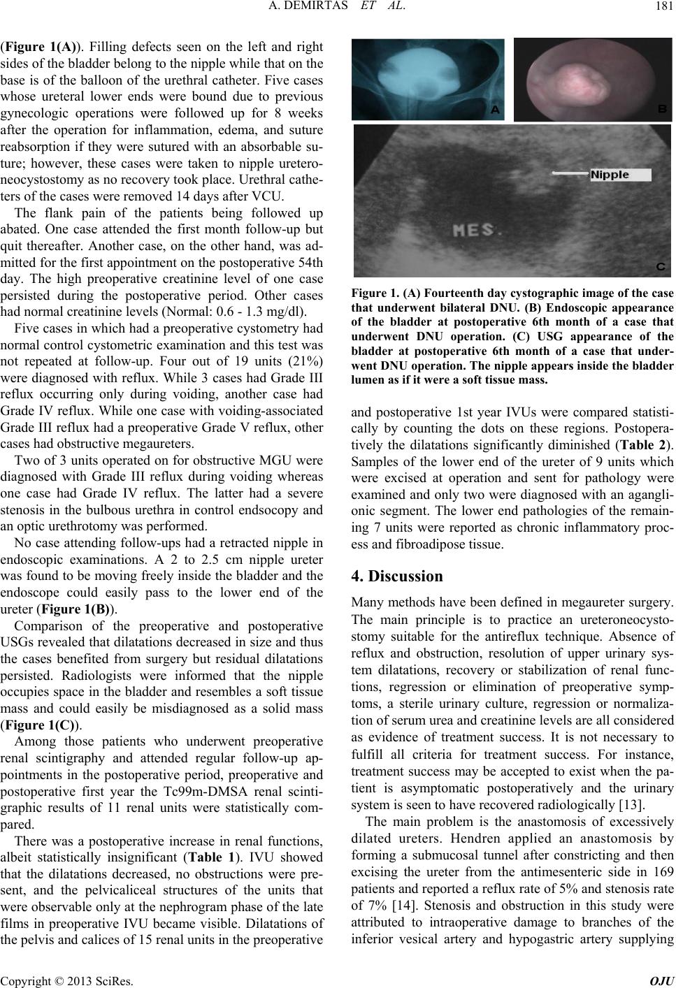 A. DEMIRTAS ET AL. 181 (Figure 1(A)). Filling defects seen on the left and right sides of the bladder belong to the nipple while that on the base is of the balloon of the urethral catheter. Five cases whose ureteral lower ends were bound due to previous gynecologic operations were followed up for 8 weeks after the operation for inflammation, edema, and suture reabsorption if they were sutured with an absorbable su- ture; however, these cases were taken to nipple uretero- neocystostomy as no recovery took place. Urethral cathe- ters of the cases were removed 14 days after VCU. The flank pain of the patients being followed up abated. One case attended the first month follow-up but quit thereafter. Another case, on the other hand, was ad- mitted for the first appointment on the postoperative 54th day. The high preoperative creatinine level of one case persisted during the postoperative period. Other cases had normal creatinine levels (Normal: 0.6 - 1.3 mg/dl). Five cases in which had a preoperative cystometry had normal control cystometric examination and this test was not repeated at follow-up. Four out of 19 units (21%) were diagno sed with reflux. Wh ile 3 cases had Grade III reflux occurring only during voiding, another case had Grade IV reflux. While one case with voiding-associated Grade III reflux had a preoperative Grade V reflux, other cases had obstructive megaureters. Two of 3 units operated on for obstructive MGU were diagnosed with Grade III reflux during voiding whereas one case had Grade IV reflux. The latter had a severe stenosis in the bulbous urethra in control endsocopy and an optic urethrotomy was perf o r med. No case attending follow-ups had a retracted nipple in endoscopic examinations. A 2 to 2.5 cm nipple ureter was found to be moving freely inside the bladder and the endoscope could easily pass to the lower end of the ureter (Figure 1(B)). Comparison of the preoperative and postoperative USGs revealed that dilatations decreased in size and thus the cases benefited from surgery but residual dilatations persisted. Radiologists were informed that the nipple occupies space in the bladder and resembles a soft tissue mass and could easily be misdiagnosed as a solid mass (Figure 1(C)). Among those patients who underwent preoperative renal scintigraphy and attended regular follow-up ap- pointments in the postoperative period, preoperative and postoperative first year the Tc99m-DMSA renal scinti- graphic results of 11 renal units were statistically com- pared. There was a postoperative increase in renal functions, albeit statistically insignificant (Table 1). IVU showed that the dilatations decreased, no obstructions were pre- sent, and the pelvicaliceal structures of the units that were observable only at the nephrogram phase of the late films in preoperative IVU became visible. Dilatations of the pelvis and calices of 15 renal units in the preoperative Figure 1. (A) Fourteenth day cystographic image of the case that underwent bilateral DNU. (B) Endoscopic appearance of the bladder at postoperative 6th month of a case that underwent DNU operation. (C) USG appearance of the bladder at postoperative 6th month of a case that under- went DNU operation. The nipple appears inside the bladder lumen as if it were a soft tissue mass. and postoperative 1st year IVUs were compared statisti- cally by counting the dots on these regions. Postopera- tively the dilatations significantly diminished (Table 2). Samples of the lower end of the ureter of 9 units which were excised at operation and sent for pathology were examined and only two were diagnosed with an agangli- onic segment. The lower end pathologies of the remain- ing 7 units were reported as chronic inflammatory proc- ess and fibroadipose tissue. 4. Discussion Many methods have been defined in megaureter surgery. The main principle is to practice an ureteroneocysto- stomy suitable for the antireflux technique. Absence of reflux and obstruction, resolution of upper urinary sys- tem dilatations, recovery or stabilization of renal func- tions, regression or elimination of preoperative symp- toms, a sterile urinary culture, regression or normaliza- tion of serum urea and creatinine levels are all considered as evidence of treatment success. It is not necessary to fulfill all criteria for treatment success. For instance, treatment success may be accepted to exist when the pa- tient is asymptomatic postoperatively and the urinary system is seen to have recovered radiologically [13]. The main problem is the anastomosis of excessively dilated ureters. Hendren applied an anastomosis by forming a submucosal tunnel after constricting and then excising the ureter from the antimesenteric side in 169 patients and reported a reflux rate of 5% and stenosis rate of 7% [14]. Stenosis and obstruction in this study were attributed to intraoperative damage to branches of the inferior vesical artery and hypogastric artery supplying Copyright © 2013 SciRes. OJU 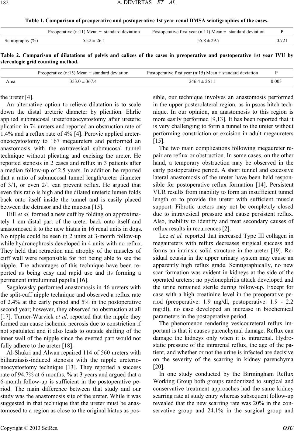 A. DEMIRTAS ET AL. Copyright © 2013 SciRes. OJU 182 Table 1. Comparison of preoperative and postoperative 1st year renal DMSA scintigraphies of the cases. Preoperative (n:11) Mean + standard deviation Postoperative first year (n:11) Mean ± standard deviation P Scintigraphy (%) 55.2 ± 26.1 55.8 ± 29.7 0.721 Table 2. Comparison of dilatations of pelvis and calices of the cases in preoperative and postoperative 1st year IVU by stereologic grid counting method. Preoperative (n:15) Mean ± standard dev i ation Postoperative first year (n:15) Mean ± standard deviation P Area 353.0 ± 367.4 246.4 ± 261.1 0.003 the ureter [4]. An alternative option to relieve dilatation is to scale down the distal ureteric diameter by plication. Ehrlic applied submucosal ureteroneocystostomy after ureteric plication in 74 ureters and repor ted an obstruction rate of 1.4% and a reflux rate of 4% [4]. Perovic applied ureter- oneocystostomy to 167 megaureters and performed an anastomosis with the extravesical submucosal tunnel technique without plicating and excising the ureter. He reported stenosis in 2 cases and reflux in 3 patients after a median follow-up of 2.5 years. In addition he reported that a ratio of submucosal tunnel length/ureter diameter of 3/1, or even 2/1 can prevent reflux. He argued that even this ratio is high and the dilated ureteric lu men fold s back onto itself inside the tunnel and is easily placed between the detrusor and the mucosa [15]. Hill et al. formed a new cuff by folding on approxima- tely 1 cm distal part of the ureter back onto itself and anastomosed it to the new hiatus in 16 renal un its in dogs. No nipple could be seen in 2 units at 3-month follow-up while hydronephrosis developed in 4 units with no reflux. They held that retraction and atrophy of the muscles of cuff wall were responsible for not being able to see the nipple. The advantages of this technique have been re- ported as being easy and rapid use and its forming a permanent intraluminal papilla [16]. Sagalowsky performed anastomosis in 46 ureters with the split-cuff nipple technique and observed a reflux rate of 2.4% at the early period and 5% in the postoperative second year; however, they observed no obstruction at all [17]. Turner-Warvick et al. reported that the nipple they formed can cause ischemic necrosis due to co nstriction if not spatulated and it also leads to outside shifting of the inner wall of the nipple since the everted part would not fully adhere to the ureter [18]. Al-Shukri and Alwan repaired 1 14 of 560 ureters with bilharziasis-induced stenosis with the nipple ureterıo- neocystostomy technique [13]. They reported a success rate of 94.7% at 6 months, % at 3 years and argued that a 6-month follow-up is sufficient in the postoperative pe- riod. The main difference between that study and our study was the anastomosis site of the ureter. While it was suggested in that technique that the ureter must be anas- tomosed to a region as close to the original hiatus as pos- sible, our technique involves an anastomosis performed in the upper posterolateral region, as in psoas hitch tech- nique. In our opinion, an anastomosis to this region is more easily performed [9,13]. It has been reported that it is very challenging to form a tunnel to the ureter without performing constriction or excision in adult megaureters [15]. The two main complications following megaureter re- pair are reflux or obstruction. In some cases, on the other hand, a temporary obstruction may be observed in the early postoperative period. A short tunnel and excessive lateral anastomosis of the ureter have been held respon- sible for postoperative reflux formation [14]. Persistent VUR results from inability to form an insufficient tunnel length or to provide the ureter with sufficient muscle support. Fibrotic ureters may not be completely closed due to intravesical pressure and cause persistent reflux. Also, inability to identify and treat secondary causes of reflux results in recurrences [2]. Lee et al. reported that increased Type III collagen in megaureters with reflux decreases surgical success and forms an intrinsic solid structure in the ureter [19]. Re- sidual ectasia in the upper urinary system may cause an apparently high reflux grade. Scintigraphically, no new scar formation was evident in kidneys at the side of the operated ureters; no pyelonephritis attack developed and the urine remained sterile during follow-up. Except for case with a high creatinine level in the preoperative pe- riod (preoperative: 1.9 mg/dl, postoperative: 1.9 - 2.2 mg/dl), no case developed an increase in biochemical parameters in the postoperative period. The phenomenon rendering vesicoureteral reflux im- portant is that it causes parenchymal damage. Reflux can damage the kidneys only when it is intrarenal. Hydro- static pressure of the intrarenal reflux, the age of the pa- tient, and whether or not the urine is infected are decisive on the severity of the scarring in kidney parenchyma [20]. In one study conducted by the Birmingham Reflux Working Group both groups randomized to surgical and conservative treatment approaches had the same kidney scarring rate at study entry whereas subsequent follow-up revealed that the new scarring rate was 20% in the con- servative group and 24.1% in the surgical group and  A. DEMIRTAS ET AL. 183 while the progressive scarring rate was 10.5 % in the sur- gical group and 11.6% in the conservative group. No significant difference was noted between the two groups in terms of infection rates [21]. In the “International Re- flux Study Group” study in which centers from America and Europe collaborated 60% of cases had scarring at the beginning, with subsequent new scarring rates of 14.6% and 14.5% in surgical and conservative groups, respec- tively [22]. It has been reported, and many authors agree, that modest refluxes following megaureter surgery may re- cover by themselves in a period of 2 - 3 months, and thus require no therapy [23]. As further surgery in patients with residual reflux carries the risk of ischemia and is technically demanding, endoscopic substance infusion is recommended. A success rate of 80% has been reported following a single endoscopic in jection to 20 u reters with ongoing reflux (Grade III or above in 13 ureters) follow- ing ureteroneocystostomy [24]. Obstruction, the other common co mplication, is the re- sult of fibrosis due to deranged ureter blood supply par- ticularly following excisional constriction or plication. Entry of the ureteric hiatus into the bladder from an ex- cessive cranial and lateral aspect also causes obstruction. In such a situation a second operation is needed. Tempo- rary obstruction seen postoperatively, on the other hand, is the result of edema at the lower end of the ureter and nephrostomy usually suffices. Although our sample size was small, we did not observe obstruction or stricture development, suggesting that a minimal trauma to the ureter ensues. No infection or worsening of renal func- tion due to residual urine in the pelvicaliceal system was observed after surgery. King reported that there is no need for constriction to repair ureter peristaltism [1]. In Whitaker test, a pressure gradient above 22 cm H2O be- tween the renal pelvis an d the bladder during administra- tion of 10 ml isotonic saline is considered diagnostic for obstruction while 15 - 22 cm H2O suggests obstruction, and below 15 cm H2O indicates no obstruction [8]. In one study it has been suggested that administration of isotonic saline at a rate of 10 ml/min may not suffice to show obstruction in dilated urinary systems and a rate of 12 - 20 ml/min may be more appropriate [25]. The nipple appears as a smooth-bordered filling defect in the bladder in postoperative cystograms and IVUs while it is observed as a soft tissue mass in the bladder in USG. Endoscopically, the nipple prolapsing into the lu- men is easily seen on the lateral superior wall of the bladder and it is easy to advance through the ureter with a cystoscope in the postoperative period. It has been cli- nically and cystometrically shown that the nipple inside the bladder does not produce symptoms of bladder irrita- tion. None of the patients had irritative symptoms in the preoperative period. 5. Conclusion As the technique is an easy-to-perform one, the learning curve is substantially short. Unlike the previously men- tioned techniques, this method does not need a submu- cosal tunnel, ureteric plication, or tapering, thus shorten- ing surgical time and anesthetic complications, and lim- iting the frequency of major surgical complications such as ureteral stenosis. Lack of the need for a ureteral stent protects the patient from stent-induced irritation and an additional endosco pic pro cedu re. A read ily seen nipple in the bladder and easily performed endoscopy in the post- operative period facilitates intervention. Its potential to be performed in obstructive and reflexive megaureter cases including ureter dilatations caused by a variety of etiologies such as iatrog enic ureteric lower end dilatation may be cause to prefer the direct nipple ureteroneo- cystostomy technique. REFERENCES [1] L. R. King, “Megaloureter: Definition, Diagnosis and Management,” The Journal of Urology, Vol. 123, No. 2, 1980, pp. 222-223. [2] A. Atala, “Vesicoureteral Reflux and Megaureter,” In: P. C. Walsh, Ed ., Campbell’s Urology, W. B. Saunders Co., Philadelphia, 2002, pp. 2059-2116. [3] H. H. Bakker, R. J. Scholtmeijer and P. J. Klopper, “Co m- parison of 2 Different Tapering Techniques in Megau- reters,” The Journal of Urology, Vol. 140, No. 5, 1988, pp. 1237-1239. [4] R. M. Ehrlich, “The Ureteral Folding Technique for Me- gaureter Surgery,” The Journal of Urology, Vol. 134, No. 4, 1985, pp. 668-670. [5] R. F. Mattingly, “Operative Injuries of the Ureter,” In: R. W. Te Linde, Ed., Te Linde’s Operative Gynecology, JB Lippincott, Philedelphia, 1995, p. 325. [6] A. Liapis, P. Bakas, V. Giannopoulos, et al., “Ureteral Injuries during Gynecological Surgery,” International Urogynecology Journal and Pelvic Floor Dysfunction, Vol. 12, No. 6, 2001, pp. 391-393. doi:10.1007/PL00004045 [7] M. Rafique and M. H. Arif, “Management of Iatrogenic Ureteric Injuries Associated with Gynecological Sur- gery,” International Urology and Nephrology, Vol. 34, No. 1, 2002, pp. 31-35. doi:10.1023/A:1021320409583 [8] R. H. Whitaker, “The Whitaker Test ,” The Urologic Clin- ics of North America, Vol. 6, No. 3, 1979, pp. 529-539. [9] A. Tatlisen and O. Ekmekcioglu, “Direct Nipple Ureter- oneocystostomy in Adults with Primary Obstructed Me- gaureter,” The Journal of Urology, Vol. 173, No. 3, 2005, pp. 877-880. doi:10.1097/01.ju.0000152533.93716.3c [10] N. Roberts, M. J. Puddephat and V. McNulty, “The Benefit of Stereology for Quantitative Radiology,” The British Journal of Radiology, Vol. 73, No. 871, 2000, pp. 679-697. [11] J. R. Nyengaard, “Stereologic Methods and Their Appli- Copyright © 2013 SciRes. OJU  A. DEMIRTAS ET AL. Copyright © 2013 SciRes. OJU 184 cation in Kidney Research,” Journal of the American So- ciety of Nephrology, Vol. 10, No, 5, 1999, pp. 1100-1123. [12] K. T. Bae, P. K. Commean and J. Lee, “Volumetric Measurement of Renal Cysts and Parenchyma Using MRI: Phantoms and Patients with Polycystic Kidney Disease,” Journal of Computer Assisted Tomography, Vol. 24, No. 4, 2000, pp. 614-619. doi:10.1097/00004728-200007000-00019 [13] S. Al-Shukri and M. H. Alwan, “Bilharzial Strictures of the Lower Third of the Ureter: A Critical Review of 560 Strictures,” British Journal of Urology, Vol. 55, No. 5, 1983, pp. 477-482. doi:10.1111/j.1464-410X.1983.tb03352.x [14] W. H. Hendren, “Complications of Megaureter Repair in Children,” The Journal of Urology, Vol. 113, No. 2, 1975, pp. 238-254. [15] S. Perovic, “Surgical Treatment of Megaureters Using Detrusor Tunneling Extravesical Ureteroneocystostomy,” The Journal of Urology, Vol. 152, No. 2, 1994, pp. 622- 625. [16] J. E. Hill, A. I. Dodson. and J. W. Hooper, “Experimental Ureteroneocystostomy Using Nipple Anastomosis Tech- nique,” The Journal of Urology, Vol. 74, No. 5, 1955, pp. 596-599. [17] A. I. Sagalowsky, “Early Results with Split-Cuff Nipple Ureteral Reimplants in Urinary Diversion,” The Journal of Urology, Vol. 154, No. 6, 1995, pp. 2028-2031. doi:10.1016/S0022-5347(01)66682-5 [18] R. T. Warwick and M. H. Ashken, “The Functional Re- sults of Partial, Subtotal and Total Cystoplasty with Spe- cial Reference to Ureterocaecocystoplasty, Selective Sphincterotomy and Cystocystoplasty,” British Journal of Urology, Vol. 39, No. 1, 1967, pp. 3-12. doi:10.1111/j.1464-410X.1967.tb11774.x [19] B. R. Lee, R. I. Silver, A. W. Partin, et al., “A Quantita- tive Histologic Analysis of Collagen Subtypes: The Pri- mary Obstructed and Refluxing Megaureter of Child- hood,” Urology, Vol. 51, No. 5, 1998, pp. 820-823. doi:10.1016/S0090-4295(98)00013-2 [20] P. G. Ransley, “Vesicoureteric Reflux: Continuing Sur- gical Dilemma,” Urology, Vol. 12, No. 3, 1978, pp. 246- 255. doi:10.1016/0090-4295(78)90387-4 [21] J. W. Duckett, R. D. Walker and R. Weiss, “Surgical Results: International Reflux Study in Children-United States Branch,” The Journal of Urology, Vol. 148, No. 5, 1992, pp. 1674-1675. [22] International Reflux Study Committee, “Medical versus Surgical Treatment of Primary Vesicoureteral Reflux: Report of the International Reflux Study Committee,” Pediatrics, Vol. 67, No. 3, 1981, pp. 392-400. [23] C. A. Peters, J. Mandell, R. L. Lebowitz, et al., “Con- genital Obstructed Megaureters in Early Infancy: Diagno- sis and Treatment,” The Journal of Urology, Vol. 142, No. 2, 1989, pp. 641-645. [24] D. Kitchens, E. Minevich, W. DeFoor, et al., “Endo- scopic Injection of Dextranomer/Hyaluronic Acid Co- polymer to Correct Vesicoureteral Reflux Following Failed Ureteroneocystostomy,” The Journal of Urology, Vol. 176, No. 4, 2006, pp. 1861-1863. doi:10.1016/S0022-5347(06)00611-2 [25] E. W. Lupton, D. Holden, N. J. George, et al., “Pressure Changes in the Dilated Upper Urinary Tract on Perfusion at Varying Flow Rates,” British Journal of Urology, Vol. 57, No. 6, 1985, pp. 622-624. doi:10.1111/j.1464-410X.1985.tb07019.x |