

Paper Menu >>
Journal Menu >>
 Journal of Cancer Therapy, 2013, 4, 7-11 http://dx.doi.org/10.4236/jct.2013.47A002 Published Online August 2013 (http://www.scirp.org/journal/jct) 7 A Multidisciplinary Approach in Management of Breast Cancer: Case Study and Literature Reviews Andee Dzulkarnaen Zakaria*, Nur Farhana Abd Salam, Wan Zainira Wan Zain, Mohd Nizam Hashim School of Medical Sciences, Universiti Sains Malaysia, Kubang Kerian, Kelantan, Malaysia. Email: *[email protected] Received May 14th, 2013; revised June 16th, 2013; accepted June 24th, 2013 Copyright © 2013 Andee Dzulkarnaen Zakaria et al. This is an open access article distributed under the Creative Commons Attribu- tion License, which permits unrestricted use, distribution, and reproduction in any medium, provided the original work is properly cited. ABSTRACT The diagnosis of breast cancer can cause a significant anxiety in someone’s life. The care of the breast cancer patient requires in many centres. The care of breast cancer patient is undertaken as a joint venture between the surgeon, medical oncologist, radiotherapist, pathologist and allied health professionals such as the clinical nurse specialist as well as counsellors. This is a case of a 39-years-old Malay woman presen ted to HUSM with a history of ulcerating breast lump with bloody pus discharge on the right breast. An initial mammogram study showed lesion suspicious of malignancy, and histopathological examination was consistent with invasive ductal carcinoma. She received neoadjuvant chemo- therapy before proceeding to right modified radical mastectomy with axillary clearance and immediate chest wall re- construction. Detailed discussion on the diagnosis, treatment, surgical intervention and progress of the p atient with ref- erence to available literature reviews are presented. Keywords: Breast Cancer; Multidisciplinary Approach; Reconstructive Surgery 1. Introduction A 39-years-old single, nulliparous Malay woman, came to our institution with a complaint of right breast lump since one year duration. She has no family history of breast cancer and other malignancies such as ovarian, endometrial and colorectal cancer. Her past medical his- tory is unremarkable. She attained menarche at the age of 12. She was initially presented with a lump located at the right upper outer quadrant, about the size of a peanut. It was described as a single lump, round in shape and soft in consistency. However, there were no complaints of tenderness, skin changes on the breast area or any nipple discharges. She only went to a tertiary centre as she felt that the breast lump is getting slightly bigger around 4 months after the initial presentation. Several investiga- tions were done for her, including mammogram study (Figure 1). The initial analysis of the imaging study was graded according to the Breast Imaging Reporting and Data System (BIRADS) as BIRADS 4. The core biopsy revealed an infiltrating ductal carcinoma (Figure 2) with positive estrogen and progesterone (ER/PR). cERB was found to be not overly-expressed. The final diagnosis of breast cancer was revealed to (a) (b) Figure 1. Mammogram in (a) medio-lateral oblique (MLO) and (b) cranio-caudal (CC) view showing lesions suspicious of malignancy located at the upper outer quadrant of the right breast. It was classifie d as BIRAD S 4. *Corresponding a uthor. Copyright © 2013 SciRes. JCT 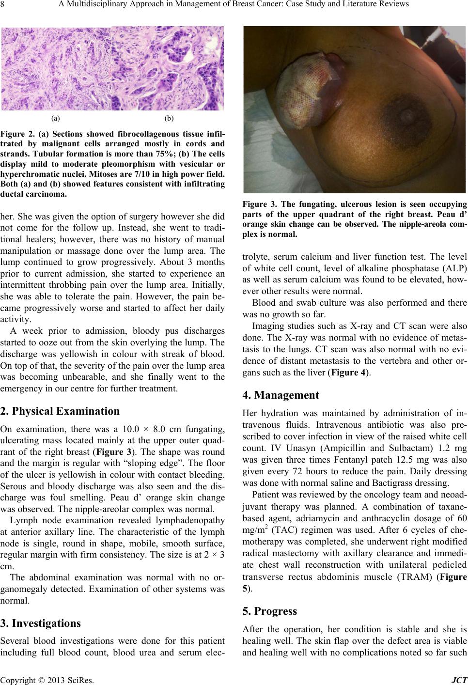 A Multidisciplinary Approach in Management of Bre a st Cancer: Ca se Study and Literature Reviews 8 (a) (b) Figure 2. (a) Sections showed fibrocollagenous tissue infil- trated by malignant cells arranged mostly in cords and strands. Tubular formation is more than 75%; (b) The cells display mild to moderate pleomorphism with vesicular or hyperchromatic nuclei. Mitoses are 7/10 in high power field. Both (a) and (b) showed features consistent with infiltrating ductal carcinoma. her. She wa s given the option of sur gery how ever she did not come for the follow up. Instead, she went to tradi- tional healers; however, there was no history of manual manipulation or massage done over the lump area. The lump continued to grow progressively. About 3 months prior to current admission, she started to experience an intermittent throbbing pain over the lump area. Initially, she was able to tolerate the pain. However, the pain be- came progressively worse and started to affect her daily activity. A week prior to admission, bloody pus discharges started to ooze out from the skin overlying the lump. The discharge was yellowish in colour with streak of blood. On top of that, the severity of the pain over the lump area was becoming unbearable, and she finally went to the emergency in our ce n tre for further treat ment. 2. Physical Examination On examination, there was a 10.0 × 8.0 cm fungating, ulcerating mass located mainly at the upper outer quad- rant of the right breast (Figure 3). The shape was round and the margin is regular with “sloping edge”. The floor of the ulcer is yellowish in colour with contact bleeding. Serous and bloody discharge was also seen and the dis- charge was foul smelling. Peau d’ orange skin change was observed. The nip ple-areolar complex was normal . Lymph node examination revealed lymphadenopathy at anterior axillary line. The characteristic of the lymph node is single, round in shape, mobile, smooth surface, regular margin with firm consistency. The size is at 2 × 3 cm. The abdominal examination was normal with no or- ganomegaly detected. Examination of other systems was normal. 3. Investigations Several blood investigations were done for this patient including full blood count, blood urea and serum elec- Figure 3. The fungating, ulcerous lesion is seen occupying parts of the upper quadrant of the right breast. Peau d’ orange skin change can be observed. The nipple-areola com- plex is normal. trolyte, serum calcium and liver function test. The level of white cell count, level of alkaline phosphatase (ALP) as well as serum calcium was found to be elevated, how- ever other results were normal. Blood and swab culture was also performed and there was no grow th so far. Imaging studies such as X-ray and CT scan were also done. The X-ray was normal with no evidence of metas- tasis to the lungs. CT scan was also normal with no evi- dence of distant metastasis to the vertebra and other or- gans such as the liver (Figure 4). 4. Management Her hydration was maintained by administration of in- travenous fluids. Intravenous antibiotic was also pre- scribed to cover infection in view of the raised white cell count. IV Unasyn (Ampicillin and Sulbactam) 1.2 mg was given three times Fentanyl patch 12.5 mg was also given every 72 hours to reduce the pain. Daily dressing was done with normal saline and Bactigrass dressing. Patient was reviewed by the oncology team and neoad- juvant therapy was planned. A combination of taxane- based agent, adriamycin and anthracyclin dosage of 60 mg/ m2 (TAC) regimen was used. After 6 cycles of che- motherapy was completed, she underwent right modified radical mastectomy with axillary clearance and immedi- ate chest wall reconstruction with unilateral pedicled transverse rectus abdominis muscle (TRAM) (Figure 5). 5. Progress After the operation, her condition is stable and she is healing well. The skin flap over the defect area is viable and healing well with no complications noted so far such Copyright © 2013 SciRes. JCT 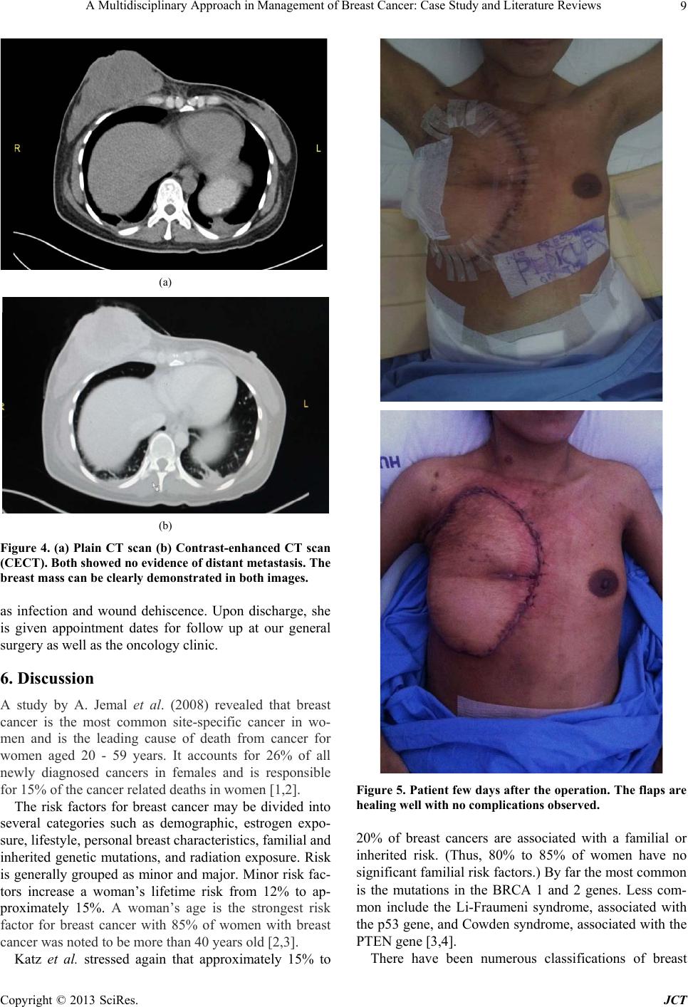 A Multidisciplinary Approach in Management of Bre a s t Cancer: Case Study and Literature Revi ews 9 (a) (b) Figure 4. (a) Plain CT scan (b) Contrast-enhanced CT scan (CECT). Both showed no evidence of distant metastasis. The breast mass can be clearly demonstrated in both images. as infection and wound dehiscence. Upon discharge, she is given appointment dates for follow up at our general surgery as well as the oncology clinic. 6. Discussion A study by A. Jemal et al. (2008) revealed that breast cancer is the most common site-specific cancer in wo- men and is the leading cause of death from cancer for women aged 20 - 59 years. It accounts for 26% of all newly diagnosed cancers in females and is responsible for 15% of the cancer related deaths in women [1,2]. The risk factors for breast cancer may be divided into several categories such as demographic, estrogen expo- sure, lifestyle, personal breast characteristics, familial and inherited genetic mutations, and radiation exposure. Risk is generally grouped as minor and major. Minor risk fac- tors increase a woman’s lifetime risk from 12% to ap- proximately 15%. A woman’s age is the strongest risk factor for breast cancer with 85% of women with breast cancer was noted to be more than 40 years old [2,3]. Katz et al. stressed again that approximately 15% to Figure 5. Patient few days after the operation. The flaps are healing wel l wi th no complications observed. 20% of breast cancers are associated with a familial or inherited risk. (Thus, 80% to 85% of women have no significant familial risk factors.) By far the most common is the mutations in the BRCA 1 and 2 genes. Less com- mon include the Li-Fraumeni syndrome, associated with the p53 gene, and Cowden syndrome, associated with the PTEN gene [3,4]. There have been numerous classifications of breast Copyright © 2013 SciRes. JCT  A Multidisciplinary Approach in Management of Bre a st Cancer: Ca se Study and Literature Reviews 10 carcinoma including histopathology classification, gene profiling and receptors present and also clinical staging. Based on histological classification, infiltrating ductal carcinoma is the most common breast malignancy, ac- counting for 75% of cases. Approximately 10% of infil- trating ductal carcinomas are of a uniform histologic pic- ture and are then classified as medullary, colloid, co- medo, tubular, or papillary carcinomas [3,4]. In order to reach a final diagnosis, the triple assess- ment method should be applied in any patient who pre- sents with breast lump or other symptoms suspicious of carcinoma. The triple assessment consists of clinical as- sessment (history taking and physical examination), im- aging studies as well as histopathology studies. This ap- proach can increase the positive predictive value (PPV) to exceed 99.9% [5]. In terms of treatment of cancer of the breast, the basic principles are to reduce the chance of local recurrence and the risk of metastatic spread. Treatment of early stage of breast cancer will usually involve surgery with or without radiotherapy. In an event where there are ad- verse prognostic factors such as lymph node involvement that increase the likelihood of metastatic relapse, sys- temic therapy such as chemotherapy or hormone therapy is added. At the other end of spectrum, locally advanced or metastatic disease is usually treated by systemic ther- apy to palliate symptoms, with surgery playing a much smaller role as mentioned by Williams et al. [5]. Kaufmann, M. et al. stated that neoadjuvant chemo- therapy (also called as primary systemic therapy or in- duction therapy) is standard management for women who have locally advanced or inflammatory breast cancer but can be applied to all women who may require postopera- tive chemotherapy for early-stage breast cancer. Dis- ease-free survival and overall survival are equivalent between patients treated with neoadjuvant chemotherapy and patients treated with the same regimen postopera- tively. There are various advantages that can be derived from this approach. Preoperative chemotherapy can offer women less morbid surgical treatment by down-staging both the primary breast tumor and axillary metastases. Finally, clinicians can be informed of the chemosensitiv- ity of the tumor by looking at the response to chemo- therapy, and can predict long-term outcome for women who have br e ast cancer [ 6]. Kaufmann, M. et al. also stated that in terms of the regimen of neoadjuvant, anthracycline-based regiment is the most extensively studied regimen. The typical ap- proach consists of at least four to six cycles of an an- thracycline-based regimen, usually adriamycin and cy- clophosphamide, with or without the addition of tax- ane-based agents [6]. Several studies including by Bear, H.D., et al. in 2003 have reported higher pathologic clini- cal response rates and rates of BCS with the addition of preoperative docetaxel. Nonetheless, taxane and anthrax- cycline regimens appear to be the most successful [7]. Mastectomy is indicated in for large tumors, multifo- cal disease, central tumors beneath or involving the nip- ple, local recurrence or patient’s reference. The radical Haslted mastectomy is no longer indicated as it caused excessive morbidity with no survival benefit. The modi- fied radical (Patey) mastectomy is more commonly per- formed. The excised mass is composed of the whole breast, a large portion of the skin, the centre which over- lies the tumor but which always include the nipple, and all the fat, fascia and lymph nodes of the axillary [5]. Few studies look at the psychological and social ad- justments to mastectomy in 160 women, who were fol- lowed at intervals of 3, 12, and 24 months after surgery. One in four women was reported to be in a state of de- pression and associated with marital and sexual problems 2 years after the initial therapy. The distress that comes from mastectomy may be more pronounced in younger women, as a result of significant treatment-related dis- tress and changes in bod y image and sexuality during th e course of the illness as observed by Fobair, P. et al. [8]. According to Nahabedian, M.Y., et al., patients under 50 years of age have a 4.3-fold greater likelihood of un- dergoing mastectomy followed by breast reconstruction than older patients [9]. The reasons behind this is de- scribed by Peppercorn, J., et al., which include the desire to achieve adequate breast cosmetic and symmetry, as well as fear of recurrence due to the fact that breast tu- mors in young women are more likely to be associated with high-risk tumor features and poorer prognosis, and tend to present with a more advanced stage at diagnosis [10]. Petit, J.Y., et al. showed that immediate breast recon- struction is now recognized as a safe procedure from the oncological point of view and is able to offer a series of advantages, such as better esthetic results compared with delayed reconstruction, attenuation of the sense of muti- lation deriving from the mastectomy and a reduction in surgical times [11]. However, extra care must be taken in-patient who needs post-operative radiotherapy. Studies done by Kronovitz, S.J., et al. showed that there is two main problems exist for performing immediate recon- struction on patients who will then undergo RT. First, RT can undo the esthetic result from immediate reconstruc- tion due to impairment of wound healing process and capsular contracture phenomena. Secondly, it can create problem especially when planning and designing the ir- radiation field. The slope of breast profile after recon- struction and the non-uniform thickness of the thoracic wall can cause under dosage radiation and non-homo- genous of dose within the fields of radiation respectively [12]. Nahabedian MY et al. emphasized that in 30% of cases, Copyright © 2013 SciRes. JCT  A Multidisciplinary Approach in Management of Bre a s t Cancer: Case Study and Literature Revi ews Copyright © 2013 SciRes. JCT 11 particularly following RT, reconstruction with autologous tissue is to be preferred [9]. The main myocutaneous flaps used in breast reconstruction are the transverse rec- tus abdominis myocutaneous (TRAM) flap introduced by Hartrampf in 1982 and used both as a pedicle flap or free (anastomosis between the inferior epigastric artery and the thoracodorsal artery, subscapular or internal breast), combined with or without prosthesis [9,13,14]. Pedicle TRAM flap uses abdominal muscle, fat and skin tissue vascularized by the rectus muscle pedicle to reconstruct the breast mound. Grossly, in the pedicle TRAM flap, excess skin, subcutaneous fat and rectus muscle from the infraumbilical area are transferred through a subcutaneous tunnel to the ipsilateral or con- tralateral mastectomy site. The flap is then rotated, shaped into a breast mound, and sutured; the umbilicus and the abdominal skin are sutured into its new position and the abdomen skin is sutured as in an abdominoplasty. Despite the loss of muscle function after a pedicle TRAM flap harvest, it is still possible for patients to become pregnant and carry a pregnancy to term, as well as to achieve a normal vaginal delivery [15] . Similar with our patient that in the free TRAM flap the skin, subcutaneous fat, deep inferior epigastric artery, and a small portion of the rectus muscle and fascia from the infraumbilical area are transferred to the chest defect, were epigastric vessels are reattached to either thora- codorsal or internal thoracic vessels via microsurgery. This technique allows the relocation of larger amounts of tissue with a lesser risk of fat necrosis. Hence, it may be a better procedure in patients with risk factors such as smoking, diabetes mellitus, and obesity [15]. 7. Conclusion Multidisciplinary approach is paramount and effective in breast cancer management. Despite the life changing di- agnosis of breast cancer, reconstructive surgery opens an avenue of possibility to encourage surgery when needed, and improves the qua lity of life, physically and mentally. 8. Consent Informed consent obtained from patient. REFERENCES [1] A. Jemal, R. Siegel, E. Ward, Y. Hao, J. Xu and T. Mur- ray, “Cancer Statistics, 2008,” A Cancer Journal for Cli- nicians, Vol. 58, No. 2, 2008, pp. 71-96. doi:10.3322/CA.2007.0010 [2] N. N. Baxter, B. A. Virnig, S. B. Durham, et al., “Trends in the Treatment of Ductal Carcinoma in Situ of the Breast,” Journal of the National Cancer Institute, Vol. 96, No. 6, 2004, pp. 443-448. doi:10.1093/jnci/djh069 [3] V. L. Katz and D. Detters, “Lentz: Comprehensive Gy- necology Mosby an Imprint of Elsevier. Chapter 15. Di- agnosis and Treatment of Benign and Malignant Breast Diseases,” 6th Edition, Elsevier, Philadelphia, 2012. [4] S. J. Katz, P. M. Lantz, N. K. Janz, et al., “Patient In- Volvement in Surgery Treatment Decisions for Breast Cancer,” Journal of Clinical Oncology, Vol. 23, No. 24, 2005, pp. 5526-5533. doi:10.1200/JCO.2005.06.217 [5] N. S. Williams, C. J. K. Bulstrode and P. R. O’Connell, “Bailey and Love’s Short Practice of Surgery Textbook,” 25th Edition, Edward Arnold, London, 2008. [6] M. Kaufmann, G. N. Hortobagyi, A. Goldhirsch, et al., “Recommendations from an International Expert Panel on the Use of Neoadjuvant (Primary) Systemic Treatment of Operable Breast Cancer: An Update,” Journal of Clinical Oncology, Vol. 24, No. 12, 2006, pp. 1940-1949. doi:10.1200/JCO.2005.02.6187 [7] H. D. Bear, S. Anderson, A. Brown, et al., “The Effect on Tumor Response of Adding Sequential Preoperative Do- cetaxel to Preoperative Doxorubicin and Cyclophos- phamide: Preliminary Results from National Surgical Adjuvant Breast and Bowel Project Protocol B-27,” Journal of Clinical Oncology, Vol. 21, No. 22, 2003, pp. 555-558. doi:10.1200/JCO.2003.12.005 [8] P. Fobair, S. L. Stewart, S. Chang, C. D’Onofrio, P. J. Banks and J. R. Bloom, “Body Image and Sexual Prob- lems in Young Women with Breast Cancer,” Psychoon- cology, Vol. 15, No. 7, 2006, pp. 579-594. doi:10.1002/pon.991 [9] M. Y. Nahabedian, “Breast Reconstruction: A Review and Rationale for Patient Selection,” Plastic and Recon- structive Surgery, Vol. 124, No. 1, 2009, pp. 55-62. doi:10.1097/PRS.0b013e31818b8c23 [10] J. Peppercorn, “Breast Cancer in Women under 40,” On- cology, Vol. 23, No. 6, 2009, pp. 465-474. [11] J. Y. Petit, M. Rietjens and C. Garusi, “Breast Recon- structive Techniques in Cancer Patients: Which Ones, When to Apply, Which Immediate and Long Term Risks?” Journal of Hematology & Oncology, Vol. 38, No. 3, 2001, pp. 231-239. [12] S. J. Kronovitz and G. L. Robb, “Controversies Regard- ing Immediate Reconstruction: Aesthetic Risks of Radia- tion,” In: S. L. Spear, Ed., Surgery of the Breast, 2nd Edi- tion, Lippincott Williams & Wilkins, Philadelphia, 2006, pp. 679-699. [13] S. Al Benna, “Female Plastic and Reconstructive Sur- geons’ Personal Decision Making for Breast Cancer Treatment and Reconstruction,” Archives of Gynecology and Obstetrics, Vol. 284, No. 3, 2011, pp. 737-741. doi:10.1007/s00404-010-1721-9 [14] A. K. Alderman, S. T. Hawley , J. Wa ljee, M. Mujahid, M. Morrow and S. J. Katz, “Understanding the Impact of Breast Reconstruction on the Surgical Decision-Making Process for Breast Cancer,” Cancer, Vol. 112, No. 3, 2008, pp. 489-494. doi:10.1002/cncr.23214 [15] M. Marín-Gutzke and A. Sánchez-Olaso, “Reconstructive Surgery in Young Women with Breast Cancer,” Breast Cancer Research and Treatment, Vol. 123, Suppl. 1, 2010, pp. 67-74. |