

Paper Menu >>
Journal Menu >>
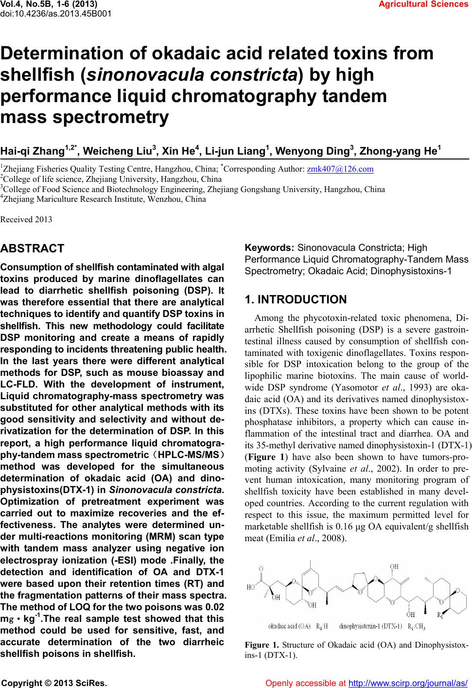 Vol.4, No.5B, 1-6 (2013) Agricultural Sciences doi:10.4236/as.2013.45B001 Determination of okadaic acid related toxins from shellfish (sinonovacula constricta) by high performance liquid chromatography tandem mass spectrometry Hai-qi Zhang1,2*, Weicheng Liu3, Xin He4, Li-jun Liang1, Wenyong Ding3, Zhong-yang He1 1Zhejiang Fisheries Quality Testing Centre, Hangzhou, China; *Corresponding Author: [email protected] 2College of life science, Zhejiang University, Hangzhou, China 3College of Food Science and Biotechnology Engineering, Zhejiang Gongshang University, Hangzhou, China 4Zhejiang Mariculture Research Institute, Wenzhou, China Received 2013 ABSTRACT Consumption of shellfish contaminated with algal toxins produced by marine dinoflagellates can lead to diarrhetic shellfish poisoning (DSP). It was therefore essential that there are analytical techniques to iden tify and quanti fy DSP toxins i n shellfish. This new methodology could facilitate DSP monitoring and create a means of rapidly responding to incidents threatening public health. In the last years there were different analytical methods for DSP, such as mouse bioassay and LC-FLD. With the development of instrument, Liquid chromatography-mass spectrometry was substituted for other analytical methods with its good sensitivity and selectivity and without de- rivatization for the determination of DSP. In this report, a high performance liquid chromatogra- phy-t andem mass spectr ometric(HPLC-MS/MS) method was developed for the simultaneous determination of okadaic acid (OA) and dino- physistoxins(DTX-1) in Sinonovacula constricta. Optimization of pretreatment experiment was carried out to maximize recoveries and the ef- fectiveness. The analytes were determined un- der multi-reactions monitoring (MRM) scan type with tandem mass analyzer using negative ion electrospray ionization (-ESI) mode .Finally, the detection and identification of OA and DTX-1 were based upon their retention times (RT) and the fragmentation patterns of their mass spectra. The method of LOQ for the t wo poisons was 0.02 mg·kg-1.The real sample test showed that this method could be used for sensitive, fast, and accurate determination of the two diarrheic shellfish poisons in shellfish. Keywords: Sinonovacula Con stri cta; High Performance Liquid Ch romatogra phy-Ta ndem Mass Spectrometry; Okadaic Acid; Dinoph ys istoxins-1 1. INTRODUCTION Among the phycotoxin-related toxic phenomena, Di- arrhetic Shellfish poisoning (DSP) is a severe gastroin- testinal illness caused by consumption of shellfish con- taminated with toxigenic dinoflagellates. Toxins respon- sible for DSP intoxication belong to the group of the lipophilic marine biotoxins. The main cause of world- wide DSP syndrome (Yasomotor et al., 1993) are oka- daic acid (OA) and its derivatives named dinophysistox- ins (DTXs). These toxins have been shown to be potent phosphatase inhibitors, a property which can cause in- flammation of the intestinal tract and diarrhea. OA and its 35-methyl derivative named dinophysistoxin-1 (DTX-1) (Figure 1) have also been shown to have tumors-pro- moting activity (Sylvaine et al., 2002). In order to pre- vent human intoxication, many monitoring program of shellfish toxicity have been established in many devel- oped countries. According to the current regulation with respect to this issue, the maximum permitted level for marketable shellfish is 0.16 μg OA equivalent/g shellfish meat (Emilia et al., 2008). Figure 1. Structure of Okadaic acid (OA) and Dinophysistox- ins-1 (DTX-1). Copyright © 2013 SciRes. Openly accessible at http://www.scirp.org/journal/as/ 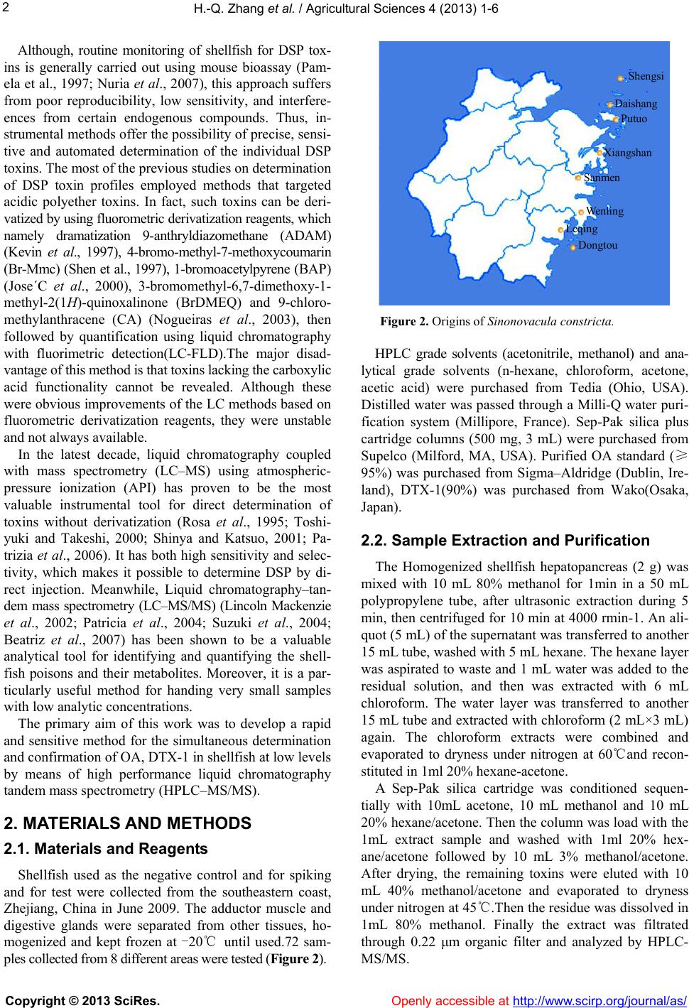 H.-Q. Zhang et al. / Agricultural Sciences 4 (2013) 1-6 2 Although, routine monitoring of shellfish for DSP tox- ins is generally carried out using mouse bioassay (Pam- ela et al., 1997; Nuria et al., 2007), this approach suffers from poor reproducibility, low sensitivity, and interfere- ences from certain endogenous compounds. Thus, in- strumental methods offer the possibility of precise, sensi- tive and automated determination of the individual DSP toxins. The most of the previous studies on determination of DSP toxin profiles employed methods that targeted acidic polyether toxins. In fact, such toxins can be deri- vatized by using fluorometric derivatization reagents, which namely dramatization 9-anthryldiazomethane (ADAM) (Kevin et al., 1997), 4-bromo-methyl-7-methoxycoumarin (Br-Mmc) (Shen et al., 1997), 1-bromoacetylpyrene (BAP) (Jose´C et al., 2000), 3-bromomethyl-6,7-dimethoxy-1- methyl-2(1H)-quinoxalinone (BrDMEQ) and 9-chloro- methylanthracene (CA) (Nogueiras et al., 2003), then followed by quantification using liquid chromatography with fluorimetric detection(LC-FLD).The major disad- vantage of this method is that toxins lacking the carboxylic acid functionality cannot be revealed. Although these were obvious improvements of the LC methods based on fluorometric derivatization reagents, they were unstable and not always available. In the latest decade, liquid chromatography coupled with mass spectrometry (LC–MS) using atmospheric- pressure ionization (API) has proven to be the most valuable instrumental tool for direct determination of toxins without derivatization (Rosa et al., 1995; Toshi- yuki and Takeshi, 2000; Shinya and Katsuo, 2001; Pa- trizia et al., 2006). It has both high sensitivity and selec- tivity, which makes it possible to determine DSP by di- rect injection. Meanwhile, Liquid chromatography–tan- dem mass spectrometry (LC–MS/MS) (Lincoln Mackenzie et al., 2002; Patricia et al., 2004; Suzuki et al., 2004; Beatriz et al., 2007) has been shown to be a valuable analytical tool for identifying and quantifying the shell- fish poisons and their metabolites. Moreover, it is a par- ticularly useful method for handing very small samples with low analytic concentrations. The primary aim of this work was to develop a rapid and sensitive method for the simultaneous determination and confirmation of OA, DTX-1 in shellfish at low levels by means of high performance liquid chromatography tandem mass spectrometry (HPLC–MS/MS). 2. MATERIALS AND METHODS 2.1. Materials and Reagents Shellfish used as the negative control and for spiking and for test were collected from the southeastern coast, Zhejiang, China in June 2009. The adductor muscle and digestive glands were separated from other tissues, ho- mogenized and kept frozen at -20℃ until used.72 sam- ples collected from 8 different areas were tested (Figure 2). Shengsi Daishang Putuo Xiangshan Sanmen Wenling Dongtou Leqing Figure 2. Origins of Sinonovacula constricta. HPLC grade solvents (acetonitrile, methanol) and ana- lytical grade solvents (n-hexane, chloroform, acetone, acetic acid) were purchased from Tedia (Ohio, USA). Distilled water was passed through a Milli-Q water puri- fication system (Millipore, France). Sep-Pak silica plus cartridge columns (500 mg, 3 mL) were purchased from Supelco (Milford, MA, USA). Purified OA standard (≥ 95%) was purchased from Sigma–Aldridge (Dublin, Ire- land), DTX-1(90%) was purchased from Wako(Osaka, Japan). 2.2. Sample Extraction and Purification The Homogenized shellfish hepatopancreas (2 g) was mixed with 10 mL 80% methanol for 1min in a 50 mL polypropylene tube, after ultrasonic extraction during 5 min, then centrifuged for 10 min at 4000 rmin-1. An ali- quot (5 mL) of the supernatant was transferred to another 15 mL tube, washed with 5 mL hexane. The hexane layer was aspirated to waste and 1 mL water was added to the residual solution, and then was extracted with 6 mL chloroform. The water layer was transferred to another 15 mL tube and extracted with chloroform (2 mL×3 mL) again. The chloroform extracts were combined and evaporated to dryness under nitrogen at 60℃and recon- stituted in 1ml 20% hexane-acetone. A Sep-Pak silica cartridge was conditioned sequen- tially with 10mL acetone, 10 mL methanol and 10 mL 20% hexane/acetone. Then the column was load with the 1mL extract sample and washed with 1ml 20% hex- ane/acetone followed by 10 mL 3% methanol/acetone. After drying, the remaining toxins were eluted with 10 mL 40% methanol/acetone and evaporated to dryness under nitrogen at 45℃.Then the residue was dissolved in 1mL 80% methanol. Finally the extract was filtrated through 0.22 μm organic filter and analyzed by HPLC- MS/MS. Copyright © 2013 SciRes. Openly accessible at http://www.scirp.org/journal/as/  H.-Q. Zhang et al. / Agricultural Sciences 4 (2013) 1-6 Copyright © 2013 SciRes. http://www.scirp.org/journal/as/Openly accessible at 3 2.3. HPLC-MS/MS Analysis HPLC-MS/MS was performed on an HP 1100 series liquid chromatograph (Agilent, Palo Alto,CA, USA), coupled to an API 3000 triple quadrupole mass spec- trometer (Applied Biosystem) with an atmospheric pres- sure ionization source and an electrospray ionization (ESI) interface. The instrumentation was controlled using Analyst v.1.2 software. Chromatographic separations of OA and DTX-1 were carried out under the following combinations of column and mobile phases: Zorbax XDB C18 (2.1 mm × 150 mm, 5 μm, Agilent) with the mobile phase, acetonitrile - 0.1% acetic acid(70:30,v/v). The column temperature and flow-rate were kept at 30℃ and 0.25 mL·min-1, re- spectively. 10 μL of sample were injected onto the col- umn at the room temperature. The mass spectrometer was operated by electrospray in negative ion mode (ESI-) with multiple reaction moni- toring (MRM) for the detection of OA, DTX-1. The monitored ions were the [M−H]− precursor ions at m/z of 803.6 (OA), 817.4 (DTX-1), respectively and the most abundant product ion observed for each toxin. The MS parameters were optimized for the ionization of standard toxins using flow injection analysis. Two different prod- uctions were used to verify the selectivity for determi- nation of OA and DTX-1 as shown in Table1. The opti- mized MRM experiment was established for the concur- rent determination of the aforementioned toxins using the following conditions: Ionspray Voltage -4500 V, Auxil- iary Gas Speed 7 L·min-1, Turbo Ionspray Source Tem- perature 500℃, Nebulizer Gas 9 psi, Curtain Gas 8 psi, Collision Gas 8 psi, Focusing Potential -260 V, Entrance Potential -9 V, and Cell Exit Potential -13 V. All gases in the MRM experiment were high-purity nitrogen gas. Other optimization of MS conditions as shown in Table 1. 2.4. HPLC-MS/MS Ass ay Stock solutions (10 mg·L-1) of individual shellfish toxin standards (OA, DTX-1) were prepared by dissolving in methanol. A mixed stock solution (1 mg·L-1) contain- ing two standards was prepared from stock solutions of individual standards by mixing and diluting with metha- nol. Different calibration standards (20, 50, 100, 200, 500, 800 μg·L-1) were prepared by appropriate dilution of the mixed stock solution with methanol. The standards were injected directly into the HPLC-MS/MS system. The calibration curve was obtained by the peak area (y-axis) plotted against the concentration of toxins standard (χ-axis). The qualitative analysis of OA and DTX-1 of the experimental samples were performed based on the retention time and the ion ration of standard solution. 2.5. The Experiment of Recovery, Precision and Accuracy Homogenized negative shellfish hepatopancreas (2 g), which spiked with 0.02, 0.1, 0.2 and 0.4 mg·kg-1 mixed standard solution of OA and DTX-1 respectively, were pretreated as section 2.2 and then analyzed by HPLC- MS/MS. Three replicate samples at each concentration were analyzed on the same day. The percentage of re- covery was calculated by comparing the concentration obtained according to the calibration curve with the ac- tual spiked concentration of standard solution (OA, DTX-1). The precision was evaluated by coefficients of variation (CV %) and the accuracy was estimated based on the average percentage of recovery. 3. RESULTS AND DISCUSSION 3.1. HPLC-MS/MS Condition Analysis The mass spectra of each compound were measured in the positive and the negative ion modes for the precursor ion full-scan of toxins standard (1 mg·L-1). It was found that the detection sensitivity for the toxins studied was better in negative rather than in positive mode with the precursor ion [M−H]− at m/z 803.6 for OA, m/z 817.4 for DTX-1, respectively. The fragmentation of the target toxins was optimized to efficiently generate several product ions from each precursor ion by collision-in- duced dissociation (CID) and shown in Figure 3. Select- ing two precursor/product ion combinations (Q1/Q3 pairs) as monitor ions to verify the selectivity and deter- mination of toxins, which were m/z 803.6/255.0, 803.6/563.4 for OA, m/z 817.4/255.0 and 817.4/113.1 for DTX-1 and shown in table 1. To achieve optimum sensi- tivity and selectivity, MRM was implemented and the optimization of MS conditions as shown in the afore- mentioned experiment. Table 1. Optimization of the partial MS condition. Analyte Precursor ion(m/z) Product ions (m/z)Declustering Potential(V) Collision Energy(V) Retention time(min) Okadaic Acid (OA) 803.6 255.1* 563.1 -70 -60 -68 3.38 Dinophysistoxins-1 (DTX-1) 817.4 255.1* 113.1 -110 -68 -94 5.70 *Quantificational ion.  H.-Q. Zhang et al. / Agricultural Sciences 4 (2013) 1-6 4 The polar solvent (methanol, acetonitrile) usually is used as mobile phase for reverse phase column C18. It was found that the efficiency of ionization with 70% metha- nol was inferior to that obtained in 70% acetonitriler; meanwhile ionization efficiency would be intensified with 0.1% acetic acid (Toshiyuki and Takeshi, 2000). So 70% acetonitrile containing 0.1% acetic acid was se- lected as the mobile phase of LC-MS/MS. 3.2. Sample Extraction and Purification Ta b l e 2 listed the recovery of the preliminary extrac- tion and elution with different solutions at the spiked level of 0.1 mg·kg-1 from standard toxins. The recovery of toxins from 80% methanol extracts were 94.2% for OA and 90.6% for DTX-1, slightly lower than 80% ace- tontril extracts. Besides the 90% methanol (Hirofumi et al., 2001) gave least residue than other organic solvent (methanol, acetone) as extractant. Considering that the toxicity of acetontrile was more harmful than methanol and wasted more time during evaporation under nitrogen. And there is no obvious difference for the recovery be- tween 80% and 90% for methanol-water. Thus, experi- ment chose 80% methanol as extractant insuring against good recovery. Figure 3. Product ions full-scan negative-ion ESI mass spec- trums of OA (up) and DTX-1(down). Purification of extractant (OA and DTX-1) from the shellfish hepatopancreas was carried out by Sep-pak sil- ica cartridge as previously described (Patrizia et al., 2006) due to the significant suppression of ionization by con- taminants. In test of elution for the toxins using three different rates of acetone–methanol, it shows that the recovery in 40% methanol/acetone was the highest (99% for OA, 97.8% for DTX-1) than others. 3.3. Method Evaluation (Recovery, Precision and Accuracy) Figure 4 showed the chromatogram of the toxins (OA, DTX-1) standard with 200 μg·L-1. From which it could Table 2. Comparison of extract and elution from different solu- tions at the spiked level of 0.1 mg·kg-1. Recovery (%, n = 6) SolutionSolution component OA DTX-1 80% Methanol- water 94.2 90.6 Extract solution 80% Acetontrile- water 96.0 92.5 40% Methanol-acetone 99.0 97.8 50% Methanol-acetone 93.2 95.2 Elution solution 60% Methanol-acetone 91.5 84.4 Figure 4. Chromatograms of the OA and DTX-1 standard solution (200 μg·l-1)(up) and the negative spiked sample (0.1 mg· kg-1) (down). Copyright © 2013 SciRes. Openly accessible at http://www.scirp.org/journal/as/  H.-Q. Zhang et al. / Agricultural Sciences 4 (2013) 1-6 5 be seen there was a single and symmetric peak and the retention time is 3.4 min for OA with the monitor ion pairs m/z 803.6/255.0, 803.6/563.4 and 5.7 min for DTX-1 with m/z 817.4/255.0, 817.4/113.1.The down of Figure 4 showed the chromatogram of negative sample was spiked with 0.1mg·kg-1. And it could be seen there was no obvious interferential peaks when the sample was pretreated as described above to the section 2.2 experi- ment and detected with HPLC-MS/MS. The good linear- ity of the peak area plotted against concentrations for the toxins with the linear (OA:у = 20-241, r = 0.9995; DTX-1: у = 141χ-85, r = 0.9997) over concentration ranging from 20 μg·L-1 to 800 μg·L-1. Table 3 gives the recovery and coefficients of varia- tion (CV %) data corresponding to negative samples that were spiked each with 0.02,0.1,0.2 and 0.4 mg OA and DTX-1 per 1kg of the hepatopancreas . The average re- covery of OA and DTX-1 were decreased with the spiked level from 0.02 to 0.4 mg·kg-1 because of the matrix ef- fect. This suggested that interferential compounds of the matrix reduced ionization efficiency of toxins. It is ac- ceptant that the mean recoveries were within the range from 79.0% to 92.2%, and the CV% was lower than 11.6%. The signal to noise(S/N) was calculated from the ratio between analyte peak signal to base line and peak-to- peak noise signal. The S/N was above 10 for the mixed standard solution (20 μg·L-1). The LOQ of the method at an S/N ratio of 10 were estimated to 0.02 mg·kg-1 for the both of OA and DTX-1. 3.4. Method Application The developed method was applied to the analysis of 72 batches of that were collected from 8 different areas in Zhejiang province. Ta b le 4 showed the results of the 14 postive samples. 2 samples were found OA, with the concentration of 25.6 µg·kg-1 and 33.0 µg·kg-1, repec- tively. 12 samples were found DTX-1, with the concen- tration from 84.1 µg·kg-1 to 293.0 µg·kg-1. Table 3. Recovery and CV% for the OA and DTX-1. Analyte Spiked level (mg·kg-1) Recovery (%,n = 6) (mean ± S.D) CV (%) 0.02 89.4 ± 7.9 8.87 0.1 87.9 ± 10.2 11.6 0.2 84.7 ± 7.3 8.57 OA 0.4 79.0 ± 8.5 10.8 0.02 92.2 ± 3.8 4.09 0.1 90.8 ± 4.1 4.51 0.2 89.9 ± 6.3 6.98 DTX-1 0.4 84.2 ± 4.7 5.54 Table 4. Results of the postive samples. No.Sampling time Sampling location Concentration (µg·kg-1) 1 August,2007 Shensi DTX-1:293.0 2 Spetember,2007 Shensi DTX-1:208.4 3 December,2007 Shensi OA:25.6 4 March,2008 Putuo DTX-1:155.0 5 June,2008 Putuo DTX-1:192.4 6 Spetember,2007 Xiangshan DTX-1:246.0 7 October,2007 Xiangshan DTX-1:168.5 8 August,2007 Shanmen OA:33.0 9 Spetember,2007 Shanmen DTX-1:257.2 10 June,2008 Wenling DTX-1:84.1 11 Spetember,2007 Leqing DTX-1:176.2 12 Spetember,2008 Leqing DTX-1:105.0 13 July,2007 Dongtou DTX-1:144.3 14 Spetember,2008 Dongtou DTX-1:97.8 4. CONCLUSIONS In conclusion, the proposed method, which offers a rather newly and rapid developed extraction, clean-up of the sample, was found to have acceptable reproducibility, high specificity and sensitivity and a detection capability which allows the detection of the diarrheic shellfish poi- sons (OA and DTX-1) by means of HPLC-MS/MS under multi-reactions monitoring (MRM) scan with tandem mass analyzer using negative ion electrospray ionization (-ESI) mode and identification based upon their retention times and the fragmentation patterns of their mass spec- tra. 5. ACKNOWLEDGEMENTS We are grateful to the financial support from the Key Scientific Re- search Projects of Zhejiang Province (No.2007C23081).We also thank all the members of the Zhejiang Fisheries Quality Testing Center and Center of Analysis and Measurement(Zhejiang University) for their helpful discussions. REFERENCES [1] Paz, B., Daranas, A. H., Cruz, P.G., Franco, J.M., Pizarro, G.,Souto, M.L., Norte, M. and Fernandez, J.J.(2007) Characterisation of okadaic acid related toxins by liquid chromatography coupled with mass spectrometry. Toxi- con, 50, 225-235. doi:10.1016/j.toxicon.2007.03.015 [2] Manerio, E., Rodas,V.L., Costas,E. and Hernandez, J.M. (2008) Shellfish consumption: A major risk factor for co- lorectal cancer. Medical Hypotheses, 70, 409-412. Copyright © 2013 SciRes. Openly accessible at http://www.scirp.org/journal/as/  H.-Q. Zhang et al. / Agricultural Sciences 4 (2013) 1-6 Copyright © 2013 SciRes. http://www.scirp.org/journal/as/Openly accessible at 6 doi:10.1016/j.mehy.2007.03.041 [3] Goto, H., Igarashi, T., Yamamoto, M.,Yasuda, M., Se- kiguchi, R., Watai, M., Tanno, K. and Yasumoto, T.(2001) Quantitative determination of marine toxins associated with diarrhetic shellfish poisoning by liquid chromatog- raphy coupled with mass spectrometry. Journal of Chro- matography A, 907, 181-189. doi:10.1016/S0021-9673(00)01047-5 [4] Gonzalez, J.C., Leira,F., Vieytes, M.R., Vieites, J.M., Botana, A.M. and Botana, L.M.(2000) Development and validation of a high- performance liquid chromatographic method using fluorimetric detection for the determination of the diarrhetic shellfish poisoning toxin okadaic acid without chlorinated solvents. Journal of Chromatography A, 876, 117-125. doi:10.1016/S0021-9673(00)00193-X [5] James, K.J., Bishop, A.G., Gillman, M., Kelly, S.S., Ro- den, C., Draisci, R., Lucentini, Glannetti, L. and Boria, P. (1997) Liquid chromatography with fluorimetric,mass spectrometric and tandem spectrometric detection for the investigation of the seafood-toxin-producing phytoplank- ton, Dinophysis acuta. Journal of Chromatography A, 777, 213-221. doi:10.1016/S0021-9673(97)00158-1 [6] Mackenzie, L., Holland, P., McNabb, P., Beuzenberg, V., Selwood, A. and Suzuki, T. (2002) Complex toxin pro- files in phytoplankton and Greenshell mussels (Perna ca- naliculus ), revealed by LC–MS/MS analysis. Toxicon, 40, 1321-1330. doi:10.1016/S0041-0101(02)00143-5 [7] Llamas, N.M., Stewart, L., Fodey, T., Higgins, H.C., Velasco, M.L.R., Botana, L.M. and Elliott C.T.(2007) Development of a novel immunobiosensor method for the rapid detection of okadaic acid contamination in shellfish extracts. Analytical and Bioanalytical Chemistry, 389, 581-587. doi:10.1007/s00216-007-1444-3 [8] Nogueiras, M.J., Ana, G.M., Antonio, I.P., Marian, T., Kevin, J.J., James, F.L. kjgjh(2003) Comparison of dif- ferent fluorimetric HPLC methods for analysis of acidic polyether toxins in marine phytoplankton. Analytical and Bioanalytical Chemistry, 377, 1202-1206. doi:10.1007/s00216-003-2221-6 [9] Pamela, E.N., Anne, C.S. (1997). Comparison of a pro- tein phosphatase inhibition assay, HPLC assay and en- zyme- linked immunosorbent assay with the mouse bio- assay for the detection of diarrhetic shellfish poisoning toxins in European shellfish. Int. J. Food Microbiol, 36, 39-48. [10] Ciminiello, P., Dell’Aversano, C., Fattorusso, E., Forino, M., Magno, S., Santelia, F. and Tsoukatou, M.(2006) In- vestigation of the toxin profile of Greek mussels Mytilus galloprovincialis by liquid chromatography-mass spec- trometry. Toxicon,. 47, 174-181. doi:10.1016/j.toxicon.2005.10.012 [11] Patricia, F.P., Mar´ıa, J.F.S, Brett, H., Mary, L., Hanne, R., Ambrose, F. and Kevin, J.J. (2004). Rapid determination of polyether marine toxins using liquid chromatogra- phy–multi- ple tandem mass spectrometry. J. Chromatogr. A, 1056, 77-82. [12] Draisci, R., Lucentini, L., Giannetti, L., Boria and P., Stacchini, A. (1995) Detection of diarrhoetic shellfish toxins in mussels from Italy by ionspray liquid chroma- tography-mass spectrometry. Toxicon, 33, 1591-1603. doi:10.1016/0041-0101(95)00105-0 [13] Sylvaine, C., Christine, M., Laurence, M., Sylvia, R., Catherine, B.(2002) Ecological analysis of digestive can- cer mortality related to contamination by diarrhetic shell- fish poisoning toxins along the coasts of France. Environ Res, 84, 145-150. [14] Shen, J.L., Christian, H. and Bernd, L.(1997) Sensitive HPLC- fluorometric and HPLC-MS determination of diarrhetic shellfish poisoning (DSP)-toxins as4-bromome thyl-7-methoxycoumarin esters. Fresenius’Journal of Analttical Chemistry, 357, 101-104. doi:10.1007/s002160050120 [15] Shinya, I. and Katsuo, T. (2001) Matrix effect and correc- tion by standard addition in quantitative liquid chroma- tographic- mass spectrometric analysis of diarrhetic shell- fish poisoning toxins. J. Chromatogr A, 943, 39-46. [16] Suzuki, T., Beuzenberg, V., Mackenzie, L., Quilliam, M. A.(2004) Discovery of okadaic acid esters in the toxic dinoflagellate Dinophysis acuta from New Zealand using liquid chromatography/tandem mass spectrometry. Rapid Commun. Mass Spectrom, 18, 1131-1138. doi:10.1002/rcm.1455 [17] Toshiyuki S, Takeshi Y.2000. Liquid chromatography– electrospray ionization mass spectrometry of the diar- rhetic shellfish-poisoning toxins okadaic acid, dinophy- sistoxin-1 and pectenotoxin-6 in bivalves. J. Chromatogr. A, 874, 199-206. doi:10.1016/S0021-9673(00)00094-7 [18] Yasumoto T, Murata M. 1993. Marine toxins. Chemical Reviews, 93, 1897-1909. doi:10.1021/cr00021a011 |