

Paper Menu >>
Journal Menu >>
 World Journal of Cardiovascular Surgery, 2013, 3, 101-105 http://dx.doi.org/10.4236/wjcs.2013.33018 Published Online July 2013 (http://www.scirp.org/journal/wjcs) Root Reconstruction with Total Replacement of Ascending Aorta Using Hypothermic Circulatory Arrest and Selective Cerebral Perfusion for Moderately Dilated Distal Ascending Aorta Satoshi Yamashiro*, Ryoko Arakaki, Yuya Kise, Hitoshi Inafuku, Yukio Kuniyoshi Department of Thoracic and Cardiovascular Surgery, University of the Ryukyus, Nishihara, Okinawa, Japan Email: *[email protected] Received June 3, 2013; revised July 3, 2013; accepted July 10, 2013 Copyright © 2013 Satoshi Yamashiro et al. This is an open access article distributed under the Creative Commons Attribution Li- cense, which permits unrestricted use, distribution, and reproduction in any medium, provided the original work is properly cited. ABSTRACT Objective: We have occasionally encountered a moderately dilated distal ascending aorta while reconstructing an aortic root. We describe reconstruction of an extended root and ascending aorta using our current strategy. Patients and Methods: Between March 2011 and December 2012, 15 (11 men; mean age, 70.1 ± 7.3 years) patients underwent root reconstruction with hemi-arch replacement under hypothermic circulatory arrest with antegrade selective cerebral per- fusion. The maximum diameter of the aortic root was 52.5 ± 4.4 mm. The distal ascending aorta just below innominate artery was moderately dilated to 41.7 ± 1.4 mm in diameter. Results: Operative outcomes excluding the diameter of the distal ascending aorta did not significantly differ from those of patients who had undergone root reconstruction under distal ascending aortic clamping during the same period. Postoperative computed tomography confirmed complete re- section of the dilated ascending aorta in the patients, and did not develop neurological dysfunction or stroke. Conclu- sion: Postoperative computed tomography confirmed complete resection of dilated ascending aortae. We considered that complete resection under hypothermic circulatory arrest and selective cerebral perfusion might help to avoid re- peated surgery to treat dilation of the distal ascending aorta over the long-term. Keywords: Annulo-Aortic Ectasia; Re-Operation 1. Introduction Ascending aorta replacement is the most common strat- egy for treating thoracic aortic pathologies. Graft repla- cement for an ascending aortic aneurysm is now a rela- tively safe and standard surgical procedure. However, management of a mildly to moderately dilated ascending aorta during cardiac surgery remains controversial. We have encountered some patients with a moderately di- lated distal ascending aorta during aortic root reconstruc- tion. Distal anastomosis of the ascending aorta usually proceeds under aortic clamping, when a dilated distal ascending aorta would remain. The most important con- sequence of an enlarged ascending aortic dimension is a proportional increase in the incidence of aortic rupture, dissection and re-operation. We have routinely wrapped borderline dilated distal ascending aortae with prosthetic grafts or felt strips to prevent dilation over the long term. However, we have recently replaced moderately dilated ascending aortae (hemi-arch replacement), using open distal anastomosis for complete resection. 2. Patients and Methods Between March 2011 and December 2012, 15 patients (11 men and 4 women; mean age, 70.1 ± 7.3 years) un- derwent root reconstruction with total replacement of a dilated ascending aorta under hypothermic circulatory arrest (HCA) with antegrade selective cerebral perfusion (ASCP), (HCA group). The ascending aorta was comple- tely resected just below the innominate artery (hemi-arch group). All patients were preoperatively diagnosed by computed tomography (CT), angiography and ultrasound cardiography as having annulo-aortic ectasia (AAE) with aortic regurgitation (AR). The maximum aortic root di- ameter was 52.5 ± 4.4 mm. The distal ascending aorta just below the innominate artery was moderately dilated *Corresponding a uthor. C opyright © 2013 SciRes. WJCS 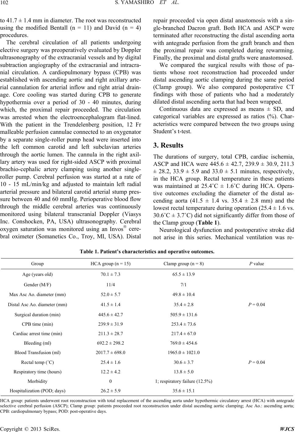 S. YAMASHIRO ET AL. 102 to 41.7 ± 1.4 mm in diameter. The root was reconstructed using the modified Bentall (n = 11) and David (n = 4) procedures. The cerebral circulation of all patients undergoing elective surgery was preoperatively evaluated by Doppler ultrasonogr aphy of the extracr anial vessels and by digital subtraction angiography of the extracranial and intracra- nial circulation. A cardiopulmonary bypass (CPB) was established with ascending aortic and right axillary arte- rial cannulation for arterial inflow and right atrial drain- age. Core cooling was started during CPB to generate hypothermia over a period of 30 - 40 minutes, during which, the proximal repair proceeded. The circulation was arrested when the electroencephalogram flat-lined. With the patient in the Trendelenberg position, 12 Fr malleable perfusion cannulae connected to an oxygenator by a separate single-roller pump head were inserted into the left common carotid and left subclavian arteries through the aortic lumen. The cannula in the right axil- lary artery was used for righ t-sided ASCP with proximal brachio-cephalic artery clamping using another single- roller pump. Cerebral perfusion was started at a rate of 10 - 15 mL/min/kg and adjusted to maintain left radial arterial pressure and bilateral carotid arterial stump pres- sure between 40 and 60 mmHg. Perioperative blood flow through the middle cerebral arteries was continuously monitored using bilateral transcranial Doppler (Viasys Inc. Conshocken, PA, USA) ultrasonography. Cerebral oxygen saturation was monitored using an Invos cere- bral oximeter (Somanetics Co., Troy, MI, USA). Distal repair proceeded via open distal anastomosis with a sin- gle-branched Dacron graft. Both HCA and ASCP were terminated after reconstructing the distal ascending aorta with antegrade perfusion from the graft branch and then the proximal repair was completed during rewarming. Finally, the proximal and distal grafts were anastomosed. We compared the surgical results with those of pa- tients whose root reconstruction had proceeded under distal ascending aortic clamping during the same period (Clamp group). We also compared postoperative CT findings with those of patients who had a moderately dilated distal ascending aorta that had bee n w rapped. Continuous data are expressed as means ± SD, and categorical variables are expressed as ratios (%). Char- acteristics were compared between the two groups using Student’s t-test. 3. Results The durations of surgery, total CPB, cardiac ischemia, ASCP and HCA were 445.6 ± 42.7, 239.9 ± 30.9, 211.3 ± 28.2, 33.9 ± 5.9 and 33.0 ± 5.1 minutes, respectively, in the HCA group. Rectal temperature in these patients was maintained at 25.4˚C ± 1.6˚C during HCA. Opera- tive outcomes excluding the diameter of the distal as- cending aorta (41.5 ± 1.4 vs. 35.4 ± 2.8 mm) and the lowest rectal temperature during operation (25.4 ± 1.6 vs. 30.6˚C ± 3.7˚C) did not significantly differ from those of the Clamp group (Table 1). Neurological dysfunction and postoperative stroke did not arise in this series. Mechanical ventilation was re- Table 1. Patient’s characteristics and operative outcomes. Group HCA group (n = 15) Clamp group (n = 8) P value Age (years old) 70.1 ± 7.3 65.5 ± 13.9 Gender (M/F) 11/4 7/1 Max Asc Ao. diameter (mm) 52.0 ± 5.7 49.8 ± 10.4 Distal Asc Ao. diamet e r (mm) 41.5 ± 1.4 35.4 ± 2.8 P = 0. 04 Surgical duration (min) 445.6 ± 42.7 505.9 ± 131.6 CPB time (min) 239.9 ± 31.9 253.4 ± 73.6 Cardiac arrest time (min) 211.3 ± 28.7 217.4 ± 67.0 Bleeding (ml) 692.2 ± 298.2 769.0 ± 454.6 Blood Transfusion (ml) 2017.7 ± 698.0 1965.0 ± 1021.0 Rectal temp (˚C) 25.4 ± 1.6 30.6 ± 3.7 P = 0.04 Respiratory time (hours) 12.2 ± 4.2 13.8 ± 5.0 Morbidity 0 1; respiratory failure (12.5%) Hospitalization (POD; days) 26.2 ± 5.9 35.6 ± 15.1 HCA group: patients underwent root reconstruction with total replacement of the ascending aorta under hypothermic circulatory arrest (HCA) with antegrade selective cerebral perfusion (ASCP); Clamp group: patients proceeded root reconstruction under distal ascending aortic clamping; Asc Ao.: ascending aorta; CP: cardiopulmonary bypass; POD: post-operative days. B Copyright © 2013 SciRes. WJCS 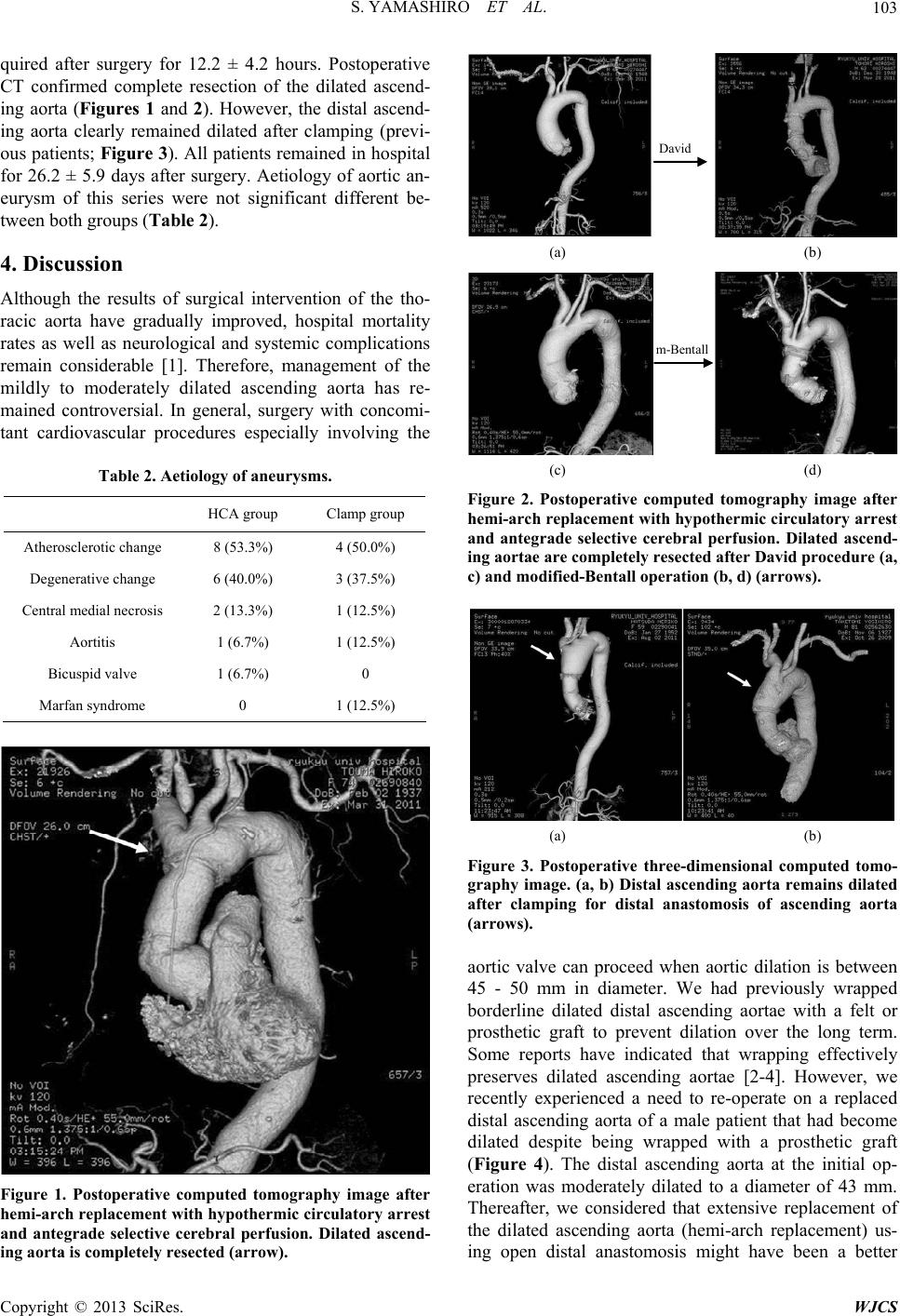 S. YAMASHIRO ET AL. 103 quired after surgery for 12.2 ± 4.2 hours. Postoperative CT confirmed complete resection of the dilated ascend- ing aorta (Figures 1 and 2). However, the distal ascend- ing aorta clearly remained dilated after clamping (previ- ous patients; Figure 3). All patients remained in hospital for 26.2 ± 5.9 days after surgery. Aetiology of aortic an- eurysm of this series were not significant different be- tween both groups (Table 2). 4. Discussion Although the results of surgical intervention of the tho- racic aorta have gradually improved, hospital mortality rates as well as neurological and systemic complications remain considerable [1]. Therefore, management of the mildly to moderately dilated ascending aorta has re- mained controversial. In general, surgery with concomi- tant cardiovascular procedures especially involving the Table 2. Aetiology of aneurysms. HCA group Clamp group Atherosclerotic change 8 (53.3%) 4 (50.0%) Degenerative change 6 (40.0%) 3 (37.5%) Central medial n e c rosis 2 (13.3%) 1 (12.5%) Aortitis 1 (6.7%) 1 (12.5%) Bicuspid valve 1 (6.7%) 0 Marfan syndrome 0 1 (12.5%) Figure 1. Postoperative computed tomography image after hemi-arch replacement with hypothermic circulatory arrest and antegrade selective cerebral perfusion. Dilated ascend- ing aorta is completely resected (arrow). David (a) (b) m-Bentall (c) (d) Figure 2. Postoer hemi-arch ry arrest perative computed tomography image aft replacement with hypothermic circulato and antegrade selective cerebral perfusion. Dilated ascend- ing aortae are completely resected after David procedure (a, c) and modified-Bentall operation (b, d) (arrows). (a) (b) Figure 3. Posed tomo- graphy ims dilated e can proceed when aortic dilation is between 5 - 50 mm in diameter. We had previously wrapped toperative three-dimensional comput age. (a, b) Distal ascending aorta remain after clamping for distal anastomosis of ascending aorta (arrows). aortic valv 4 borderline dilated distal ascending aortae with a felt or prosthetic graft to prevent dilation over the long term. Some reports have indicated that wrapping effectively preserves dilated ascending aortae [2-4]. However, we recently experienced a need to re-operate on a replaced distal ascending aorta of a male patient that had become dilated despite being wrapped with a prosthetic graft (Figure 4). The distal ascending aorta at the initial op- eration was moderately dilated to a diameter of 43 mm. Thereafter, we considered that extensive replacement of the dilated ascending aorta (hemi-arch replacement) us- ing open distal anastomosis might have been a better Copyright © 2013 SciRes. WJCS 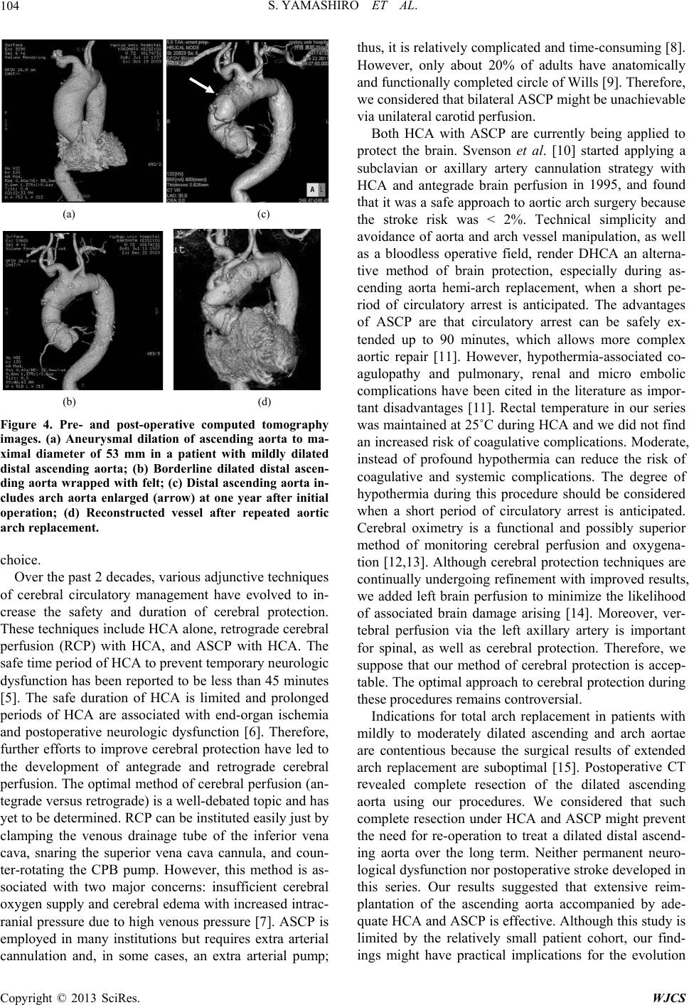 S. YAMASHIRO ET AL. 104 (a) (c) (b) (d) Figure 4. Pre- and post-operative computed tomography images. (a) Aneurysmal dilation of ascending aorta to ma- ximal diameter of 53 mm in with mildly dilated distal asceal ascen- ory management have evolved to in- rease the safety and duration of cerebral protection. niques include HCA alone, retrograde cerebral pe sion in 1995, and found th operative CT re a patient nding aorta; (b) Borderline dilated dist ding aorta wrapped w ith felt; (c) Distal ascending aorta in- cludes arch aorta enlarged (arrow) at one year after initial operation; (d) Reconstructed vessel after repeated aortic arch replacement. choice. Over the past 2 decades, various adjunctive techniques of cerebral circulat c These tech rfusion (RCP) with HCA, and ASCP with HCA. The safe time period of HCA to prevent temporary neurologic dysfunction has been reported to be less than 45 minutes [5]. The safe duration of HCA is limited and prolonged periods of HCA are associated with end-organ ischemia and postoperative neurologic dysfunction [6]. Therefore, further efforts to improve cerebral protection have led to the development of antegrade and retrograde cerebral perfusion. The optimal method of cerebral perfusion (an- tegrade versus retrograde) is a well-debated topic and has yet to be determined. RCP can be instituted easily ju st by clamping the venous drainage tube of the inferior vena cava, snaring the superior vena cava cannula, and coun- ter-rotating the CPB pump. However, this method is as- sociated with two major concerns: insufficient cerebral oxygen supply and cerebral edema with increased intrac- ranial pressure due to high venous pressure [7]. ASCP is employed in many institutions but requires extra arterial cannulation and, in some cases, an extra arterial pump; thus, it is relatively complicated and time-consuming [8]. However, only about 20% of adults have anatomically and functionally completed circle of Wills [9]. Therefor e, we considered that bilateral ASCP might be unachievable via unilateral carotid perfusion. Both HCA with ASCP are currently being applied to protect the brain. Svenson et al. [10] started applying a subclavian or axillary artery cannulation strategy with HCA and antegrade brain perfu at it was a safe approach to aortic arch surgery because the stroke risk was < 2%. Technical simplicity and avoidance of aorta and arch vessel manipulation, as well as a bloodless operative field, render DHCA an alterna- tive method of brain protection, especially during as- cending aorta hemi-arch replacement, when a short pe- riod of circulatory arrest is anticipated. The advantages of ASCP are that circulatory arrest can be safely ex- tended up to 90 minutes, which allows more complex aortic repair [11]. However, hypothermia-associated co- agulopathy and pulmonary, renal and micro embolic complications have been cited in the literature as impor- tant disadvantages [11]. Rectal temperature in our series was maintained at 25˚C during HCA and we did not find an increased risk of coagulative complications. Moderate, instead of profound hypothermia can reduce the risk of coagulative and systemic complications. The degree of hypothermia during this procedure should be considered when a short period of circulatory arrest is anticipated. Cerebral oximetry is a functional and possibly superior method of monitoring cerebral perfusion and oxygena- tion [12,13]. Although cerebral protection techniques are continually undergo ing refinement with improved results, we added left brain perfusion to minimize the likelihood of associated brain damage arising [14]. Moreover, ver- tebral perfusion via the left axillary artery is important for spinal, as well as cerebral protection. Therefore, we suppose that our method of cerebral protection is accep- table. The optimal approach to cerebral protection during these procedures remains controversial. Indications for total arch replacement in patients with mildly to moderately dilated ascending and arch aortae are contentious because the surgical results of extended arch replacement are suboptimal [15]. Post vealed complete resection of the dilated ascending aorta using our procedures. We considered that such complete resection under HCA and ASCP might prevent the need for re-operation to treat a dilated distal ascend- ing aorta over the long term. Neither permanent neuro- logical dysfunction nor postoperative stroke developed in this series. Our results suggested that extensive reim- plantation of the ascending aorta accompanied by ade- quate HCA and AS CP is effective. Although this study is limited by the relatively small patient cohort, our find- ings might have practical implications for the evolution Copyright © 2013 SciRes. WJCS  S. YAMASHIRO ET AL. Copyright © 2013 SciRes. WJCS 105 D. Spielvogel, A. Apaygin, S. L. Lansman, J. N. McCullough, J. D. Galla and R. B. Griepp, “Surgical Treatment of a: When and How?” The Ary, Vol. 67, No. 6 of controversial cases. Aetiology of aortic aneurysm of this series was not significant different between both groups. We considered that it is important to the distal aortic diameter. REFERENCES [1] M. A. Ergin, the Dilated Ascending Aort nnals of Thoracic Surge, 1999, pp. 1834-1839. doi:10.1016/S0003-4975(99)00439-7 [2] O. Cohen, J. Odim, D. D. L. Zerda, C. Ukatu, R. Vyas, N. Vyas, K. Oaratnik and H. Laks, “Long Term Experience of Girdling the Ascending Aorta with Dacron Mesh as Definitive Treatment for Aneurismal Dilatation,” The Annals of Thoracic Surgery, Vol. 83, No. 2, 2007, pp. S780-S784. doi:10.1016/j.athoracsur.2006.10.086 [3] G. I. Tagarakis, D. Karangelis, A. Baddour, M. E. Daska- lopouls, V. T. Liouras, D. Papadopoulos, K. Stamoulis, S. S. Lampoura and N. B. Tsilimingas, “An Alternate Solu- tion for the Treatment of Ascending Aortic Aneurysms: The Wrapping Technique,” Journal of Cardiothoracic Surgery, Vol. 5, 2010, p. 100. doi:10.1186/1749-8090-5-100 [4] E. Tappainer, V. Fiorani, A. Nocchi, E. Likaj, S. Mem- ishaj and M. Zogno, “Safe Wrapping of the Borderline Dilated Ascending Aorta during Aortic Valve Replace- ment,” Journal of Cardiothoracic Surgery, Vol. 2, 2007, p. 15. doi:10.1186/1749-8090-2-15 [5] A. Gega, J. A. Rizzo, M. H. Johnson, M. Tranquilli, E. A. Farkas and J. A. Elefteriades, “Straight Deeo Hypother- mic Arrest: Experience in 394 Patients Supports Its Ef- fectiveness as a Sole Means of Brain Preservation,” The Annals of Thoracic Surgery, Vol. 84, No. 3, 2007, pp. 759-767. doi:10.1016/j.athoracsur.2007.04.107 [6] M. A. Ergin, J. D. Galla, S. L. Lansman, C. Quintana, C. Bodian and R. B. Gripp, “Hypothermic Circulatory Arrest in Operations on the Thoracic Aorta. Determinants of Operative Mortality and Neurologic Outcome,” The Jour- nal of Thoracic and Cardiovascular Surgery, Vol. 107, No. 3, 1994, pp. 788-797. [7] A. Usui, K. Oohara, T. L. Liu, M. Murase, M. Tanaka , E. Takeuchi and T. Abe, “Determination of Optimum Ret- rograde Cerebral Perfusion Conditions,” The Journal of Thoracic and Cardiovascular Surgery, Vol. 107, No. 1, 1994, pp. 300-308. [8] H. Ogino, H. Sasaki, K. Minatoya, H. Matsuda, H. Ta- naka, H. Watanuki, M. Ando and S. Kitamura, “Evolving Arch Surgery Using Integrated Selective Cerebral Perfu- sion: Impact of Axillary Artery Perfusion,” The Journal of Thoracic and Cardiovascular Surgery, Vol. 136, No. 3, 2008, pp. 641-648. doi:10.1016/j.jtcvs.2008.02.089 [9] S. Morita, M. Yasaka, K. Yasumori, Y. Oishi, T. Takase- ya, H. Sonoda and T. Kawara, “Transcranial Doppler Study to Assess Intracranial Arterial Communication be- fore Aortic Arch Operation,” The Annals of Thoracic Surgery, Vol. 86, No. 2, 2008, pp. 448-451. doi:10.1016/j.athoracsur.2008.04.024 [10] L. G. Svenson, E. M. Nadolny, D. L. Penney, J. W. A. Kimmel, M. H. Entrup and Jacobson, R. S. D’Agostino, “Prospective Randomized Neurocognitive and S-100 Study of Hypothermic Circulatory Arrest, Retrograde Brain Perfusion, and Antegrade Brain Perfusion for Aor- tic Arch Operations,” The Annals of Thoracic Surgery, Vol. 71, No. 6, 2001, pp. 1905-1912. doi:10.1016/S0003-4975(01)02570-X [11] M. Eusanio, R. M. J. Wesselink, W. Dossche and M. A. A. M. Schpens, “D Morsunis, K. M. eep Hypothermic Circulatory Arrest and Antegrade Selective Cerebral Per- fusion during Ascending Aorta-Hemiarch Replacement: A Retrospective Comparative Study,” The Journal of Thoracic and Cardiovascular Surgery, Vol. 125, No. 4, 2003, pp. 849-854. doi:10.1067/mtc.2003.8 [12] N. Fassiadis, H. Zayed, H. Rashid and D. W. Green, “In- vos Cerebral Oximeter Compared with the Transcranial k and J. C. Stanley, “Evaluation of a Cerebral Oxi- Doppler for Monitoring Adequacy of Cerebral Perfusion in Patients Undergoing Carotid Endarterectomy,” Inter- national Journal of Angiology, Vol. 25, 2006, pp. 401- 406. [13] S. K. Samra, E. A. Dy, K. Welch, P. Dorje, G. B. Ze- lenoc meter as a Monitor of Cerebral Ischemia during Carotid Endarterectomy,” Anesthesiology, Vol. 93, No. 4, 2000, pp. 964-970. doi:10.1097/00000542-200010000-00015 [14] S. Yamashiro, Y. Kuniyoshi, K. Arakaki, H. Inafuku, Y. Morishima and Y. Kise, “Total Arch Replacement Using ns for Elective Surgi- Bilateral Axillary Antegrade Selective Cerebral Perfu- sion,” Annals of Thoracic and Cardiovascular Surgery, Vol. 16, No. 4, 2010, pp. 216-219. [15] R. Codecasa, M. A. Mariani, A. D’Alfonso, C. Nardi and J. G. Grandjean, “Current indicatio cal Treatment of Dilated Ascending Aorta: A New For- mula,” Annals of Thoracic and Cardiovascular Surger, Vol. 125, No. 6, 2003, pp. 1528-1530. doi:10.1016/S0022-5223(03)00059-X |