

Paper Menu >>
Journal Menu >>
 Journal of Environmental Protection, 2013, 4, 38-42 http://dx.doi.org/10.4236/jep.2013.46A005 Published Online June 2013 (http://www.scirp.org/journal/jep) Analysis of Sheep Lymphocyte Chromosomal Aberrations after Exposition to Chlortetracycline Irena Šutiaková1, Václav Šutiak2, Mária Tulenková1 1Department of Biology, Faculty of Humanities and Natural Sciences, University of Prešov, Prešov, Slovak Republic; 2St. Elizabeth University College of Health and Social Work, Bratislava, Slovak Republic. Email: [email protected] Received April 19th, 2013; revised May 17th, 2013; accepted June 7th, 2013 Copyright © 2013 Irena Šutiaková et al. This is an open access article distributed under the Creative Commons Attribution License, which permits unrestricted use, distribution, and reproduction in any medium, provided the original work is properly cited. ABSTRACT Tetracyclines are broad spectrum antibiotics and one type of various their compounds—chlortetracycline has been suc- cessfully used for our cytogenetic studies. We have selected for these studies the chromosomal assay because it is well- known cytogenetic biomarker, which has been used to assess DNA damage at the chromosomal level. We analyzed the chromosomal aberrations and mitotic index in peripheral lymphocytes of sheep exposed by chlortetracycline chloride in pure tap water. To the food of the experimental group of animals (à n = 6) were added also 0.35 g of preparation Aureovit 12 C 80 plv. a. u. v. per kg b. w. and day (i.e. 168 mg of chlortetracycline hydrochloride/kg b. w.) daily in food. The frequencies of aberrant cells (ABC) in the experimental and control groups of sheep were stated to differ sig- nificantly (P = < 0.001). In experimental group, chromatid breaks were the dominant type of chromosomal aberrations. No statistical differences in mitotic index values were found in the both groups (P > 0.05). Increased frequencies of chromosome aberrations in peripheral lymphocytes of sheep exposed by chlortetracycline in food, suggested a potential hazard which needs the attention from the viewpoint of human and animal health. This knowledge is useful for all, for the human and veterinary medicine, as well as for the aquaculture. Many of these chlortetracycline substances for their environmental persistence and chemical unstability are present in the environment for various periods, and as a result, they are responsible for several effects on human and animal health through food chain. Keywords: Antibiotics; Ovine Lymphocytes; Chromosomal Assay; Genotoxicity; Cytotoxicity; Health; Environment 1. Introduction Tetracyclines (TCs) are broad spectrum antibiotics that have been successfully used worldwide in human and ve- terinary medicine (mainly for medical therapy of dis- eases) and in aquaculture [1-4]. However their effect may be significantly limited [5]. Many of these substances for their environmental persistence and chemical unstability, are present in the environment for long period, and as a result they are responsible for several effects on human and animal health through food chain [6]. TCs are known to possess limited stability on aqueous media and abiotic degradation products or reversible epimers may be formed through hydrolysis or photolysis, including epi-TCs and anhydro-TCs, as well as iso-TCs for chlortetracycline (CTC), [7,8]. Capleton et al. [9] proposed a method for veterinary medicine active ingredients according to esti- mates of their potential for indirect human exposure via the environment and their toxicity profile. Greater em- phasis was given to more serious health endpoints, such as carcinogenity and mutagenicity. Genotoxicity is the genetic damage, due to DNA damaging agents including drugs by different mechanisms. Cytotoxic and genotoxic properties of tetracycline. HCl (TC) have been studied in human fibroblast experiments and human peripheral lym- phocytes [10-12]. Cytogenetic endpoints (chromosomal aberrations, mi- cronuclei and sister chromatid exchanges) are most fre- quently used in hazard identification assays as the crite- rion in the risk assessment process [13-16]. It is neces- sary to investigate how TCs may affect target organ- isms-microorganisms and also non-target organisms. Ac- cording to van der Schalie et al. [17], the use of non- human organisms, as early warning systems for human health risk is not new. Sentinel animal models could in- volve mammalian or non-mammalian species, domestic animals, or wildlife species. Sentinel animal populations would be exposed to a single chemical or a complex Copyright © 2013 SciRes. JEP  Analysis of Sheep Lymphocyte Chromosomal Aberrations after Exposition to Chlortetracycline 39 mixture, or to different media (e.g. air, water, soil, and sediment) in various locations. CTC is usually administered in the feed or water for prophylactic and therapeutic purposes. To provide addi- tional genotoxicity data, we studied the effect of chlor- tetracycline chloride (preparation Aureovit 12 C 80 plv. a.u.v.) in peripheral blood lymphocytes of sheep after its subchronical administration on the induction chromoso- mal aberrations, as cytogenetic endpoint. 2. Materials and Methods 2.1. Experimental Animals and Nutrition In the experiment two-months old Slovak merino ewes weighing 15.2 ± 1.05 kg were used. The animals were purchased from a private breeder. Prior to their inclusion in the experiments, they were nursed by mothers in such a way that they had limited access to drinking water and received no additional feed. Two weeks before the expe- riment the lambs were adapted to experimental housing conditions. The animals were fed with the ČOJ-2 feeding mixture ration for animals and with meadow hay at doses of 250 - 300 g of feeding mixture and 350 g dose of hay per each animal, respectively. Water was obtained at the “Výcho- doslovenské vodárne a kanalizácie” enterprise in Košice, where chlorine levels and water pH were determined ac- cording to the valid standard STN 75 7111 [18]. For 30 days the lambs in the control group were given the same amounts of feed and water, they had been receiving dur- ing the adaptation period. 2.2. Scheme of the Experiment The animals of the experimental group (n = 6) received the ČOJ-2 feeding mixture for animals and it was sup- plemented in this group only with chlortetracycline hy- drochloride (preparation Aureovit 12 C 80 plv. a.u.v., containing 8% of the ingredient, 0.5 kg packaging) in such a way that each lamb consumed 0.35 g of the pre- paration per kg live weight (i.e. 168 mg of chlortetra- cycline hydrochloride/kg b.w./day, added to the feeding mixture. The total quantity of the feeding mixture and meadow hay supplied was the same also in the control group (also n = 6 animals), however without CTC. 2.3. Lymphocyte Cultures After 30 days, blood samples were taken from jugular vein (v. jugularis) from each animal individually into glass tubes containing heparin (100 IU per ml blood). Heparinized blood (0.4 ml) was cultured at 37.5˚C for 48 h with 7.0 ml chromosome medium S-chromo-cell sup- plemented with FCS, PHA, and L-glutamine (PAN Sys- tems GmbH, Biotechnologische Produkte, Germany). Antibiotics were added to the culturing medium as fol- lows: penicillin G 100 IU/ml, streptomycin 100 µg/ml and 7.5% NaHCO3. Two hours prior to harvest the cul- tures were treated with the spindle inhibitor colchicin (Fluka Biochemika) at the concentration of 10 µg/ml to arrest cells in metaphase. The squashes were stained with a 10% solution of Giemsa-Romanovski stain in phosphate buffer (pH = 7.0). 2.4. Cytogenetic Assay For each donor, 100 well spread metaphases were scored for chromosomal aberrations studies (magnification 1000×) using microscope (Nikon). Images were taken by imaging microscopy (Nikon) using CCD-100 camera system (Mitsubishi). The images were processed using the Animal and Photostyler software system. The chro- mosomal aberrations were scored according to the classi- fication criteria suggested by Savage [19]. The identifi- cation of the individual chromosome pairs was carried out according to the standardized karyotyping of the do- mestic sheep [20]. Chromatid breaks were distinguished from gaps when the centric piece was displaced with re- spect to the chromosome axis, or the size of the discon- tinuity exceeded the width of the chromatid. Gap aberra- tions were excluded from the total number of aberrations and considered separately. Chromosome exchanges were classified as stable (translocations, when detection was possible) and unstable (dicentric, ring and fragments). 2.5. Mitotic Index In the chromosomal aberration study the mitotic index (MI) was evaluated by counting at least 1000 cells per treatment and dividing the number of splitting cells (me- taphases) by the total number of cells. 2.6. Statistical Evaluation of the Results The Sigma Stat program (Statistical software™, Jandel Scientific) was employed for statistical evaluation of the results. The statistically significant differences between experimental and control groups were determined using analysis of variance (ANOVA) followed by Tukey test at the 95% confidence level. 3. Results and Discussion The effect of CTC on the frequency of chromosome ab- errations in peripheral lymphocytes of ewe lambs is summarized in Table 1. The experimental group of ani- mals exhibited a significantly higher frequency of genetic damage (20.50% ± 5.24% aberrant cells) (ABC) com- pared to control group animals (4.00% ± 1.095% ABC; P =< 0.001, ANOVA followed by Tukey test). In the ex- perimental group, chromatid breaks were the dominant Copyright © 2013 SciRes. JEP 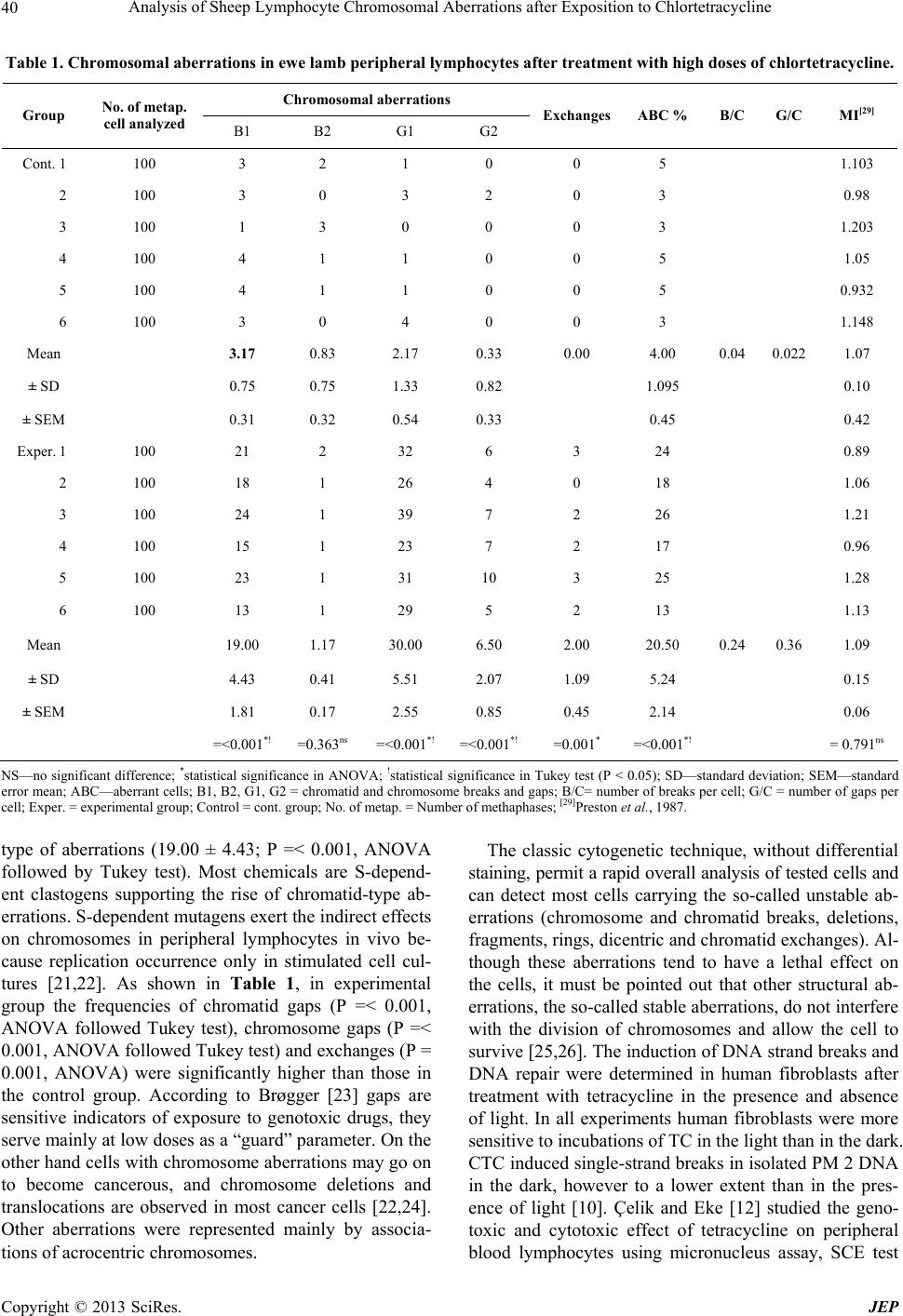 Analysis of Sheep Lymphocyte Chromosomal Aberrations after Exposition to Chlortetracycline Copyright © 2013 SciRes. JEP 40 Table 1. Chromosomal aberrations in ewe lamb peripheral lymphocytes after treatment with high doses of chlortetracycline. Chromosomal aberrations Group No. of metap. cell analyzed B1 B2 G1 G2 ExchangesABC % B/C G/C MI[29] Cont. 1 100 3 2 1 0 0 5 1.103 2 100 3 0 3 2 0 3 0.98 3 100 1 3 0 0 0 3 1.203 4 100 4 1 1 0 0 5 1.05 5 100 4 1 1 0 0 5 0.932 6 100 3 0 4 0 0 3 1.148 Mean 3.17 0.83 2.17 0.33 0.00 4.00 0.04 0.022 1.07 ± SD 0.75 0.75 1.33 0.82 1.095 0.10 ± SEM 0.31 0.32 0.54 0.33 0.45 0.42 Exper. 1 100 21 2 32 6 3 24 0.89 2 100 18 1 26 4 0 18 1.06 3 100 24 1 39 7 2 26 1.21 4 100 15 1 23 7 2 17 0.96 5 100 23 1 31 10 3 25 1.28 6 100 13 1 29 5 2 13 1.13 Mean 19.00 1.17 30.00 6.50 2.00 20.50 0.24 0.36 1.09 ± SD 4.43 0.41 5.51 2.07 1.09 5.24 0.15 ± SEM 1.81 0.17 2.55 0.85 0.45 2.14 0.06 =<0.001*! =0.363ns =<0.001*! =<0.001*! =0.001* =<0.001*! = 0.791ns NS—no significant difference; *statistical significance in ANOVA; !statistical significance in Tukey test (P < 0.05); SD—standard deviation; SEM—standard error mean; ABC—aberrant cells; B1, B2, G1, G2 = chromatid and chromosome breaks and gaps; B/C= number of breaks per cell; G/C = number of gaps per cell; Exper. = experimental group; Control = cont. group; No. of metap. = Number of methaphases; [29]Preston et al., 1987. type of aberrations (19.00 ± 4.43; P =< 0.001, ANOVA followed by Tukey test). Most chemicals are S-depend- ent clastogens supporting the rise of chromatid-type ab- errations. S-dependent mutagens exert the indirect effects on chromosomes in peripheral lymphocytes in vivo be- cause replication occurrence only in stimulated cell cul- tures [21,22]. As shown in Table 1, in experimental group the frequencies of chromatid gaps (P =< 0.001, ANOVA followed Tukey test), chromosome gaps (P =< 0.001, ANOVA followed Tukey test) and exchanges (P = 0.001, ANOVA) were significantly higher than those in the control group. According to Brøgger [23] gaps are sensitive indicators of exposure to genotoxic drugs, they serve mainly at low doses as a “guard” parameter. On the other hand cells with chromosome aberrations may go on to become cancerous, and chromosome deletions and translocations are observed in most cancer cells [22,24]. Other aberrations were represented mainly by associa- tions of acrocentric chromosomes. The classic cytogenetic technique, without differential staining, permit a rapid overall analysis of tested cells and can detect most cells carrying the so-called unstable ab- errations (chromosome and chromatid breaks, deletions, fragments, rings, dicentric and chromatid exchanges). Al- though these aberrations tend to have a lethal effect on the cells, it must be pointed out that other structural ab- errations, the so-called stable aberrations, do not interfere with the division of chromosomes and allow the cell to survive [25,26]. The induction of DNA strand breaks and DNA repair were determined in human fibroblasts after treatment with tetracycline in the presence and absence of light. In all experiments human fibroblasts were more sensitive to incubations of TC in the light than in the dark. CTC induced single-strand breaks in isolated PM 2 DNA in the dark, however to a lower extent than in the pres- ence of light [10]. Çelik and Eke [12] studied the geno- toxic and cytotoxic effect of tetracycline on peripheral blood lymphocytes using micronucleus assay, SCE test  Analysis of Sheep Lymphocyte Chromosomal Aberrations after Exposition to Chlortetracycline 41 and measuring mitotic activity and proliferation index (PRI) or/and nuclear division index (NDI) respectively in vitro. Their results indicated that tetracycline is able to induce both cytotoxic and moderate genotoxic effects in cultured human blood lymphocytes in vitro. No statistical differences were found in MI values in the groups (Table 1; P > 0.05). Cellular proliferation is a phenomenon that may be causally associated with the induction of chromo- somal damage for a number of compounds which are non-DNA reactive and thus are threshold in vitro clasto- gens [27]. Changes in biochemical biomarkers (antioxi- dant enzymes, catalase and superoxide dismutase) and DNA damage in soil on the earthworm Eisenia fetida ex- posed to tetracycline and chlortetracycline were investi- gated. Compared to enzyme activities, DNA damage as a biomarker was more sensitive and more suitable for de- tection low concentration exposure the genotoxicity of contaminants in terestrial environment [28]. The both an- tibiotics induced significant genotoxicity on earthworm Eisenia fetida in a dose—dependent manner (P < 0.01). 4. Conclusion On the whole, when we consider our results together with other published data our results demonstrate that chlortetracycline chloride (preparation Aureovit 12 C 80 plv. a.u.v.) induced a significant increase in chromoso- mal aberrations. These findings suggest a potential geno- toxic hazard of this antibacterial drug as it may become capable of attacking the animal genetic material. In our experimental group of animals, chromatid breaks were the dominant type of chromosomal aberrations. Our ex- periences demonstrated that chromosomal damage stud- ies may be used as the sensitive and effective biomarker procedure for detecting the genotoxicity effect of hazar- dous agents from the viewpoint of animal health. This is the argument also for the further studies and other exa- mination especially in connection with the human health state because residues of hazardous agents in animal food- stuffs may be also their hazard factor. 5. Acknowledgements This work was supported by the Grant Vega ME SR No. 1/0545/08. REFERENCES [1] J. Sokol and E. Matisova, “Determination of Tetracycline Antibiotics in Animal Tissues of Food-Producing Ani- mals by High-Performance Liquid Chromatography Us- ing Solid-Phase Extraction,” Journal of Chromatography, Vol. 669, No. 1-2, 1994, pp. 75-80. doi:10.1016/0021-9673(94)80338-2 [2] B. F. Kania, “Farmakologia Weterynaryjna Z Elementami Terapii. (Ed.), Wydawnictvo Lekarskie PZWL,” Wars- zava, 2001. [3] W. C. Andersen, J. E. Roybal, S. A. Gonzales, S. B. Tur- nipseed, A. P. Pfenning and L. R. Kuck, “Determination of Tetracycline Residues in Shrimp and Whole Milk Us- ing Liquid Chromatography with Ultraviolet Detection and Residue Confirmation by Mass Spectrometry,” Ana- lytica Chimica Acta, Vol. 529, No. 1-2, 2005, pp. 145- 150. doi:10.1016/j.aca.2004.08.012 [4] D. Lin, Q. Zhou, Y. Xu, Ch. Chen and Y. Li, “Physio- logical and Molecular Responses of the Earthworm (Ei- senia fetida) to Soil Chlortetracycline Contamination,” En- vironmental P ollution, Vol. 171, 2012, pp. 46-51. doi:10.1016/j.envpol.2012.07.020 [5] V. Šutiak and I. Šutiaková, “Principal, Harmful and Side Effects of Drugs in Animals,” Veterinary Journal Czech, Vol. 49, No. 2, 1999, pp. 77-81. [6] E. Martínez-Carbalo, C. Gonzáles-Barreiro, S. Scharf and O. Gans, “Environmental Monitoring Study of Selected Veterinary Antibiotics in Animal Manure and Soils in Austria,” Environmental Pollution, Vol. 148, No. 2, 2007, pp. 570-579. doi:10.1016/j.envpol.2006.11.035 [7] B. Halling-Sørensen, G. Sengeløv and J. Tjørnelund, “To- xicity of Tetracyclines and Tetracycline Degradation Pro- ducts to Environmentally Relevant Bacteria, Including Se- lected Tetracycline-Resistant Bacteria,” Archives of Envi- ronmental Contamination and Toxicology, Vol. 42, No. 3, 2002, pp. 263-271. doi:10.1007/s00244-001-0017-2 [8] B. Halling-Sørensen, A. Lykkeberg, F. Ingerslev, P. Black- well and J. Tjørnelund, “Characterisation of the Abiotic Degradation Pathways of Oxytetracyclines in Soil Inter- stitial Water Using LC-MS-MS,” Chemosphere, Vol. 50, No. 10, 2003, pp. 1331-1342. doi:10.1016/S0045-6535(02)00766-X [9] A. C. Capleton, C. Courage, P. Rumsby, Ph. Holmes, E. Stutt, A. B. A. Boxall and L. S. Levy, “Priorisiting Veteri- nary Medicines According to Their Potential Indirect Hu- man Exposure and Toxicity Profile,” Toxicology Letters, Vol. 163, No. 3, 2006, pp. 213-223. doi:10.1016/j.toxlet.2005.10.023 [10] J. Witte, G. Oetken, C. Buschhfort and A. Hartman, “A Comparison of the DNA-Damaging, the Cytotoxic and Ge- notoxic Properties of Tetracycline in Human Fibrablasts in the Presence and Absence of Light,” Mutation Re- search, Vol. 315, No. 1, 1994, pp. 33-40. doi:10.1016/0921-8777(94)90025-6 [11] A. Hartman, D. Wess and I. Witte, “Enhanced Cyto- and Genotoxicity of Tetracycline in Wilson Disease Fibro- blasts,” Mutation Research, Vol. 348, No. 1, 1995, pp. 7- 12. doi:10.1016/0165-7992(95)90014-4 [12] A. Çelik and D. Eke, “The Assessment of Cytotoxicity and Genotoxicity of Tetracycline Antibiotic in Human Blood Lymphocytes Using CBMN and SCE Analysis, in Vitro,” International Journal of Human Genetics, Vol. 11, No. 1, 2011, pp. 23-29. [13] J. D. Tucker and R. J. Preston, “Chromosome Aberrations, Micronuclei, Aneuploids Sister Chromatid Changes and Cancer Risk Assessment,” Mutation Research, Vol. 365, No. 1-3, 1996, pp. 147-159. doi:10.1016/S0165-1110(96)90018-4 Copyright © 2013 SciRes. JEP  Analysis of Sheep Lymphocyte Chromosomal Aberrations after Exposition to Chlortetracycline Copyright © 2013 SciRes. JEP 42 [14] H. Norppa, S. Bonassi, I. L. Hansteen, L. Hagmar, U. Strömberg, P. Rössner, P. Boffetta, C. Lindholm, S. Gun- dy, J. Lazutka, A. Cebulska-Wasilewska, A. Fabiánová, R. J. Srám, L. E. Knudsen, L. Barale and A. Fucic, “Chro- mosomal Aberrations and SCEs as Biomarkers of Cancer Risk,” Mutation Research, Vol. 600, No. 1-2, 2006, pp. 37-45. doi:10.1016/j.mrfmmm.2006.05.030 [15] M. Fenech, “Cytokinesis-Block Micronucleus Cytome As- says,” Nature Protocols, Vol. 2, No. 5, 2007, pp. 1084- 1104. doi:10.1038/nprot.2007.77 [16] C. Bolognesi, A. Creus, P. Ostrosky-Wegman and R. Mar- cos, “Review Micronuclei and Pescticide Exposure,” Mu- tagenesis, Vol. 26, No. 1, 2011, pp. 19-26. doi:10.1093/mutage/geq070 [17] W. A. Van der Schalie, J. H. Gardner, J. A. Buntle, C. H. T. De Rosa, R. A. Finch, J. S. Reif, R. H. Reuter, L. C. Bac- ker, J. Burger, L. C. Folmar and W. S. Stokes, “Animals as Sentinels of Human Health Hazards of Environmental Chemicals,” Environmental Health Perpectives, Vol. 107, No. 4, 1999, pp. 309-315. doi:10.1289/ehp.99107309 [18] A. Laciaková, E. Bekeová, V. Hendrichovský, P. Turek and V. Laciak, “K Problematikevýskytu Chlóru v Pitnej Vode a Jeho Vplyv na Obsah Chloridov V Krvnom Sére,” Hygiena, Vol. 44, 1999, pp. 78-84. [19] J. R. K. Savage, “Classification and Relationships of In- duced Chromosomal Structural Changes,” Journal of Me- dical Genetics, Vol. 12, 1975, pp. 103-122. [20] D. Di Berardino, G. P. Di Moe, D. S. Gullagher, H. Hayes and I. Iannuzii, “ISCNDB International System for Chro- mosome Nomenclature of Domestic Bovids (Coordina- tor),” Cytogenetics Cell Genetics, Vol. 92, No. 3-4, 2001, pp. 283-299. doi:10.1159/000056917 [21] M. Sorsa, J. Wilbourn and H. Vainio, “Human Cytoge- netic Damage as a Predictor of Cancer Risk,” In: H. Vainio, P. N. Magee, D. B. McGregor and A. B. Mc Mi- chael, Eds., Mechanisms of Carcinogenesis in Risk Iden- tification, International Agency for Research on Cancer, DARC, Lyon, 1992, pp. 543-554. [22] G. Obe, P. Pfeiffer, J. R. K. Savage and J. C. Johannes, W. Goedecke, P. Jeppesen, A. T. Natarajan, W. Martinez-Ló- pez, G. A. Folle and M. E. Drets, “Chromosomal Aberra- tions: Formation, Identification and Distribution,” Muta- tion Research, Vol. 504, No. 1-2, 2002, pp. 17-36. doi:10.1016/S0027-5107(02)00076-3 [23] A. Brøger, “The Chromatid Gap—A Useful Parameter in Genotoxicology?” Cytogenetetics Cell Genetics, Vol. 33, No. 1-2, 1982, pp. 14-19. doi:10.1159/000131720 [24] N. McGranahan, R. A. Burell, D. Endesfelder, M. R. No- velli and Ch. Swanton, “Cancer Chromosomal Instability: Therapeutic and Diagnostic Challenges,” EMBO Reports, Vol. 13, No. 6, 2012, pp. 528-538. doi:10.1038/embor.2012.61 [25] W. W. Au, “Monitoring Human Populations for Effects of Radiation and Chemical Exposures Using Cytogenetic Techniques,” Occupational Medicine, Vol. 6, 1991, pp. 597-611. [26] A. T. Natarajan and F. Palitti, “DNA Repair and Chromo- somal Alterations,” Mutation Research, Vol. 657, No. 1, 2008, pp. 3-7. doi:10.1016/j.mrgentox.2008.08.017 [27] L. Műller and P. Kasper, “Human Biological Relevance and the Use of Treshold-Arguments in Regulatory Geno- toxicity Assessment: Experience with Pharmaceutical,” Mu- tation Research, Vol. 464, No. 1, 2000, pp. 19-34. doi:10.1016/S1383-5718(99)00163-1 [28] L. Dong, J. Gao, X. Xie and Q. Zhou, “DNA Damage and Biochemical Toxicity of Antibiotics in Soil on the Earth- worm Eisenia fetida,” Chemosphere, Vol. 89, No. 1, 2012, pp. 44-51. doi:10.1016/j.chemosphere.2012.04.010 [29] R. J. Preston, B. J. Dean, S. Galloway, H. Holden, A. F. McFee and H Shelby, “Mammalian in Vivo Cytogenetic Assays: Analysis of Chromosome Aberration in Bone Mar- row Cells,” Mutation Research, Vol. 189, No. 2, 1987, pp. 157-165. doi:10.1016/0165-1218(87)90021-8 |