

Paper Menu >>
Journal Menu >>
 American Journal of Plant Sciences, 2013, 4, 36-41 http://dx.doi.org/10.4236/ajps.2013.46A006 Published Online June 2013 (http://www.scirp.org/journal/ajps) Identification of Alternaria spp. as Pathogenic on the Native Species Terminalia australis and Salvia guaranitica Mariana Kameniecki1,2, Eduardo R. Wright1*, Marta C. Rivera1 1Plant Pathology, School of Agronomy, University of Buenos Aires, Buenos Aires, Argentina; 2Asociación Ribera Norte, Buenos Aires, Argentina. Email: *[email protected] Received April 16th, 2013; revised May 16th, 2013; accepted June 1st, 2013 Copyright © 2013 Mariana Kameniecki et al. This is an open access article distributed under the Creative Commons Attribution License, which permits unrestricted use, distribution, and reproduction in any medium, provided the original work is properly cited. ABSTRACT This work is the result of activities included in the cooperative project between Asociación Ribera Norte (ARN) and the Department of Plant Pathology, School of Agronomy, University of Buenos Aires. ARN is a non-governmental organi- zation created in 1993 mainly for the management of the Reserva Ecológica Municipal Ribera Norte, a preservation area located in San Isidro (North of Buenos Aires), within the Provincia Paranaense region. The aims were to identify diseases in native plant species growing there. Light brown leaf spots with thin brown margins and narrow yellow halos were observed on Terminalia australis (mean disease severity: 25%). The pathogen was isolated, inoculated on healthy plants, reisolated from infected leaves and identified as Alternaria tenuissima. Similarly, another leaf-spot disease was observed on Salvia guaranitica, characterized by reddish brown large spots developed from the apexes and margins towards the centre of the leaves, reaching the veins in some cases (mean severity: 20%). Pathogen isolation, inoculation and reisolation led to the identification of A. alternata as the causal agent of the disease. Unusual heavy rain is analyzed as disease conductive. Keywords: Alternaria tenuissima; Terminalia australis; A. alternata; Salvia guaranitica; Diagnosis 1. Introduction Terminalia australis Cambess., belonging to Combreta- ceae, is known as “palo amarillo” or “ta n i m b ú”. It is a tree or shrub with grayish red cortex [1] and deciduous elliptical leaves, native to the Northeast of Argentina [2], and restricted in range to the more northerly provinces [3]. It is traditionally used to obtain wood [4] that is yel- low, compact, elastic, with remarkable durability [5] be- cause of the astringent properties of boiled leaves [6]. This species is also used as an antiseptic agent, as it was shown to possess antifungal properties against species of Aspergillus and Candida [7,8]. Salvia guaranitica A. St.-Hil. ex Benth. is a rhizome- tous species belonging to the Lamiaceae [2], usually re- ferred to as “salvia azul” [9]. It is a semi-woody peren- nial subshrub that exhibits a bushy, somewhat open habit with upright, branching, square, dark green stems. The leaves are dark green, ovate, wrinkled, pointed and ligh- tly-toothed. This species blooms from mid-summer to fall, producing long terminal spikes with bilabiate, tu- bular, deep blue flowers with purple-blue calyxes. It is an ornamental and medicinal plant originally from South Brazil, Paraguay, Uruguay and Argentina [10], tradition- ally used in Latin America as sedative [11]. Sedative and hypnotic properties of the ethanolic extracts [12] have been associated with the presence of high concentrations of the flavonoid cirsiliol and caffeic acid ethyl ester [13]. Aerial parts also contain essential oils [9,14]. Extracts from this species are innocuous to honey bees [15]. Salvia guaranitica has ornamental value [16] that has led to the evaluation of procedures for its propagation and domestication [17]. T. australis is also considered to have ornamental characteristics [18]. T. australis has been recorded in the provinces of Buenos Aires, Corrien- tes, Entre Ríos y Misiones; and S. guaranitica in Buenos Aires, Corrientes, Entre Ríos, Misiones, Salta and Tucumán [19]. Although there is scarce information on conserva- tion status of the flora in Argentina, both species have been found to be included in a list of endangered trees and shrubs in the province of Entre Ríos (Northeast of Argentina) [20]. These species were originated in the ecological region called Provincia Paranaense [21], *Corresponding author. Copyright © 2013 SciRes. AJPS 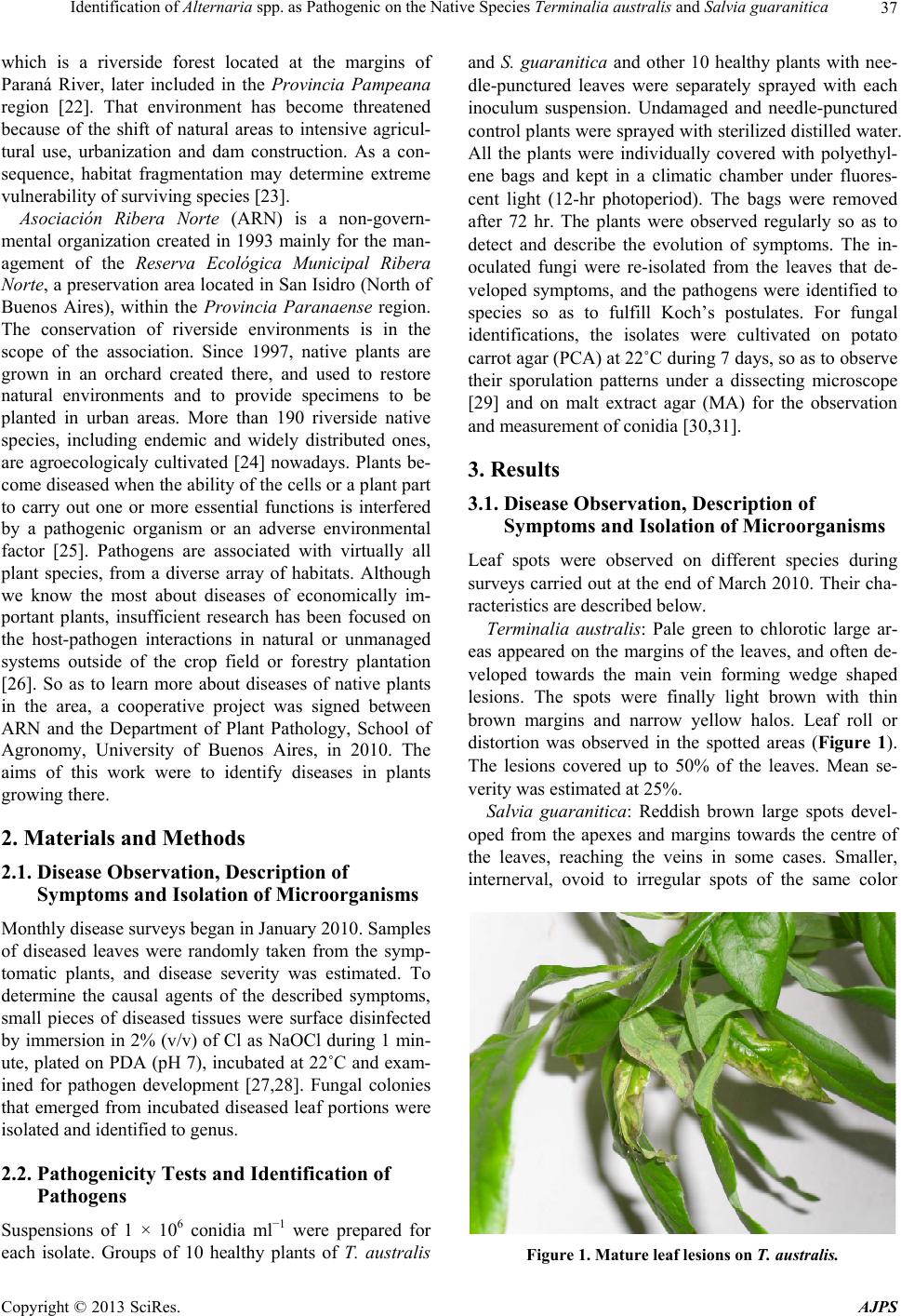 Identification of Alternaria spp. as Pathogenic on the Native Species Terminalia australis and Salvia guaranitica 37 which is a riverside forest located at the margins of Paraná River, later included in the Provincia Pampeana region [22]. That environment has become threatened because of the shift of natural areas to intensive agricul- tural use, urbanization and dam construction. As a con- sequence, habitat fragmentation may determine extreme vulnerability of surviving species [23]. Asociación Ribera Norte (ARN) is a non-govern- mental organization created in 1993 mainly for the man- agement of the Reserva Ecológica Municipal Ribera Norte, a preservation area located in San Isidro (North of Buenos Aires), within the Provincia Paranaense region. The conservation of riverside environments is in the scope of the association. Since 1997, native plants are grown in an orchard created there, and used to restore natural environments and to provide specimens to be planted in urban areas. More than 190 riverside native species, including endemic and widely distributed ones, are agroecologicaly cultivated [24] nowadays. Plants be- come diseased when the ability of the cells or a plant part to carry out one or more essential functions is interfered by a pathogenic organism or an adverse environmental factor [25]. Pathogens are associated with virtually all plant species, from a diverse array of habitats. Although we know the most about diseases of economically im- portant plants, insufficient research has been focused on the host-pathogen interactions in natural or unmanaged systems outside of the crop field or forestry plantation [26]. So as to learn more about diseases of native plants in the area, a cooperative project was signed between ARN and the Department of Plant Pathology, School of Agronomy, University of Buenos Aires, in 2010. The aims of this work were to identify diseases in plants growing there. 2. Materials and Methods 2.1. Disease Observation, Description of Symptoms and Isolation of Microorganisms Monthly disease surveys began in January 2010. Samples of diseased leaves were randomly taken from the symp- tomatic plants, and disease severity was estimated. To determine the causal agents of the described symptoms, small pieces of diseased tissues were surface disinfected by immersion in 2% (v/v) of Cl as NaOCl during 1 min- ute, plated on PDA (pH 7), incubated at 22˚C and exam- ined for pathogen development [27,28]. Fungal colonies that emerged from incubated diseased leaf portions were isolated and identified to genus. 2.2. Pathogenicity Tests and Identification of Pathogens Suspensions of 1 × 10⁶ conidia ml−1 were prepared for each isolate. Groups of 10 healthy plants of T. australis and S. guaranitica and other 10 healthy plants with nee- dle-punctured leaves were separately sprayed with each inoculum suspension. Undamaged and needle-punctured control plants were sprayed with sterilized distilled water. All the plants were individually covered with polyethyl- ene bags and kept in a climatic chamber under fluores- cent light (12-hr photoperiod). The bags were removed after 72 hr. The plants were observed regularly so as to detect and describe the evolution of symptoms. The in- oculated fungi were re-isolated from the leaves that de- veloped symptoms, and the pathogens were identified to species so as to fulfill Koch’s postulates. For fungal identifications, the isolates were cultivated on potato carrot agar (PCA) at 22˚C during 7 days, so as to observe their sporulation patterns under a dissecting microscope [29] and on malt extract agar (MA) for the observation and measurement of conidia [30,31]. 3. Results 3.1. Disease Observation, Description of Symptoms and Isolation of Microorganisms Leaf spots were observed on different species during surveys carried out at the end of March 2010. Their cha- racteristics are described below. Terminalia australis: Pale green to chlorotic large ar- eas appeared on the margins of the leaves, and often de- veloped towards the main vein forming wedge shaped lesions. The spots were finally light brown with thin brown margins and narrow yellow halos. Leaf roll or distortion was observed in the spotted areas (Figure 1). The lesions covered up to 50% of the leaves. Mean se- verity was estimated at 25%. Salvia guaranitica: Reddish brown large spots devel- oped from the apexes and margins towards the centre of the leaves, reaching the veins in some cases. Smaller, internerval, ovoid to irregular spots of the same color Figure 1. Mature leaf lesions on T. australis. Copyright © 2013 SciRes. AJPS 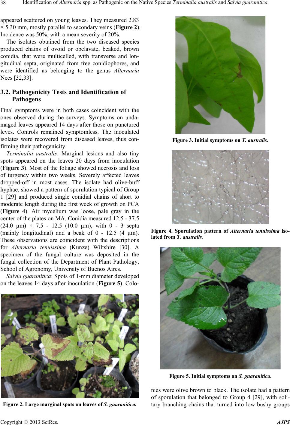 Identification of Alternaria spp. as Pathogenic on the Native Species Terminalia australis and Salvia guaranitica 38 appeared scattered on young leaves. They measured 2.83 × 5.30 mm, mostly parallel to secondary veins (Figure 2). Incidence was 50%, with a mean severity of 20%. The isolates obtained from the two diseased species produced chains of ovoid or obclavate, beaked, brown conidia, that were multicelled, with transverse and lon- gitudinal septa, originated from free conidiophores, and were identified as belonging to the genus Alternaria Nees [32,33]. 3.2. Pathogenicity Tests and Identification of Pathogens Final symptoms were in both cases coincident with the ones observed during the surveys. Symptoms on unda- maged leaves appeared 14 days after those on punctured leves. Controls remained symptomless. The inoculated isolates were recovered from diseased leaves, thus con- firming their pathogenicity. Terminalia australis: Marginal lesions and also tiny spots appeared on the leaves 20 days from inoculation (Figure 3). Most of the foliage showed necrosis and loss of turgency within two weeks. Severely affected leaves dropped-off in most cases. The isolate had olive-buff hyphae, showed a pattern of sporulation typical of Group 1 [29] and produced single conidial chains of short to moderate length during the first week of growth on PCA (Figure 4). Air mycelium was loose, pale gray in the center of the plates on MA. Conidia measured 12.5 - 37.5 (24.0 µm) × 7.5 - 12.5 (10.0 µm), with 0 - 3 septa (mainly longitudinal) and a beak of 0 - 12.5 (4 µm). These observations are coincident with the descriptions for Alternaria tenuissima (Kunze) Wiltshire [30]. A specimen of the fungal culture was deposited in the fungal collection of the Department of Plant Pathology, School of Agronomy, University of Buenos Aires. Salvia guaranitica : Spots of 1-mm diameter developed on the leaves 14 days after inoculation (Figure 5). Colo- Figure 2. Large marginal spots on leaves of S. guaranitica. Figure 3. Initial symptoms on T. australis. Figure 4. Sporulation pattern of Alternaria tenuissima iso- lated from T. australis. Figure 5. Initial symptoms on S. guaranitica. nies were olive brown to black. The isolate had a pattern of sporulation that belonged to Group 4 [29], with soli- tary branching chains that turned into low bushy groups Copyright © 2013 SciRes. AJPS  Identification of Alternaria spp. as Pathogenic on the Native Species Terminalia australis and Salvia guaranitica 39 of branched chains of spores (Figure 6). Conidia measured 17.0 - 40.0 (25.0 µm) × 5.0 - 17.0 (11.0 µm), with 0 - 4 septa (mainly longitudinal) and a beak of 0 - 7.5 (2.0 µm). The morphological characteristics of the conidia and conidiophores fit the description of A. alternata (Fr.) Keissl. [31]. A specimen of the fungal culture was deposited in the fungal collection of the De- partment of Plant Pathology, School of Agronomy, Uni- versity of Buenos Aires. 4. Discussion The pathogenic species identified in this work show cosmopolitan distribution. A total of 336 fungus-host combinations, and 568 records of A tenuissima and its synonyms can be found in the literature. Similarly, there are 914 fungus-host combinations, and 1496 records for A. alternata around the world [34]. No citations have been found on T. australis and S. guaranitica; only A. alternata on S. nemorosa in Poland [35, cited by 34]. As a result of this work, A. tenuissima and A. alternata are reported on T. australis and S. guaranitica, respectively, as causal agents of leaf spots. To our knowledge, it is the first report of the pathogens on these species, and might be the first pathogens identified on them. The period of incubation differed between the two diseases; 20 and 14 days for the pathosystems T. aus- tralis-A. tenuissima and S. guaranitica-A. alternata, re- spectively. Wounds accelerated symptom appeareance by two weeks for both pathologies. Local wounds are known to increase infection by all Alternaria pathogens [36]. How- ever, foliage injury cannot be considered a prerequisite for fungal penetration for these diseases. In fact, both pathogens succeeded to infect unwounded leaves, al- though with extended incubation period when compared with the infection on punctured foliage. It is generally accepted that vigorously growing plants are more resistant to infection by Alternaria pathogens than poorly developing ones, and that soil moisture is a major factor affecting the conditions of growth [36]. Figure 6. Sporulation pattern of Alternaria alternata iso- lated from S. guaranitica. Both diseases identified in this work were observed after unusual rainy conditions that occurred in February 2010, when accumulated rain reached 419.6 mm. Consequently, soil moisture should be considered as conductive for disease development in these cases. In addition, rain may have facilitated spore germination and penetration by supplying free moisture on leaves or by increasing rela- tive humidity [37]. Alternaria spores are dispersed by wind currents and are eventually deposited on the surface of susceptible tissues [38]. The germination of spores under high humidity conditions may take only few hours [39]. Our results constitute the first step in the management of plant diseases, that is the identification of their ethiology and the estimation of damages. Infectious dis- ease is a major causal factor in the demography of human, plant and animal populations [40]. An intensive work has to be done in order to identify the causal agents of new diseases on the studied hosts or other ones, and to understand the conducive factors, so as to evaluate tools to preserve native species. 5. Acknowledgements This work was supported by the University of Buenos Aires (FAUBA). REFERENCES [1] A. L. Cabrera and E. H. Zardini, “Manual de la Flora de los Alrededores de Buenos Aires,” ACME, Buenos Aires, 1978. [2] M. J. Dimitri, “Enciclopedia Argentina de Agricultura y Jardinería. Vol. I,” ACME, Buenos Aires, 1987. [3] A. W. Exell, “The Combretaceae of Argentina,” Lilloa, Vol. 5, 1940, pp. 123-130. [4] J. N. Cardoso Marchiori, “Descriptive Wood Anatomy of Terminalia australis Camb. (Combretaceae),” Revista do Centro de Ciências Rurais, Vol. 16, No. 4, 1886, pp. 329- 340. [5] M. P. Corréa, “Diccionário das Plantas Úteis do Brasil— E das Exóticas Cultivadas. Vol. 1,” Imprensa Nacional, Rio de Janeiro, 1926. [6] A. Bertucci, F. Haretche, C. Olivaro and A. Vázquez, “Phytochemical Screening of the Native Forest of Uru- guay River,” Revista Brasileira de Farmacognosia. Bra- zilian Journal of Pharmacognosy, Vol. 18, No. 1, 2008, pp. 21-25. [7] S. M. Carpano, E. D. Spegazzini, J. S. Rossi, M. T. Castro and S. L. Debenedetti, “Antifungal Activity of Terminalia australis,” Fitoterapia, Vol. 74, No. 3, 2003, pp. 294-297. doi:10.1016/S0367-326X(03)00026-1 [8] L. Muschietti, M. Derita, V. Sülsen, J. D. Muñoz, G. Ferraro, S. Zacchino and V. Martino, “In Vitro Antifungal Assay of Traditional Argentine Medicinal Plants,” Journal of Ethnopharmacology, Vol. 102, No. 2, 2005, pp. 233-238. Copyright © 2013 SciRes. AJPS  Identification of Alternaria spp. as Pathogenic on the Native Species Terminalia australis and Salvia guaranitica 40 doi:10.1016/j.jep.2005.06.017 [9] A. Lombardo, “Flora Montevidensis. Vol II. Gamopéta- las,” Intendencia Municipal de Montevideo, Montevideo, 1983. [10] C. Pereira, “Flora do Estado do Paraná,” Família Labiatae, Leandra, Vol. 1, 1971, pp. 13-33. [11] H. Viola, C. Wasowski, M. Marder, C. Wolfman, A. C. Paladini and J. H. Medina, “Sedative and Hypnotic Properties of Salvia guaranitica St. Hil. and of Its Active Principle, Cirsiliol,” Phytomedicine, Vol. 4, No. 1, 1997, pp. 47-62. doi:10.1016/S0944-7113(97)80027-X [12] J. H. Medina, C. Peña, M. Levi de Stein, C. Wolfman and A. C. Paladini, “Benzodiazepine-Like Molecules as Well as other Ligands for the Brain Benzodiazepine Receptor are Relative Common Constituents of Plants,” Biochemi- cal and Biophysical Research Communications, Vol. 165, No. 2, 1989, pp. 547-553. doi:10.1016/S0006-291X(89)80001-4 [13] M. Mader, H. Viola, C. Wasowski, C. Wolfman, P. G. Waterman, J. H. Medina and A. C. Paladini, “Cirsiliol and Caffeic Acid Ethyl Ester, Isolated from Salvia guara- nitica, are Competitive Ligands for the Central Benzodi- azepine Receptors,” Phytomedicine, Vol. 3, No. 1, 1996, pp. 29-31. doi:10.1016/S0944-7113(96)80006-7 [14] C. Vallverdú, R. Vila, D. Lorenzo, D. Paz, E. Dellacassa, P. Davies, J. Villamil, F. Tomi, J. Casanova and S. Ca- ñigueral, “Composition of the Essential Oil of Cultivated Salvia guaranitica from Uruguay,” Flavour and Fragance Journal, Vol. 20, No. 4, 2005, pp. 421-424. doi:10.1002/ffj.1450 [15] L. Castillo, A. González-Coloma, A. González, M. Díaz, E. Santos, E. Alonso-Paz, M. J. Bassagoda and C. Rossini, “Screening of Uruguayan Plants for Deterrent Activity against Insects,” Industrial Crops and Products, Vol. 29, No. 1, 2009, pp. 235-240. doi:10.1016/j.indcrop.2008.05.004 [16] INIA, “Estudios en Domesticación y Cultivo de Especies Medicinales y Aromáticas Nativas. Serie FPTA-INIA 11,” Instituto Nacional de Investigación Agropecuaria Uruguay, Montevideo, 2004. [17] S. Echeverrigaray, R. P. Carrer and L. B. Andrade, “Mi- cropropagation of Salvia guaranitica Benth. through Axil- lary Shoot Proliferation,” Brazilian Archives of Biology and Technology, Vol. 53, No. 4, 2010, pp. 883-888. doi:10.1590/S1516-89132010000400018 [18] E. Haene and G. Aparicio, “100 Árboles Argentinos,” Albatros, Buenos Aires, 2001. [19] Administración de Parques Nacionales, “Sistema de In- formación de Biodiversidad,” Administración de Parques Nacionales, 2013. http://www.sib.gov.ar/ [20] Sistema Provincial de Áreas Naturales Protegidas, “Lis- tado de Árboles y Arbustos de Entre Ríos y Especies Ve- getales en Peligro de Extinción de la Provincia,” Provin- cia de Entre Ríos, Entre Ríos, 1993. [21] A. W. Cabrera, “Fitogeografía. Vol. 3,” In: F. de Aparicio and H. A. Difrieri, Eds., La Argentina. Suma de Geo- grafía, Peuser, Buenos Aires, 1958, pp. 101-207. [22] A. L. Cabrera, “Regiones Fitogeográficas Argentinas,” In: W. F. Kugler, Ed., Enciclopedia Argentina de Agricultura y Jardinería, Vol. 2, ACME, Buenos Aires, 1994, pp. 1- 85. [23] M. S. Di Bitetti, G. Placci and L. A. Dietz, “Una Visión de Biodiversidad para la Ecorregión del Bosque Atlántico del Alto Paraná: Diseño de un Paisaje para la Conser- vación de la Biodiversidad y Prioridades para las Ac- ciones de Conservación,” World Wildlife Fund, Wash- ington DC, 2003. [24] M. A. Altieri, “Agroecology: The Science of Sustainable Agriculture,” Westview Press, Colorado, 1995. [25] G. N. Agrios, “Plant Pathology,” Elsevier Academic Press, New York, 2005. [26] H. M. Alexander, “Disease in Natural Plant Populations, Communities, and Ecosystems: Insights into Ecological and Evolutionary Processes,” Plant Disease, Vol. 94, No. 5, 2010, pp. 492-503. doi:10.1094/PDIS-94-5-0492 [27] A. L. Cheheid, E. R. Wright, S. E. Lopez and A. M. R. Codazzi, “First Report of Alternaria Leaf Spot of Spa- thiphyllum sp. in Argentina,” Plant Disease, Vol. 84, No. 3, 2000, p. 370. doi:10.1094/PDIS.2000.84.3.370A [28] E. R. Wright, M. C. Rivera, J. Esperón, A. Cheheid and A. R. Codazzi, “Alternaria Leaf Spot, Twig Blight, and Fruit Rot of Highbush Blueberry in Argentina,” Plant Disease, Vol. 88, No. 12, 2004, p. 1383. doi:10.1094/PDIS.2004.88.12.1383B [29] E. G. Simmons, “Alternaria Themes and Variations (73),” Mycotaxon, Vol. 48, 1993, pp. 109. 140. [30] P. Neergard, “Danish Species of Alternaria and Stem- phylium,” Oxford University Press, London, 1945. [31] E. G. Simmons, “Alternaria: an Identification Manual,” CBS Fungal Biodiversity Center, Utrercht, 2007. [32] M. B. Ellis, “Dematiaceous Hyphomycetes,” Common- wealth Mycological Institute, Kew, 1971. [33] S. Andrews, “Differentiation of Alternaria Species Iso- lated from Serials on Dichloran Malt Extract Agar,” In: R. A. Samson, A. D. Hocking, J. I. Pitt and A. D. King, Ed., Modern Methods in Food Mycology, Elsevier, Amster- dam, 1992, pp. 351-355. [34] D. F. Farr and A. Y. Rossman, “Fungal Databases,” Sys- tematic Mycology and Microbiology Laboratory, ARS, USDA, 2013. http://nt.ars-grin.gov/fungaldatabases/ [35] W. Mulenko, T. Majewski and M. Ruszkiewicz-Michal- ska, “A Preliminary Checklist of Micromycetes in Po- land,” Polish Academy of Sciences, W. Szafer Institute of Botany, Krakow, 2008. [36] J. Rotem, “The Genus Alternaria, Biology, Epidemiology and Pathogenicity,” The American Phytopathological So- ciety, Minnesota, 1994. [37] J. Lacey, “Water Availability and Fungal Reproduction: Patterns of Spore Production, Liberation and Dispersal,” In: P. G. Ayres, L. Boddy, Eds., Water, Fungi and Plants, Cambridge University Press, Cambridge, 1986, pp. 65-86. [38] L. W. Timmer, T. L. Peever, Z. V. I. Solel and K. Akimitsu, “Alternaria Diseases of Citrus—Novel Pathosystems,” Phytopathologia Mediterranea, Vol. 42, No. 2, 2003, pp. 99-112. Copyright © 2013 SciRes. AJPS  Identification of Alternaria spp. as Pathogenic on the Native Species Terminalia australis and Salvia guaranitica Copyright © 2013 SciRes. AJPS 41 [39] S. Slavov, S. Mayama and A. Atanassov, “Some Aspects of Epidemiology of Alternaria alternata Tobacco Patho- type,” Biotechnology & Biotechnological Equipment, Vol. 18, No. 3, 2004, pp. 85-89. [40] J. J. Burdon and P. H. Thrall, “Pathogen Evolution across the Agroecological Interface: Implications for Disease Management,” Evolutionary Applications, Vol. 1, No. 1, 2008, pp. 57-65. doi:10.1111/j.1752-4571.2007.00005.x |