

Paper Menu >>
Journal Menu >>
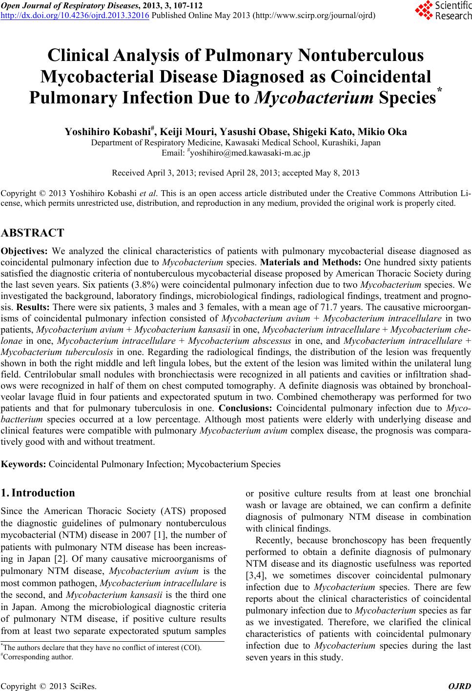 Open Journal of Respiratory Diseases, 2013, 3, 107-112 http://dx.doi.org/10.4236/ojrd.2013.32016 Published Online May 2013 (http://www.scirp.org/journal/ojrd) Clinical Analysis of Pulmonary Nontuberculous Mycobacterial Disease Diagnosed as Coincidental Pulmonary Infection Due to Mycobacterium Species* Yoshihiro Kobashi#, Keiji Mouri, Yasushi Obase, Shigeki Kato, Mikio Oka Department of Respiratory Medicine, Kawasaki Medical School, Kurashiki, Japan Email: #[email protected] Received April 3, 2013; revised April 28, 2013; accepted May 8, 2013 Copyright © 2013 Yoshihiro Kobashi et al. This is an open access article distributed under the Creative Commons Attribution Li- cense, which permits unrestricted use, distribution, and reproduction in any medium, provided the original work is properly cited. ABSTRACT Objectives: We analyzed the clinical characteristics of patients with pulmonary mycobacterial disease diagnosed as coincidental pulmonary infection due to Mycobacterium species. Materials and Methods: One hundred sixty patients satisfied the diagnostic criteria of nontuberculous mycobacterial disease proposed by American Thoracic Society during the last seven years. Six patients (3.8%) were coincidental pulmonary infection due to two Mycobacterium species. We investigated the background, laboratory findings, microbiological findings, radiological findings, treatment and progno- sis. Results: There were six patients, 3 males and 3 females, with a mean age of 71.7 years. The causative microorgan- isms of coincidental pulmonary infection consisted of Mycobacterium avium + Mycobacterium intracellulare in two patients, Mycobacterium avium + Mycobacterium kansasii in one, Mycobacterium intracellulare + Mycobacterium che- lonae in one, Mycobacterium intracellulare + Mycobacterium abscessus in one, and Mycobacterium intracellulare + Mycobacterium tuberculosis in one. Regarding the radiological findings, the distribution of the lesion was frequently shown in both the right middle and left lingula lobes, but the extent of the lesion was limited within the unilateral lung field. Centrilobular small nodules with bronchiectasis were recognized in all patients and cavities or infiltration shad- ows were recognized in half of them on chest computed tomography. A definite diagnosis was obtained by bronchoal- veolar lavage fluid in four patients and expectorated sputum in two. Combined chemotherapy was performed for two patients and that for pulmonary tuberculosis in one. Conclusions: Coincidental pulmonary infection due to Myco- bactterium species occurred at a low percentage. Although most patients were elderly with underlying disease and clinical features were compatible with pulmonary Mycobacterium a vium complex disease, the prognosis was compara- tively good with and without treatment. Keywords: Coincidental Pulmonary Infection; Mycobacterium Species 1. Introduction Since the American Thoracic Society (ATS) proposed the diagnostic guidelines of pulmonary nontuberculous mycobacterial (NTM) disease in 2007 [1], the number of patients with pulmonary NTM disease has been increas- ing in Japan [2]. Of many causative microorganisms of pulmonary NTM disease, Mycobacterium avium is the most common pathogen, Mycobacterium intracellulare is the second, and Mycobacterium kansasii is the third one in Japan. Among the microbiological diagnostic criteria of pulmonary NTM disease, if positive culture results from at least two separate expectorated sputum samples or positive culture results from at least one bronchial wash or lavage are obtained, we can confirm a definite diagnosis of pulmonary NTM disease in combination with clinical findings. Recently, because bronchoscopy has been frequently performed to obtain a definite diagnosis of pulmonary NTM disease and its diagnostic usefulness was reported [3,4], we sometimes discover coincidental pulmonary infection due to Mycobacterium species. There are few reports about the clinical characteristics of coincidental pulmonary infection due to Mycobacterium species as far as we investigated. Therefore, we clarified the clinical characteristics of patients with coincidental pulmonary infection due to Mycobacterium species during the last seven years in this study. *The authors declare that they have no conflict of interest (COI). #Corresponding author. C opyright © 2013 SciRes. OJRD 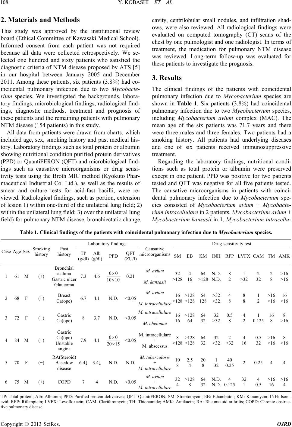 Y. KOBASHI ET AL. 108 2. Materials and Methods This study was approved by the institutional review board (Ethical Committee of Kawasaki Medical School). Informed consent from each patient was not required because all data were collected retrospectively. We se- lected one hundred and sixty patients who satisfied the diagnostic criteria of NTM disease proposed by ATS [5] in our hospital between January 2005 and December 2011. Among these patients, six patients (3.8%) had co- incidental pulmonary infection due to two Mycobacte- rium species. We investigated the backgrounds, labora- tory findings, microbiological findings, radiological find- ings, diagnostic methods, treatment and prognosis of these patients and the remaining patients with pulmonary NTM disease (154 patients) in this study. All data from patients were drawn from charts, which included age, sex, smoking history and past medical his- tory. Laboratory findings such as total protein or albumin showing nutritional condition purified protein derivatives (PPD) or QuantiFERON (QFT) and microbiological find- ings such as causative microorganisms or drug sensi- tivity tests using the Broth MIC method (Kyokuto Phar- maceutical Industrial Co. Ltd.), as well as the results of smear and culture tests for acid-fast bacilli, were re- viewed. Radiological findings, such as portion, extension of lesion 1) within one-third of the unilateral lung field; 2) within the unilateral lung field; 3) over the unilateral lung field) for pulmonary NTM disease, bronchiectatic change, cavity, centrilobular small nodules, and infiltration shad- ows, were also reviewed. All radiological findings were evaluated on computed tomography (CT) scans of the chest by one pulmologist and one radiologist. In terms of treatment, the medication for pulmonary NTM disease was reviewed. Long-term follow-up was evaluated for these patients to investigate the prognosis. 3. Results The clinical findings of the patients with coincidental pulmonary infection due to Mycobacterium species are shown in Table 1. Six patients (3.8%) had coincidental pulmonary infection due to two Mycobacterium species, including Mycobacterium avium complex (MAC). The mean age of the six patients was 71.7 years and there were three males and three females. Two patients had a smoking history. All patients had underlying diseases and one of six patients received immunosuppressive treatment. Regarding the laboratory findings, nutritional condi- tions such as total protein or albumin were preserved except in one patient. PPD was positive for two patients tested and QFT was negative for all five patients tested. The causative microorganisms in patients with coinci- dental pulmonary infection due to Mycobacterium spe- cies consisted of Mycobacterium avium + Mycobacte- rium intracellulare in 2 patients, Mycobacterium avium + Mycobacterium kansasii in 1, Mycobacterium intracellu- Table 1. Clinical findings of the patients with coincidental pulmonar y infe c tion due to Mycobacterium species. Laboratory findings Drug-sensitivity test Case Age Sex Smoking history Past history TP (g/dl) Alb (g/dl) PPD QFT (ZU/l) Causative microorganisms SMEBKMINH RFP LVFX CAMTMAMK 1 61 M (+) Bronchial asthma Gastric ulcer Glaucoma 7.3 4.6 00 10 10 0.21 M. avium + M. kansasii 32 >128 4 16 64 >128 N.D. N.D. 8 2 1 >32 2 32 2 8 >16 >16 2 68 F (−) Breast Ca(ope) 6.7 4.1 N.D. <0.05 M. avium + M. intracellulare 16 >128 >128 >128 64 128 >32 >32 4 8 8 8 1 2 >16 >16 16 >16 3 72 F (−) Gastric Ca(ope) 8 3.7 N.D. <0.05 M. intracellulare + M. chelonae 16 16 >128 64 64 32 32 >32 0.5 8 4 2 1 0.125 16 8 8 >16 4 84 M (−) Gastric Ca(ope) Unstable angina 7.9 4.1 00 20 15 <0.05 M. intracellulare + M. abscessus 8 >128 >128 >128 64 32 32 >32 2 >32 4 16 0.5 32 >16 >16 8 >16 5 70 F (−) RA(Steroid) Basedow disease 6.4↓ 3.4↓ N.D. N.D. M. tuberculosis + M. intracellulare 10 8 2.5 4 20 8 1 32 40 0.25 2 0.25 44 6 75 M (+) COPD 7 4 N.D. <0.05 M. avium + M. intracellulare 32 4 >128 8 64 32 N.D. N.D. 4 0.125 32 1 4 0.5 >16 16 >16 4 TP: Total protein; Alb: Albumin; PPD: Purified protein delivatives; QFT: QuantiFERON; SM: Streptomycin; EB: Ethambutol; KM: Kanamycin; INH: Isoni- azid; RFP: Rifampicin; LVFX: Levofloxacin; CAM: Clarithromycin; TH: Thionamide; AMK: Amikacin; RA: Rheumatoid arthritis; COPD: Chronic obstruc- ive pulmonary disease. t Copyright © 2013 SciRes. OJRD 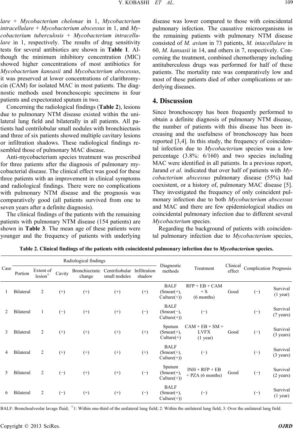 Y. KOBASHI ET AL. 109 lare + Mycobacterium chelonae in 1, Mycobacterium intracellulare + Mycobacterium abscessus in 1, and My- cobacterium tuberculosis + Mycobacterium intracellu- lare in 1, respectively. The results of drug sensitivity tests for several antibiotics are shown in Table 1. Al- though the minimum inhibitory concentration (MIC) showed higher concentrations of most antibiotics for Mycobacterium kansasii and Mycobacterium abscessus, it was preserved at lower concentrations of clarithromy- cin (CAM) for isolated MAC in most patients. The diag- nostic methods used bronchoscopic specimens in four patients and expectorated sputum in two. Concerning the radiological findings (Table 2), lesions due to pulmonary NTM disease existed within the uni- lateral lung field and bilaterally in all patients. All pa- tients had centrilobular small nodules with bronchiectasis and three of six patients showed multiple cavitary lesions or infiltration shadows. These radiological findings re- sembled those of pulmonary MAC disease. Anti-mycobacterium species treatment was prescribed for three patients after the diagnosis of pulmonary my- cobacterial disease. The clinical effect was good for these three patients with an improvement in clinical symptoms and radiological findings. There were no complications with pulmonary NTM disease and the prognosis was comparatively good (all patients survived from one to seven years after a definite diagnosis). The clinical findings of the patients with the remaining patients with pulmonary NTM disease (154 patients) are shown in Table 3. The mean age of these patients were younger and the frequency of patients with underlying disease was lower compared to those with coincidental pulmonary infection. The causative microorganisms in the remaining patients with pulmonary NTM disease consisted of M. avium in 73 patients, M. intacellulare in 60, M. kansasii in 14, and others in 7, respectively. Con- cerning the treatment, combined chemotherapy including antituberculous drugs was performed for half of these patients. The mortality rate was comparatively low and most of these patients died of other complications or un- derlying diseases. 4. Discussion Since bronchoscopy has been frequently performed to obtain a definite diagnosis of pulmonary NTM disease, the number of patients with this disease has been in- creasing and the usefulness of bronchoscopy has been reported [3,4]. In this study, the frequency of coinciden- tal infection due to Mycobacterium species was a low percentage (3.8%: 6/160) and two species including MAC were identified in all patients. In a previous report, Jarand et al. indicated that over half of patients with My- cobacterium abscessus pulmonary disease (55%) had coexistent, or a history of, pulmonary MAC disease [5]. They investigated the frequency of only coincident pul- monary infection due to both Mycobacterium abscessus and MAC and there are few epidemiological studies on coincidental pulmonary infection due to different several Mycobacterium species. Regarding the background of patients with coinciden- tal pulmonary infection due to Mycobacterium species, Table 2. Clinical findings of the patients with coincidental pulmonar y infe c tion due to Mycobacterium species. Radiological findings Case Portion Extent of lesion※ Cavity Bronchiectatic change Centriliobular small nodules Infiltration shadow Diagnostic methods Treatment Clinical effect Complication Prognosis 1 Bilateral 2 (+) (+) (+) (+) BALF (Smear(+), Culture(+)) RFP + EB + CAM + S (6 months) Good (−) Survival (1 year) 2 Bilateral 1 (−) (+) (+) (−) BALF (Smear(−), Culture(+)) (−) (−) Survival (7 years) 3 Bilateral 2 (+) (+) (+) (+) Sputum (Smear(+), Culture(+) CAM + EB + SM + LVFX (1 year) Good (−) Survival (3 years) 4 Bilateral 2 (+) (+) (+) (+) BALF (Smear(+), Culture(+)) (−) (−) Survival (3 years) 5 Bilateral 2 (−) (+) (+) (−) Sputum (Smear(+), Culture(+)) INH + RFP + EB + PZA (6 months) Good (−) Survival (2 years) 6 Bilateral 2 (−) (+) (+) (−) BALF (Smear(+), Culture(+)) (−) (−) Survival (1 year) B ALF: Bronchoalveolar lavage fluid; ※1: Within one-third of the unilateral lung field; 2: Within the unilateral lung field; 3: Over the unilateral lung field. Copyright © 2013 SciRes. OJRD 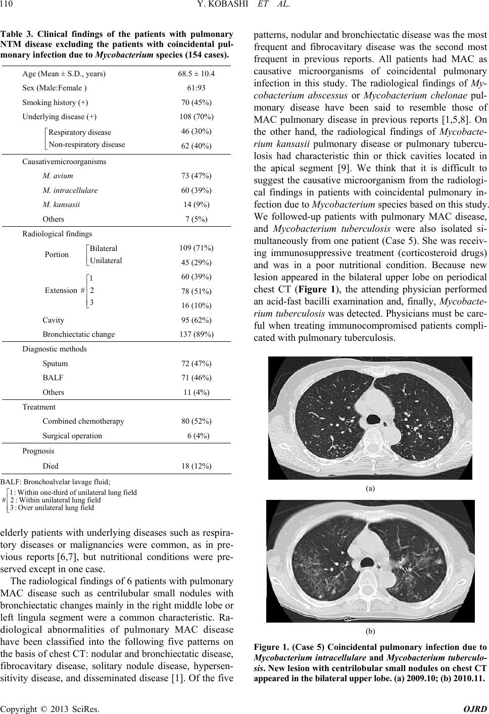 Y. KOBASHI ET AL. 110 Table 3. Clinical findings of the patients with pulmonary NTM disease excluding the patients with coincidental pul- monary infection due to Mycobacterium species (154 cases). Age (Mean ± S.D., years) 68.5 ± 10.4 Sex (Male:Female ) 61:93 Smoking history (+) 70 (45%) Underlying disease (+) 108 (70%) 46 (30%) Respiratory disease Non-respiratory disease 62 (40%) Causativemicroorganisms M. avium 73 (47%) M. intracellulare 60 (39%) M. kansasii 14 (9%) Others 7 (5%) Radiological findings 109 (71%) Bilateral Portion Unilateral 45 (29%) 60 (39%) 78 (51%) 1 Extension #2 3 16 (10%) Cavity 95 (62%) Bronchiectatic change 137 (89%) Diagnostic methods Sputum 72 (47%) BALF 71 (46%) Others 11 (4%) Treatment Combined chemotherapy 80 (52%) Surgical operation 6 (4%) Prognosis Died 18 (12%) BALF: Bronchoalvelar lavage fluid; 1:Within one-third of unilateral lung field #2:Within unilateral lung field 3:Over unilateral lung field elderly patients with underlying diseases such as respira- tory diseases or malignancies were common, as in pre- vious reports [6,7], but nutritional conditions were pre- served except in one case. The radiological findings of 6 patients with pulmonary MAC disease such as centrilubular small nodules with bronchiectatic changes mainly in the right middle lobe or left lingula segment were a common characteristic. Ra- diological abnormalities of pulmonary MAC disease have been classified into the following five patterns on the basis of chest CT: nodular and bronchiectatic disease, fibrocavitary disease, solitary nodule disease, hypersen- sitivity disease, and disseminated disease [1]. Of the five patterns, nodular and bronchiectatic disease was the most frequent and fibrocavitary disease was the second most frequent in previous reports. All patients had MAC as causative microorganisms of coincidental pulmonary infection in this study. The radiological findings of My- cobacterium abscessus or Mycobacterium chelonae pul- monary disease have been said to resemble those of MAC pulmonary disease in previous reports [1,5,8]. On the other hand, the radiological findings of Mycobacte- rium kansasii pulmonary disease or pulmonary tubercu- losis had characteristic thin or thick cavities located in the apical segment [9]. We think that it is difficult to suggest the causative microorganism from the radiologi- cal findings in patients with coincidental pulmonary in- fection due to Mycobacterium species based on this study. We followed-up patients with pulmonary MAC disease, and Mycobacterium tuberculosis were also isolated si- multaneously from one patient (Case 5). She was receiv- ing immunosuppressive treatment (corticosteroid drugs) and was in a poor nutritional condition. Because new lesion appeared in the bilateral upper lobe on periodical chest CT (Figure 1), the attending physician performed an acid-fast bacilli examination and, finally, Mycobacte- rium tuberculosis was detected. Physicians must be care- ful when treating immunocompromised patients compli- cated with pulmonary tuberculosis. (a) (b) Figure 1. (Case 5) Coincidental pulmonary infection due to Mycobacterium intracellulare and Mycobacterium tuberculo- sis. New lesion with centrilobular small nodules on chest CT appea r ed i n th e b i l a tera l u p p e r l obe . ( a ) 2 009. 10; (b) 2010.11. Copyright © 2013 SciRes. OJRD  Y. KOBASHI ET AL. 111 Concerning the diagnostic methods, the bronchoscopic procedure is important to acquire accurate information about coincidental pulmonary infection due to Mycobac- terium species. In this study, four of six patients were given an accurate diagnosis using bronchoalveolar lavage fluid (BALF). Although bronchoscopy is an invasive technique, we think it is necessary to consider a differen- tial diagnosis of pulmonary NTM disease. Regarding the selection of treatment, ATS/IDSA pro- posed a guideline for treatment of pulmonary NTM dis- ease in 2007 [1]. The therapy for pulmonary MAC dis- ease was recommended as combined chemotherapy in- cluding a macrolide (clarithromycin (CAM) or azithro- mycin (AZM)), ethambutol (EB), rifampicin (RFP) ± aminoglycoside depending on disease status and/or se- verity. On the other hand, therapy for pulmonary Myco- bacterium abscessus or Mycobacterium chelonae disease was recommended as combined chemotherapy consisting of a macrolide, aminoglycoside, imipenem and cefoxitin and that for pulmonary Mycobacterium kansasii disease was combined chemotherapy including isoniazid (INH), RFP and EB. There is a little difference in the treatment between Mycobacterium species. Therefore, it is impor- tant to identify the species in NTM. However, the clinical effect of combined chemotherapy for pulmonary NTM disease except for pulmonary Mycobacterium kansasii disease was not good (clinical symptoms and/or abnor- mal radiological findings) [10]. There are no guidelines for initiating or ending treatment or the adaptation of treatment for elderly patients. Consequently, combined chemotherapy was performed for three of six patients because there were mainly elderly patients in this study; multi-drug chemotherapy including CAM for two pa- tients and antituberculous treatment for Mycobacterium tuberculosis for one. The clinical effect due to combined chemotherapy was good and the prognosis was also good despite their general condition. We do not think there was a relationship between coincidental pulmonary in- fection and clinical effect or prognosis of the subjects in this study. There are a few limitations in this study. Firstly, there were only a few patients with coincidental pulmonary infection due to Mycobacterium species among patients with pulmonary mycobacterial disease in one hospital in a restricted area, so we need to perform a large-scale study including several hospitals in Japan to get an accu- rate of coincidental mycobacterial infection. Secondly, the rate of coincidental mycobacterial infection was lower than in other reports [5]. This is because the diagnostic accuracy of the acid-fast bacilli culture was poor in our laboratory when patients with coincidental infection due to Mycobacterium species were tested. In conclusion, the rate of coincidental pulmonary in- fection due to Mycobacterium species was a low per- centage and clinical characteristics were similar to those of pulmonary MAC disease mainly on the radiological findings. Although most patients were elderly with un- derlying diseases, the clinical effect and prognosis were comparatively good. REFERENCES [1] D. E. Griffith, T. Aksamit, B. A. Brown-Elliot, et al., “An Official ATS/IDSA Statement: Diagnosis, Treatment, and Prevention of Nontuberculous Mycobacterial Diseases,” American Journal of Respiratory and Critical Care Medi- cine, Vol. 175, No. 4, 2007, pp. 367-416. doi:10.1164/rccm.200604-571ST [2] The Nontuberculous Mycobacteriosis Control Committee of the Japanese Society for Tuberculosis, The Scientific Assembly for Infection and Tuberculosis of the Japanese Respiratory Society, “Guideline for the Diagnosis of Pul- monary Nontuberculous Mycobacterial Diseases-2008,” Kekkaku, Vol. 86, No. 10, 2011, pp. 37-39. [3] E. Sugihara, N. Hirota, T. Nizeki, et al., “Usefulness of Bronchial Lavage for the Diagnosis of Pulmonary Disease Caused by Mycobacterium avium-intracellulare Complex (MAC) Infection,” Journal of Infection and Chemother- apy, Vol. 9, No. 4, 2003, pp. 328-332. doi:10.1007/s10156-003-0267-1 [4] E. Tanaka, R. Amitani, A. Niimi, et al., “Yield of Com- puted Tomography and Bronchoscopy for the Diagnosis of Mycobacterium avium Complex Pulmonary Disease,” American Journal of Respiratory and Critical Care Me- dicine, Vol. 155, No. 6, 1997, pp. 2041-2046. doi:10.1164/ajrccm.155.6.9196113 [5] J. Jarand, A. Levin, L. Zhang, et al., “Clinical and Micro- biologic Outcomes in Patients Receiving Treatment for Mycobacterium abscessus Pulmonary Disease,” Clinical Infectious Disease, Vol. 52, No. 5, 2011, pp. 565-571. doi:10.1093/cid/ciq237 [6] R. J. O’Brien, L. J. Geiter and D. E. Snider, “The Epide- miology of Nontuberculous Mycobacterial Diseases in the United States: Results from a National Survey,” Ameri- can Review Respiratory Disease, Vol. 135, No. 5, 1987, pp. 1007-1014. [7] D. E. Griffith, W. M. Girard and R. J. Wallace, Jr., “Cli- nical Features of Pulmonary Disease Caused by Rapidly Growing Mycobacteria: An Analysis of 154 Patients,” American Review Respiratory Disease, Vol. 147, No. 5, 1993, pp. 1271-1278. doi:10.1164/ajrccm/147.5.1271 [8] D. Han, K. S. Lee, W. J. Koh, et al., “Radiographic and CT Findings of Nontuberculous Mycobacterial Pulmo- nary Infection Caused by Mycobacterium abscessus,” American Journal of Roentgenology, Vol. 181, No. 2, 2003, pp. 513-517. doi:10.2214/ajr.181.2.1810513 [9] D. Shitrit, G. L. Baum, R. Pricess, et al., “Pulmonary Mycobacterium kansasii Infection in Israel, 1999-2004. Clinical Features, Drug Susceptibility, and Outcome,” Chest, Vol. 129, No. 3, 2006, pp. 771-776. doi:10.1378/chest.129.3.771 [10] Y. Kobashi and T. Matsushima, “The Microbiological and Copyright © 2013 SciRes. OJRD  Y. KOBASHI ET AL. Copyright © 2013 SciRes. OJRD 112 Clinical Effects of Combined Therapy According to Guide- lines on the Treatment of Pulmonary Mycobacterium avium Complex Disease in Japan—Including a Follow- Up Study,” Respiration, Vol. 74, No. 4, 2007, pp. 394- 400. doi:10.1159/000095674 |