

Paper Menu >>
Journal Menu >>
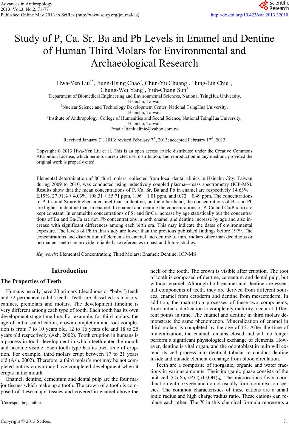 Advances in Anthropology 2013. Vol.3, No.2, 71-77 Published Online May 2013 in SciRes (http://www.scirp.org/journal/aa) http://dx.doi.org/10.4236/aa.2013.32010 Copyright © 2013 SciRes. 71 Study of P, Ca, Sr, Ba and Pb Levels in Enamel and Dentine of Human Third Molars for Environmental and Archaeological Research Hwa-Yen Liu1*, Jiunn-Hsing Chao2, Chun-Yu Chuang1, Hung-Lin Chiu3, Chung-Wei Yang1, Yuh-Chang Sun1 1Department of Biomedical Engineering and Environmental Sciences, National TsingHua University, Hsinchu, Taiwan 2Nuclear Science and Technology Development Center, National TsingHua University, Hsinchu, Taiwan 3Institute of Anthropology, College of Humanities and Social Science, National TsingHua University, Hsinchu, Taiwan Email: *[email protected] Received January 7th, 2013; revised February 7th, 2013; accepted February 17th, 2013 Copyright © 2013 Hwa-Yen Liu et al. This is an open access article distributed under the Creative Commons Attribution License, which permits unrestricted use, distribution, and reproduction in any medium, provided the original work is properly cited. Elemental determination of 80 third molars, collected from local dental clinics in Hsinchu City, Taiwan during 2009 to 2010, was conducted using inductively coupled plasma—mass spectrometry (ICP-MS). Results show that the mean concentrations of P, Ca, Sr, Ba and Pb in enamel are respectively 14.63% ± 2.19%, 27.91% ± 4.03%, 108.31 ± 35.71 ppm, 1.96 ± 1.01 ppm, and 0.72 ± 0.49 ppm. The concentrations of P, Ca and Sr are higher in enamel than in dentine, on the other hand, the concentrations of Ba and Pb are higher in dentine than in enamel. In enamel and dentine the concentrations of P, Ca and Ca/P ratio are kept constant. In enamelthe concentrations of Sr and Sr/Ca increase by age statistically but the concentra- tions of Ba and Ba/Ca are not. Pb concentrations in both enamel and dentine increase by age and also in- crease with significant differences among each birth era. This may indicate the dates of environmental exposure. The levels of Pb in this study are lower than the previous published findings before 1979. The concentrations and distribution of elements in enamel and dentine of third molars other than deciduous or permanent teeth can provide reliable base references to past and future studies. Keywords: Elemental Concentration; Third Molars; Enamel; Dentine; ICP-MS Introduction The Properties of Teeth Humans usually have 20 primary (deciduous or “baby”) teeth and 32 permanent (adult) teeth. Teeth are classified as incisors, canines, premolars and molars. The development timeline is very different among each type of tooth. Each tooth has its own development stage time line. For example, for third molars, the age of initial calcification, crown completion and root comple- tion is from 7 to 10 years old, 12 to 16 years old and 18 to 25 years old respectively (Ash, 2002). Tooth eruption in humans is a process in tooth development in which teeth enter the mouth and become visible. Each tooth type has its own time of erup- tion. For example, third molars erupt between 17 to 21 years old (Ash, 2002). Therefore, a third molar’s root may be not com- pleted but its crown may have completed development when it erupts in the mouth. Enamel, dentine, cementum and dental pulp are the four ma- jor tissues which make up a tooth. The crown of a tooth is com- posed of these major tissues and covered in enamel above the neck of the tooth. The crown is visible after eruption. The root of tooth is composed of dentine, cementum and dental pulp, but without enamel. Although both enamel and dentine are essen- tial components of teeth, they are derived from different sour- ces, enamel from ectoderm and dentine from mesectoderm. In addition, the maturation processes of these two components, from initial calcification to completely maturity, occur at differ- rent points in time. The enamel and dentine in third molars de- monstrate the same phenomenon. Mineralization of enamel in third molars is completed by the age of 12. After the time of mineralization, the enamel remains closed and will no longer perform a significant physiological exchange of elements. How- ever, dentine is vital organ, and the odontoblast in pulp will ex- tend its cell process into dentinal tubular to conduct dentine inside and outside element exchange from blood circulation. Teeth are a composite of inorganic, organic and water frac- tions in various amounts. Their inorganic phase consists of the unit cell (Ca,X)10(P,C)6(O,OH)26. The microcations favor coor- dination with oxygen and do not usually form complex ion spe- cies. The common characteristics of these cations are a small ionic radius and high charge/radius ratio. These cations can re- place each other. The X in this chemical formula represents a *Corresponding author. 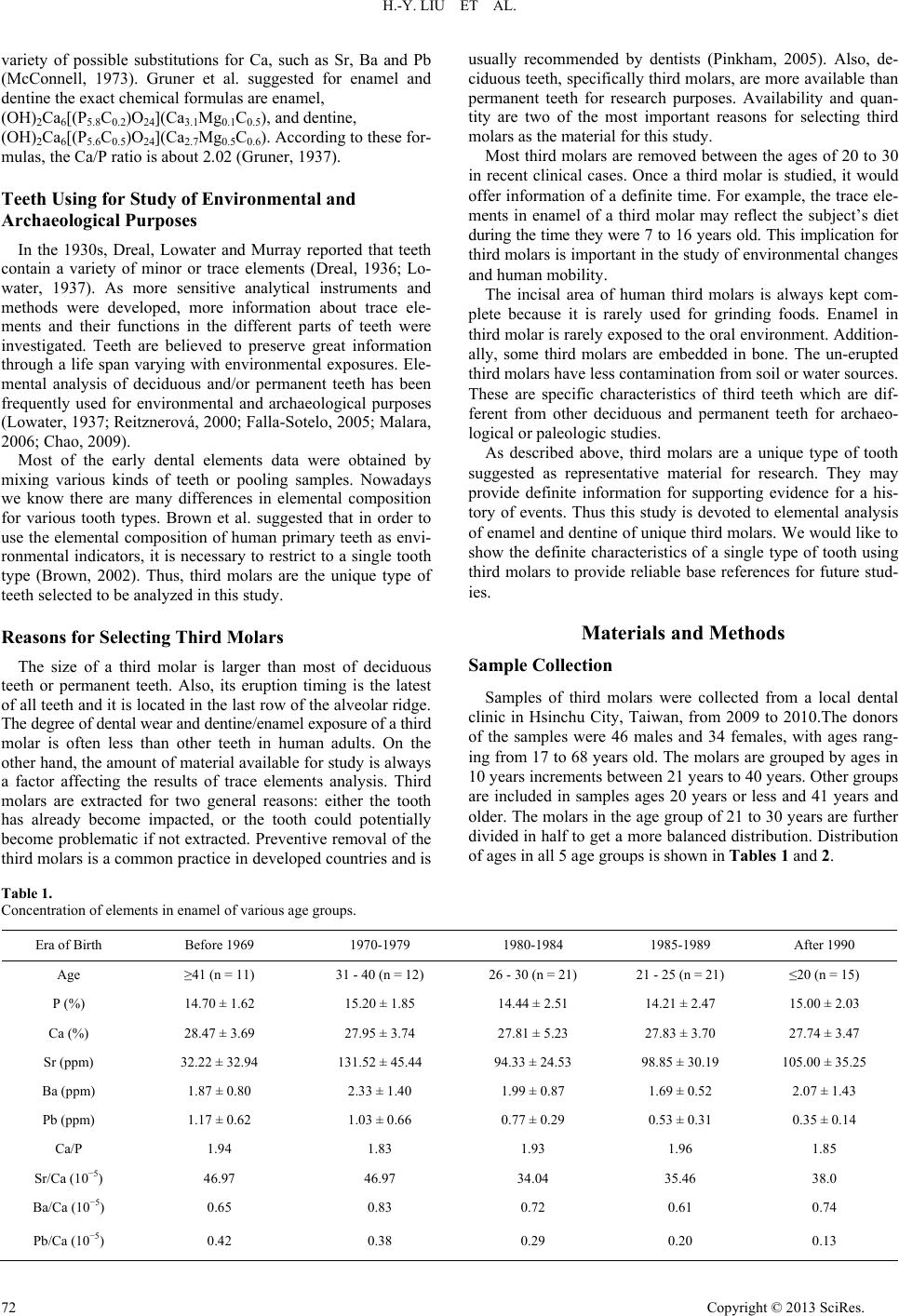 H.-Y. LIU ET AL. variety of possible substitutions for Ca, such as Sr, Ba and Pb (McConnell, 1973). Gruner et al. suggested for enamel and dentine the exact chemical formulas are enamel, (OH)2Ca6[(P5.8C0.2)O24](Ca3.1Mg0.1C0.5), and dentine, (OH)2Ca6[(P5.6C0.5)O24](Ca2.7Mg0.5C0.6). According to these for- mulas, the Ca/P ratio is about 2.02 (Gruner, 1937). Teeth Using for Study of Environmental and Archaeological Purposes In the 1930s, Dreal, Lowater and Murray reported that teeth contain a variety of minor or trace elements (Dreal, 1936; Lo- water, 1937). As more sensitive analytical instruments and methods were developed, more information about trace ele- ments and their functions in the different parts of teeth were investigated. Teeth are believed to preserve great information through a life span varying with environmental exposures. Ele- mental analysis of deciduous and/or permanent teeth has been frequently used for environmental and archaeological purposes (Lowater, 1937; Reitznerová, 2000; Falla-Sotelo, 2005; Malara, 2006; Chao, 2009). Most of the early dental elements data were obtained by mixing various kinds of teeth or pooling samples. Nowadays we know there are many differences in elemental composition for various tooth types. Brown et al. suggested that in order to use the elemental composition of human primary teeth as envi- ronmental indicators, it is necessary to restrict to a single tooth type (Brown, 2002). Thus, third molars are the unique type of teeth selected to be analyzed in this study. Reasons for Selecting Third Molars The size of a third molar is larger than most of deciduous teeth or permanent teeth. Also, its eruption timing is the latest of all teeth and it is located in the last row of the alveolar ridge. The degree of dental wear and dentine/enamel exposure of a third molar is often less than other teeth in human adults. On the other hand, the amount of material available for study is always a factor affecting the results of trace elements analysis. Third molars are extracted for two general reasons: either the tooth has already become impacted, or the tooth could potentially become problematic if not extracted. Preventive removal of the third molars is a common practice in developed countries and is usually recommended by dentists (Pinkham, 2005). Also, de- ciduous teeth, specifically third molars, are more available than permanent teeth for research purposes. Availability and quan- tity are two of the most important reasons for selecting third molars as the material for this study. Most third molars are removed between the ages of 20 to 30 in recent clinical cases. Once a third molar is studied, it would offer information of a definite time. For example, the trace ele- ments in enamel of a third molar may reflect the subject’s diet during the time they were 7 to 16 years old. This implication for third molars is important in the study of environmental changes and human mobility. The incisal area of human third molars is always kept com- plete because it is rarely used for grinding foods. Enamel in third molar is rarely exposed to the oral environment. Addition- ally, some third molars are embedded in bone. The un-erupted third molars have less contamination from soil or water sources. These are specific characteristics of third teeth which are dif- ferent from other deciduous and permanent teeth for archaeo- logical or paleologic studies. As described above, third molars are a unique type of tooth suggested as representative material for research. They may provide definite information for supporting evidence for a his- tory of events. Thus this study is devoted to elemental analysis of enamel and dentine of unique third molars. We would like to show the definite characteristics of a single type of tooth using third molars to provide reliable base references for future stud- ies. Materials and Methods Sample Collection Samples of third molars were collected from a local dental clinic in Hsinchu City, Taiwan, from 2009 to 2010.The donors of the samples were 46 males and 34 females, with ages rang- ing from 17 to 68 years old. The molars are grouped by ages in 10 years increments between 21 years to 40 years. Other groups are included in samples ages 20 years or less and 41 years and older. The molars in the age group of 21 to 30 years are further divided in half to get a more balanced distribution. Distribution of ages in all 5 age groups is shown in Tables 1 and 2. Table 1. Concentration of elements in enamel of various age groups. Era of Birth Before 1969 1970-1979 1980-1984 1985-1989 After 1990 Age ≥41 (n = 11) 31 - 40 (n = 12) 26 - 30 (n = 21) 21 - 25 (n = 21) ≤20 (n = 15) P (%) 14.70 ± 1.62 15.20 ± 1.85 14.44 ± 2.51 14.21 ± 2.47 15.00 ± 2.03 Ca (%) 28.47 ± 3.69 27.95 ± 3.74 27.81 ± 5.23 27.83 ± 3.70 27.74 ± 3.47 Sr (ppm) 32.22 ± 32.94 131.52 ± 45.44 94.33 ± 24.53 98.85 ± 30.19 105.00 ± 35.25 Ba (ppm) 1.87 ± 0.80 2.33 ± 1.40 1.99 ± 0.87 1.69 ± 0.52 2.07 ± 1.43 Pb (ppm) 1.17 ± 0.62 1.03 ± 0.66 0.77 ± 0.29 0.53 ± 0.31 0.35 ± 0.14 Ca/P 1.94 1.83 1.93 1.96 1.85 Sr/Ca (10−5) 46.97 46.97 34.04 35.46 38.0 Ba/Ca (10−5) 0.65 0.83 0.72 0.61 0.74 Pb/Ca (10−5) 0.42 0.38 0.29 0.20 0.13 Copyright © 2013 SciRes. 72 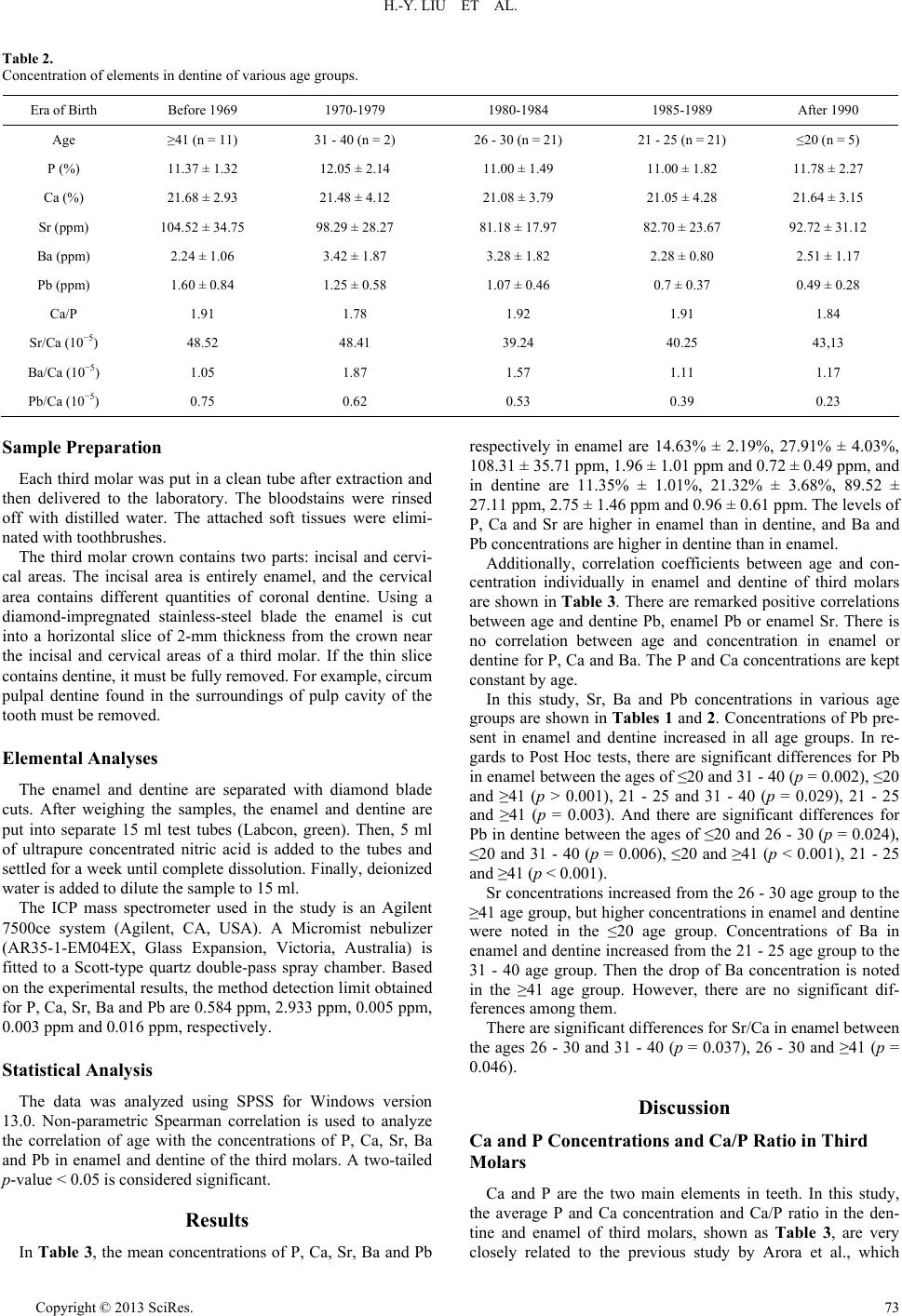 H.-Y. LIU ET AL. Table 2. Concentration of elements in dentine of various age groups. Era of Birth Before 1969 1970-1979 1980-1984 1985-1989 After 1990 Age ≥41 (n = 11) 31 - 40 (n = 2) 26 - 30 (n = 21) 21 - 25 (n = 21) ≤20 (n = 5) P (%) 11.37 ± 1.32 12.05 ± 2.14 11.00 ± 1.49 11.00 ± 1.82 11.78 ± 2.27 Ca (%) 21.68 ± 2.93 21.48 ± 4.12 21.08 ± 3.79 21.05 ± 4.28 21.64 ± 3.15 Sr (ppm) 104.52 ± 34.75 98.29 ± 28.27 81.18 ± 17.97 82.70 ± 23.67 92.72 ± 31.12 Ba (ppm) 2.24 ± 1.06 3.42 ± 1.87 3.28 ± 1.82 2.28 ± 0.80 2.51 ± 1.17 Pb (ppm) 1.60 ± 0.84 1.25 ± 0.58 1.07 ± 0.46 0.7 ± 0.37 0.49 ± 0.28 Ca/P 1.91 1.78 1.92 1.91 1.84 Sr/Ca (10−5) 48.52 48.41 39.24 40.25 43,13 Ba/Ca (10−5) 1.05 1.87 1.57 1.11 1.17 Pb/Ca (10−5) 0.75 0.62 0.53 0.39 0.23 Sample Preparation Each third molar was put in a clean tube after extraction and then delivered to the laboratory. The bloodstains were rinsed off with distilled water. The attached soft tissues were elimi- nated with toothbrushes. The third molar crown contains two parts: incisal and cervi- cal areas. The incisal area is entirely enamel, and the cervical area contains different quantities of coronal dentine. Using a diamond-impregnated stainless-steel blade the enamel is cut into a horizontal slice of 2-mm thickness from the crown near the incisal and cervical areas of a third molar. If the thin slice contains dentine, it must be fully removed. For example, circum pulpal dentine found in the surroundings of pulp cavity of the tooth must be removed. Elemental Analyses The enamel and dentine are separated with diamond blade cuts. After weighing the samples, the enamel and dentine are put into separate 15 ml test tubes (Labcon, green). Then, 5 ml of ultrapure concentrated nitric acid is added to the tubes and settled for a week until complete dissolution. Finally, deionized water is added to dilute the sample to 15 ml. The ICP mass spectrometer used in the study is an Agilent 7500ce system (Agilent, CA, USA). A Micromist nebulizer (AR35-1-EM04EX, Glass Expansion, Victoria, Australia) is fitted to a Scott-type quartz double-pass spray chamber. Based on the experimental results, the method detection limit obtained for P, Ca, Sr, Ba and Pb are 0.584 ppm, 2.933 ppm, 0.005 ppm, 0.003 ppm and 0.016 ppm, respectively. Statistical Analysis The data was analyzed using SPSS for Windows version 13.0. Non-parametric Spearman correlation is used to analyze the correlation of age with the concentrations of P, Ca, Sr, Ba and Pb in enamel and dentine of the third molars. A two-tailed p-value < 0.05 is considered significant. Results In Table 3, the mean concentrations of P, Ca, Sr, Ba and Pb respectively in enamel are 14.63% ± 2.19%, 27.91% ± 4.03%, 108.31 ± 35.71 ppm, 1.96 ± 1.01 ppm and 0.72 ± 0.49 ppm, and in dentine are 11.35% ± 1.01%, 21.32% ± 3.68%, 89.52 ± 27.11 ppm, 2.75 ± 1.46 ppm and 0.96 ± 0.61 ppm. The levels of P, Ca and Sr are higher in enamel than in dentine, and Ba and Pb concentrations are higher in dentine than in enamel. Additionally, correlation coefficients between age and con- centration individually in enamel and dentine of third molars are shown in Table 3. There are remarked positive correlations between age and dentine Pb, enamel Pb or enamel Sr. There is no correlation between age and concentration in enamel or dentine for P, Ca and Ba. The P and Ca concentrations are kept constant by age. In this study, Sr, Ba and Pb concentrations in various age groups are shown in Tables 1 and 2. Concentrations of Pb pre- sent in enamel and dentine increased in all age groups. In re- gards to Post Hoc tests, there are significant differences for Pb in enamel between the ages of ≤20 and 31 - 40 (p = 0.002), ≤20 and ≥41 (p > 0.001), 21 - 25 and 31 - 40 (p = 0.029), 21 - 25 and ≥41 (p = 0.003). And there are significant differences for Pb in dentine between the ages of ≤20 and 26 - 30 (p = 0.024), ≤20 and 31 - 40 (p = 0.006), ≤20 and ≥41 (p < 0.001), 21 - 25 and ≥41 (p < 0.001). Sr concentrations increased from the 26 - 30 age group to the ≥41 age group, but higher concentrations in enamel and dentine were noted in the ≤20 age group. Concentrations of Ba in enamel and dentine increased from the 21 - 25 age group to the 31 - 40 age group. Then the drop of Ba concentration is noted in the ≥41 age group. However, there are no significant dif- ferences among them. There are significant differences for Sr/Ca in enamel between the ages 26 - 30 and 31 - 40 (p = 0.037), 26 - 30 and ≥41 (p = 0.046). Discussion Ca and P Concentrations and Ca/P Ratio in Third Molars Ca and P are the two main elements in teeth. In this study, the average P and Ca concentration and Ca/P ratio in the den- tine and enamel of third molars, shown as Table 3, are very closely related to the previous study by Arora et al., which Copyright © 2013 SciRes. 73 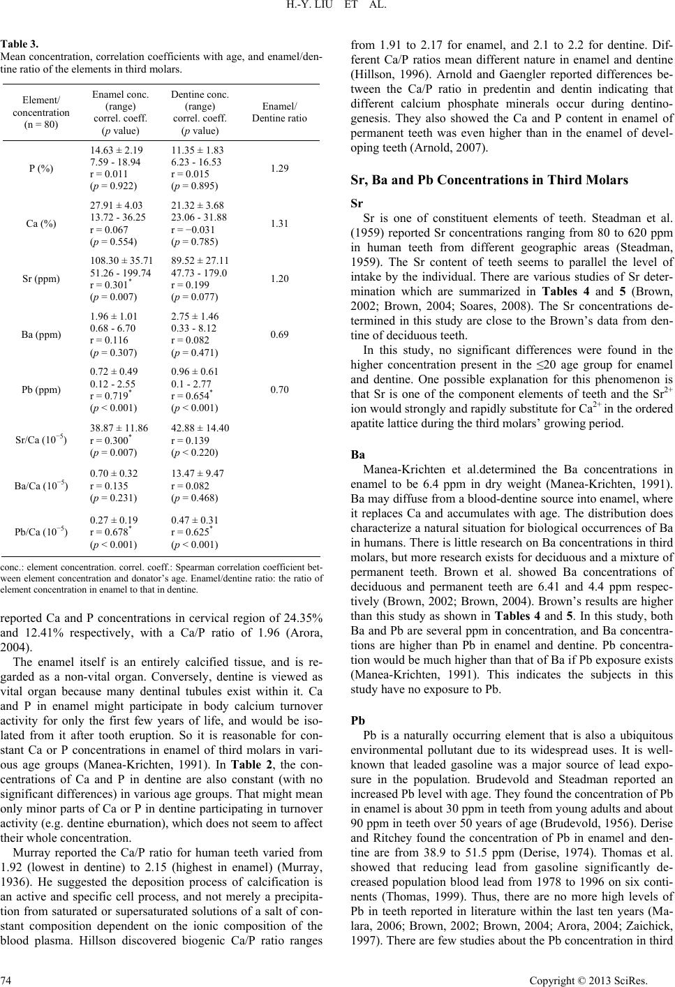 H.-Y. LIU ET AL. Table 3. Mean concentration, correlation coefficients with age, and enamel/den- tine ratio of the elements in third molars. Element/ concentration (n = 80) Enamel conc. (range) correl. coeff. (p value) Dentine conc. (range) correl. coeff. (p value) Enamel/ Dentine ratio P (%) 14.63 ± 2.19 7.59 - 18.94 r = 0.011 (p = 0.922) 11.35 ± 1.83 6.23 - 16.53 r = 0.015 (p = 0.895) 1.29 Ca (%) 27.91 ± 4.03 13.72 - 36.25 r = 0.067 (p = 0.554) 21.32 ± 3.68 23.06 - 31.88 r = −0.031 (p = 0.785) 1.31 Sr (ppm) 108.30 ± 35.71 51.26 - 199.74 r = 0.301* (p = 0.007) 89.52 ± 27.11 47.73 - 179.0 r = 0.199 (p = 0.077) 1.20 Ba (ppm) 1.96 ± 1.01 0.68 - 6.70 r = 0.116 (p = 0.307) 2.75 ± 1.46 0.33 - 8.12 r = 0.082 (p = 0.471) 0.69 Pb (ppm) 0.72 ± 0.49 0.12 - 2.55 r = 0.719* (p < 0.001) 0.96 ± 0.61 0.1 - 2.77 r = 0.654* (p < 0.001) 0.70 Sr/Ca (10−5) 38.87 ± 11.86 r = 0.300* (p = 0.007) 42.88 ± 14.40 r = 0.139 (p < 0.220) Ba/Ca (10−5) 0.70 ± 0.32 r = 0.135 (p = 0.231) 13.47 ± 9.47 r = 0.082 (p = 0.468) Pb/Ca (10−5) 0.27 ± 0.19 r = 0.678* (p < 0.001) 0.47 ± 0.31 r = 0.625* (p < 0.001) conc.: element concentration. correl. coeff.: Spearman correlation coefficient bet- ween element concentration and donator’s age. Enamel/dentine ratio: the ratio of element concentration in enamel to that in dentine. reported Ca and P concentrations in cervical region of 24.35% and 12.41% respectively, with a Ca/P ratio of 1.96 (Arora, 2004). The enamel itself is an entirely calcified tissue, and is re- garded as a non-vital organ. Conversely, dentine is viewed as vital organ because many dentinal tubules exist within it. Ca and P in enamel might participate in body calcium turnover activity for only the first few years of life, and would be iso- lated from it after tooth eruption. So it is reasonable for con- stant Ca or P concentrations in enamel of third molars in vari- ous age groups (Manea-Krichten, 1991). In Table 2, the con- centrations of Ca and P in dentine are also constant (with no significant differences) in various age groups. That might mean only minor parts of Ca or P in dentine participating in turnover activity (e.g. dentine eburnation), which does not seem to affect their whole concentration. Murray reported the Ca/P ratio for human teeth varied from 1.92 (lowest in dentine) to 2.15 (highest in enamel) (Murray, 1936). He suggested the deposition process of calcification is an active and specific cell process, and not merely a precipita- tion from saturated or supersaturated solutions of a salt of con- stant composition dependent on the ionic composition of the blood plasma. Hillson discovered biogenic Ca/P ratio ranges from 1.91 to 2.17 for enamel, and 2.1 to 2.2 for dentine. Dif- ferent Ca/P ratios mean different nature in enamel and dentine (Hillson, 1996). Arnold and Gaengler reported differences be- tween the Ca/P ratio in predentin and dentin indicating that different calcium phosphate minerals occur during dentino- genesis. They also showed the Ca and P content in enamel of permanent teeth was even higher than in the enamel of devel- oping teeth (Arnold, 2007). Sr, Ba and Pb Concentrations in Third Molars Sr Sr is one of constituent elements of teeth. Steadman et al. (1959) reported Sr concentrations ranging from 80 to 620 ppm in human teeth from different geographic areas (Steadman, 1959). The Sr content of teeth seems to parallel the level of intake by the individual. There are various studies of Sr deter- mination which are summarized in Tables 4 and 5 (Brown, 2002; Brown, 2004; Soares, 2008). The Sr concentrations de- termined in this study are close to the Brown’s data from den- tine of deciduous teeth. In this study, no significant differences were found in the higher concentration present in the ≤20 age group for enamel and dentine. One possible explanation for this phenomenon is that Sr is one of the component elements of teeth and the Sr2+ ion would strongly and rapidly substitute for Ca2+ in the ordered apatite lattice during the third molars’ growing period. Ba Manea-Krichten et al.determined the Ba concentrations in enamel to be 6.4 ppm in dry weight (Manea-Krichten, 1991). Ba may diffuse from a blood-dentine source into enamel, where it replaces Ca and accumulates with age. The distribution does characterize a natural situation for biological occurrences of Ba in humans. There is little research on Ba concentrations in third molars, but more research exists for deciduous and a mixture of permanent teeth. Brown et al. showed Ba concentrations of deciduous and permanent teeth are 6.41 and 4.4 ppm respec- tively (Brown, 2002; Brown, 2004). Brown’s results are higher than this study as shown in Tables 4 and 5. In this study, both Ba and Pb are several ppm in concentration, and Ba concentra- tions are higher than Pb in enamel and dentine. Pb concentra- tion would be much higher than that of Ba if Pb exposure exists (Manea-Krichten, 1991). This indicates the subjects in this study have no exposure to Pb. Pb Pb is a naturally occurring element that is also a ubiquitous environmental pollutant due to its widespread uses. It is well- known that leaded gasoline was a major source of lead expo- sure in the population. Brudevold and Steadman reported an increased Pb level with age. They found the concentration of Pb in enamel is about 30 ppm in teeth from young adults and about 90 ppm in teeth over 50 years of age (Brudevold, 1956). Derise and Ritchey found the concentration of Pb in enamel and den- tine are from 38.9 to 51.5 ppm (Derise, 1974). Thomas et al. showed that reducing lead from gasoline significantly de- creased population blood lead from 1978 to 1996 on six conti- nents (Thomas, 1999). Thus, there are no more high levels of Pb in teeth reported in literature within the last ten years (Ma- lara, 2006; Brown, 2002; Brown, 2004; Arora, 2004; Zaichick, 1997). There are few studies about the Pb concentration in third Copyright © 2013 SciRes. 74  H.-Y. LIU ET AL. Copyright © 2013 SciRes. 75 Table 4. Concentration of elements in dentine of human teeth reported in previous studies. Authors Method Teeth types No P (%) Ca (%) Sr (ppm) Ba (ppm) Pb (ppm) this work ICP-MS third molar n = 90 11.37 ± 1.91 21.26 ± 3.74 89.27 ± 28.42 2.78 ± 1.58 1.09 ± 0.91 Soares INAA permanent deciuous 10 8 21.40 ± 1.79 25.0 ± 2.2 174.2 ± 118.7 92.1 ± 25.4 Shi EMPA deciduous I 37 34.33 ± 1.33 Liang EMPA deciduous 38 17.42 ± 0.59 Arnold EDX third molar 3 17.2 ± 2.2 37.7 ± 5.9 Zenóbio ICP/AES third molar 30 10.98 ± 0.5 23.10 ± 0.5 Soremark INAA 13.5 ± 2.8 28.2 ± 1.2 Arora ICP-MS deciduous molar canine incisor 5 4 7 Cervical 12.41 ± 0.53 12.56 ± 0.33 11.86 ± 0.22 Cervical 24.35 ± 1.29 23.84 ± 1.14 22.56 ± 0.43 Cervical 1.11 ± 0.30 0.87 ± 0.41 1.23 ± 0.39 Arora ICP-MS deciduous molar canine Incisor 5 4 7 Apical 11.97 ± 0.54 12.52 ± 0.32 11.28 ± 0.20 Apical 24.21 ± 1.38 24.22 ± 0.68 22.45 ± 0.31 Apical 2.95 ± 2.06 2.17 ± 0.70 3.32 ± 1.56 Malara AAS permanent incisor canine pre-molar molar 33 38 43 41 root 55.27 ± 8.52 58.11 ± 10.84 56.90 ± 8.12 57.03 ± 9.73 root 14.08 ± 2.74 9.97 ± 3.57 10.39 ± 2.85 10.11 ± 2.14 Brown ICP-MS deciduous whole teeth UK 27 UG 21 31.4 ± 19.6 31.5 ± 13.7 118 ± 37.8 175 ± 38.0 6.41 ± 2.92 9.87 ± 4.43 1.33 ± 0.89 1.21 ± 0.44 AAS: atomic absorption spectrometer. EDX: energy-dispersive X-ray analysis. EDXRF: energy dispersive X-ray fluorescent analysis. EMPA: electron microprobe X-ray mMicroanalyzer. ICP/AES: ductively coupled plasma spectrometer. ICP-MS: inductively coupled plasma mass spectrometer. INAA: instrument neutron activation analy- sis. Table 5. Concentration of elements in enamel of human teeth from various studies. Authors Method Teeth No P (%) Ca (%) Sr (ppm) Ba (ppm) Pb (ppm) this work ICP-mass third molar n = 90 14.63 ± 2.21 27.87 ± 4.08 107.57 ± 37.31 1.91 ± 1.01 0.76 ± 0.54 Soares INAA permanent deciduous 10 8 31.20 ± 4.76 29.0 ± 5.2 285.8 ± 181.7 84.8 ± 16.7 Shi EMPA deciduous 38.74 ± 1.31 Liang EMPA deciduous 38 18.29 ± 0.44 Arnold EDX third molar 3 19.9 ± 1.8 42.7 ± 5.1 Zenóbio ICP-AES third molar 30 17.5 ± 0.5 36 ± 0.5 Lakomaa INAA 18.6 ± 0 34 ± 2.0 Zaichick EDXRF permanent 35 36.3 ± 0.8 240 ± 40 ≤3 Brown ICP-MS permanent 8 78% ± 5% 4.4% ± 6% 6.0% ± 5% AAS: atomic absorption spectrometer. EDX: energy-dispersive X-ray analysis. EDXRF: energy dispersive X-ray fluorescent analysis. EMPA: Electron microprobe X-ray mMicroanalyzer. ICP/AES: ductively coupled plasma spectrometer. ICP-MS: inductively coupled plasma mass spectrometer. INAA: instrument neutron activation analy- sis. molars. The present results of third molars compare with other studies of permanent teeth are shown in Tables 4 and 5. Pb Concentrations in Various Age Groups Indicate Eras of Exposure There are significant positive correlations between age and Pb in enamel and dentine. Pb is believed to have a cumulative effect. This study shows the Pb concentration in teeth increases from younger groups to older groups. This means that Pb is deposited in teeth year by year. The various age groups may be looked upon as eras of birth groups. Eras of birth indicate the eras of environmental expo- sure. Initial calcification of human third molars occurs between  H.-Y. LIU ET AL. the ages of 7 to 10. That means if more Pb exists in one’s third molars, he may have been exposed to Pb before 7 to 10 years ago. The eras are defined by birth year of the donor as follows: ≤20 age group is 1990 and later, 21 - 25 age group is 1985- 1989, 26 - 30 age group is 1980-1984, 31 - 40 age group is 1970-1979, and ≥41 age group is before 1969. Figure 1 shows Sr, Ba and Pb concentrations in various eras of birth. Pb concentrations are seen to increase by age group. More importantly, this shows Pb concentration increases by eras of birth. In Taiwan, unleaded gasoline was supplied start- ing in 1986, which decreased the extent of cord blood lead level decline in the Taipei area from 1985 to 2002 (Hwang, 2004). It is reasonable that donors born before 1979 will have higher concentrations of Pb in their teeth. Sr/Ca and Ba/Ca The ratio of Sr to Ca concentration is an indicator for bio- puri- fication of calcium. The Sr/Ca ratio in teeth is an interest- ing issue (Chao, 2009; Arora, 2004; Webb, 2005). Data for Sr/Ca and Ba/Ca in teeth enamel may offer much information for researchers in many fields of study, such as environmental sci- ence, ecology, biology, paleontology and archaeology (Wang, 2010). In this study, the Sr/Ca ratio is 3.86 × 10⁻⁶ and 4.20 × 10⁻⁶ in enamel and dentine of third molars, respectively. In Tables 1 and 2, Sr/Ca ratios increase with age except in the under the Period of time after 19901985-19891980- 19841970-1979befor e 1969 Concentration (ppm) 0 1 2 3 4 5 6 7 8 9 10 80 100 120 140 160 180 Enamel Sr Dentine Sr Enamel Ba Dentine Ba Enamel Pb Dentine Pb *** ** ** # # Figure 1. Sr, Ba and Pb concentration in in enamel or dentine of third molars at various eras of birth year. #: p value, 0.05 < p < 0.1. *: p value, p < 0.05. Sr concentrations are higher in enamel than that in dentine. Ba and Pb concentrations are higher in dentine than that in enamel. Sr concentra- tions in enamel or dentine are higher in after 1990, and increased grad- ually from 1985-1989 to before 1969. However there are no significant differences among each group. Pb concentrations increased gradually by age in both enamel and dentine. For enamel there are significant dif- ferences between after 1990 and 1970-1979 (p = 0.002), after 1990 and before 1969 (p < 0.001), 1985-1989 and 1970-1979 (p = 0.029), 1985- 1989 and before 1969 (p = 0.003). For dentine there are significant differences between after 1990 and 1980-1984 (p = 0.024), after 1990 and 1970-1979 (p = 0.006), after 1990and before 1969 (p < 0.001), 1985-1989 and before 1969 (p < 0.001). Ba concentrations decay in older age groups in both enamel and dentine. There are no significant differences among each group. 20-year-old age group. Chao et al. also indicated that Sr/Ca ratios in teeth are dependent on age (Chao, 2009). The informa- tion for third molars in this study provides a reference for the diet of modern adults. Element Concentration Rati o of Enamel to Dentine In Table 3, elements with enamel/dentine ratio >1 means that the element concentration in enamel is higher than that in dentine. Enamel is the hardest tissue in the human body. P and Ca are major component elements in teeth. Enamel is closed construction where no more elemental exchange since P and Ca are deposited at about the age of 16. It is reasonable that their concentrations are higher in enamel than those in dentine. In this study, both of P and Ca concentrations are always higher in enamel than those in dentine (Table 3). These results of third molars are compared with other studies of different teeth types in spite of fewer references in the past (Tables 4 and 5). Cate also showed that the composition of enamel is 96 wt% inorganic material and 4 wt% organic material and water, while 70% of dentin consists of the mineral hydroxylapatite, 20% is organic material and 10% is water (Cate, 1998). Therefore, P and Ca elements are more concentrated in enamel than in den- tine. However, for trace elements in teeth the concentrations of Pb and Ba are higher in dentine than those in enamel as shown in Table 3. The reasonable explanation is that dentine is a vital organ where trace elements, like Ba and Pb, could enter dentine from blood circulation, resulting in an increasing in concentra- tion year after year. In this study, the level of Sr is higher in enamel than in den- tine. This may be due to the fact that Sr, as well as Ca and P, are essential elements for tooth structure, which deposits during the enamel formation process. These observations in Tables 4 and 5 are consistent with the results reported by Soares et al. (Soares, 2008). Conclusions Enamel and dentine of 80 third molars were analyzed by ICP-MS in this study. The concentrations of P, Ca and Sr are higher in enamel than in dentine of third molars, and the con- centrations of Ba and Pb are higher in dentine than in enamel. In enamel and dentine, the concentrations of P, Ca and Ca/P ratio are stable and persist. In enamel the concentrations of Sr and Sr/Ca increase by age statistically but the concentrations of Ba and Ba/Ca do not. In spite of increasing with age, the con- centration of Sr is higher in the under 20-year-old age group. Pb concentrations in enamel and dentine are increased by birth eras, which may indicate the years of environmental exposure. The levels of Pb in this study are lower than the previous published findings before 1979. Teeth are believed to be a particularly rich source of infor- mation on life history and environmental exposures. A single type of teeth such, as third molars, may be suitable for demon- strating the definite characteristics of teeth. These results show some basic data that may offer information for supplemental evidence to the history of events. REFERENCES Arnold, W. H., & Gaengler, P. (2007). Quantitative analysis of the Copyright © 2013 SciRes. 76  H.-Y. LIU ET AL. Copyright © 2013 SciRes. 77 calcium and phosphorus content of developing and permanent human teeth. Annals of Anatomy, 189, 183-190. doi:10.1016/j.aanat.2006.09.008 Arora, M., Chan, S. W. Y., Kennedy, B. J., Sharma, A., Crisante, D., & Walker, D. M. (2004). Spatial distribution of lead in the roots of hu- man primary teeth. Journal of Trace Elements in Medicine and Bi- ology, 18, 135-139. doi:10.1016/j.jtemb.2004.07.001 Ash, M. M., & Nelson, S. J. (2002). Wheeler’s dental anatomy, physi- ology and occlusion (pp. 32,45,53). Philadelphia, PA: W.B. Saun- ders. Brown, C. J., Chenery, S. R. N., Smith, B., Tomkins, A., Roberts, G. J., Sserunjogi, L., & Thompson, M. (2002). A sampling and analytical methodology for dental trace element analysis. Analyst, 127, 319- 323. doi:10.1039/b109066f Brown, C. J., Chenery, S. R. N., Smith, B., Mason, C., Tomkins, A., Roberts, G. J., Sserunjogi, L., & Tiberindwa, J. V. (2004). Environ- mental influences on the trace element content of teeth—Implica- tions for disease and nutritional status. Archives of Oral Biology, 49, 705-717. doi:10.1016/j.archoralbio.2004.04.008 Brudevold F., & Steadman, L. T. (1956). The distribution of lead in human enamel. Journal of Dental Research, 35, 430-437. doi:10.1177/00220345560350031401 Cate, A. R. (1998). Oral histology: Development, structure, and func- tion (5th ed., pp. 1,150). Maryland Heights, MO: Mosby Publisher. Chao, J. H., Liu, M. T., Yeh, S. A., Huang, S. S., Wu, J. M., Chang, Y. L., Hsu, F. Y., Chuang, C. Y., Liu, H. Y., & Sun, Y. C. (2009). Using medical accelerators and photon activation to determine Sr/Ca con- centration ratios in teeth. Applied Radiation and Isotopes, 67, 1121- 1126. doi:10.1016/j.apradiso.2009.02.089 Derise, N. L., & Ritchey, S. J. (1974). Mineral composition of normal human enamel and dentin and the relation of composition to dental caries: II. Microminerals. Journal of Dental Research, 53, 853-858. doi:10.1177/00220345740530041601 Dreal, W. F. (1936). Spectrum analysis of dental tissues for trace ele- ments. Journal of Dental Research, 15, 403-406. doi:10.1177/00220345350150060401 Falla-Sotelo, F. O., Rizzutto, M. A., Tabacniks, M. H., Added, N., & Barbosa, M. D. L. (2005). Analysis and discussion of trace elements in teeth of different animal species. Brazilian Journal of Physics, 35, 761-762. doi:10.1590/S0103-97332005000500010 Gruner, J.W., McConnell, D., & Armstrong, W.D. (1937). The rela- tionship between crystal structure and chemical composition of ena- mel and dentin. The Journal of Biological Chemistry, 121, 771-781. Hillson, S. (1996). Dental anthropology (pp. 217-225). Cambridge, UK: Combridge University Press. doi:10.1017/CBO9781139170697.010 Hwang, Y. H., Ko, Y., Chiang, C. D., Hsu, S. P., Lee, Y. H., Yu, C. H., Chiou, C. H., Wang, J. D., & Chuang, H. Y. (2004). Transition of cord blood lead level, 1985-2002, in the Taipei area and its determi- nants after the cease of leaded gasoline use. Environmental Research, 96, 274-282. doi:10.1016/j.envres.2004.02.002 Lakomaa, E. L., & Rytomaa, I. (1977). Mineral composition of enamel and dentine of primary and permanent in Finland. Scandinavian Journal of Dental Research, 85, 89-95. Liang, Q., Shi, S. Z., & Liu, Y. (2005). Microanalysis of phosphorus in enamel and dentin of deciduous teeth. Journal of PractStomatol, 21, 455-459. Lowater, F., & Murray, M. M. (1937). Chemical composition of teeth. V. Spectrographic analysis. Biochemical Journal, 31, 837-841. Malara, P., Kwapulinski, J., & Malara, B. (2006). Do the levels of se- lected metals differ significantly between the roots of carious and non-carious teeth? Science of the Total Environment, 369, 59-68. doi:10.1016/j.scitotenv.2006.04.016 Manea-Krichten, M., Patterson, C., Miller, G., Settle, D., & Erel, Y. (1991). Comparative increases of lead and barium with age in human tooth enamel, rib and ulna. Science of the Total Environment, 107, 179-203. doi:10.1016/0048-9697(91)90259-H McConnell, D. (1973). Apatite, its crystal chemistry, mineralogy, utili- zation and geologic and biologic occurrences, applied mineralogy. Wien/New York: Springer-Verlag. Murray, M. M. (1936). The Chemical composition of teeth. IV. The calcium, magnesium and phosphorus contents of the teeth of differ- ent animals. A brief consideration of the mechanism of calcification (pp.1567-1571). London, UK: University of London. Pinkham, J. R. (2005). Pediatric dentistry: Infancy through adoles- cence (4th ed.). Alexandria, VA: Mosby Publisher. Reitznerová, E., Amarasiriwardena, D., Kopčáková, M., & Barnes, R. M. (2000). Determination of some trace elements in human tooth enamel. Fresenius’ Journal of Analytical Chemistry, 367, 748-754. doi:10.1007/s002160000461 Shi, S. Z, Liang, Q., & Lai, H. (2005). Study on the calcium content of enamel and dentin in deciduous teeth. Journal of Oral Science, 21, 226-229. Soares, M. A. B., Adachi, E. M., & Saiki, M. (2008). INAA of enamel and dentine samples of a group of children and adults: A comparative study. Journal of Radio Analytical and Nuclear Chemistry, 276, 49- 52. doi:10.1007/s10967-007-0408-6 Soremark, R., & Samsahl, K. (1962). Gammaray spectrometric analysis of elements in normal human dentin. Journal Dental Research, 41, 603-606. doi:10.1177/00220345620410031201 Steadman, L. T., Brudevold, F., Smith, F. A., Gardner, D. E., & Little, M. F. (1959). Trace elements in ancient indian teeth. Journal of Den- tal Research, 38, 285-292. doi:10.1177/00220345590380021001 Thomas, V. N., Socolow, R. H., Fanelli, J. J., & Spiro, T. G. (1999). Effects of reducing lead in gasoline: an analysis of the international experience. Environmental Science & Technology, 33, 3942-3948. doi:10.1021/es990231+ Wang, C. H., Hsu, C. C., Chang, C. W., You, C. F., & Tzeng, W. N. (2010). The migratory environmental history of freshwater resident flathead. Mullet mugilcephalus L. in the Tanshui river, Northern Taiwan. Zoological Studies, 49, 504-514. Webb, E., Amarasiriwardena, D., Tauch, S., Green, E. F., Jones, J., & Goodman, A. H. (2005). Inductively coupled plasma-mass (ICP-MS) and atomic emission spectrometry (ICP-AES): Versatile analytical techniques to identify the archived elemental information in human teeth. Microchemical Journa l , 81, 201-208. doi:10.1016/j.microc.2005.04.002 Zaichick, V., Ovchjarenko, N., & Zaichick, S. (1997). In vivo energy dispersive X-ray fluorescence for measuring the content of essential and toxic trace elements in teeth. Applied Raidiation and Isotopes, 50, 283-293. doi:10.1016/S0969-8043(97)10150-6 Zenobio, M. A. F., Nogueira, M. S., & Zenóbio, E. G. (2011). Chemical composition of human enamel and dentin.Preliminary results to de- termination of the effective atomic number. http://biblioteca.cdtn.br/cdtn/arpel/adobe/Art-01_Madelon_AFZenob io.pdf |