

Paper Menu >>
Journal Menu >>
 Crystal Structure Theory and Applications, 2013, 2, 1-7 http://dx.doi.org/10.4236/csta.2013.21001 Published Online March 2013 (http://www.scirp.org/journal/csta) Plasma-Assisted Chemical Vapor Deposition of TiO 2 Thin Films for Highly Hydrophilic Performance Satoshi Yamauchi1*, Yoh Imai2 1Department of Biomolecular Functional Engineering, Ibaraki University, Hitachi, Japan 2Department of Electric and Electronic Engineering, Ibaraki University, Hitachi, Japan Email: *[email protected] Received October 31, 2012; revised December 8, 2012; accepted December 21, 2012 ABSTRACT Titanium-oxide layer was grown on glass substrate by plasma-assisted chemical vapor deposition (PCVD) using oxygen gas plasma excited by radio-frequency power at 13.56 MHz in the pressure as low as 3mtorr at relatively low tempera- ture below 400˚C, and studied on the crystallographic properties with the hydrophilic behavior comparing to the layer deposited by low-pressure chemical vapor deposition (LPCVD). Raman spectra indicated anatase-phase TiO2 layer without amorphous-phase could be formed above 340˚C by simultaneous supply of plasma-cracked and non-cracked titanium-tetra-iso-propoxide (TTIP) used as preliminary precursor. Surface Scanning Electron Microscope images indi- cated the PCVD-layer consists of distinct nanometer-size plate-like columnar grains, in contrast to rugged microme- ter-size grains in the LPCVD-layer. Extremely small water contact angle about 5˚ in dark and the quick conversion to super-hydrophilicity by UV-irradiation with a light-power density as low as 50 W/cm2 were observed on the PCVD- layer grown at 380˚C, while the large initial contact angle was above 40˚ and the response for the UV-irradiation was gradual on the LPCVD-layer. Keywords: PCVD; Titanium-Oxide Films; Anatase-TiO2; Hydrophilicity 1. Introduction Titanium dioxide (TiO2) has been extensively investi- gated in view of photo-induced applications using the photo-catalytic reactions and the hydrophilicity on the surface [1,2], in addition to electronic and optoelectronic applications [3,4]. Commonly, anatase-phase TiO2 is pre- ferred for the photo-induced applications because of the efficient surface reaction by UV-irradiation compared to another crystal phases (brookite, rutile) [5]. In addition, the defects in the crystal should be also taken into ac- count to control the reaction because it is recognized that the Ti3+ sites reduced from Ti4+ at the surface by photo- excited electrons accompanying oxygen vacancies gen- erated by the photoexcited holes play an important role in the photo-induced surface reaction [6]. Therefore, a low of wet or dry process to fabricate anatase-TiO2 layer has been advanced to control the crystallinity such as stoi- chiometric composition, impurity concentration, surface morphology, crystal orientation and so on. In such proc- esses, wet process such as dip-coating, spray or sol-gel has been widely used for the photocatalytic and hydro- philic coating [7,8] with advantages of the low-cost and the wide-area coatings. In contrast, dry process such as reactive sputtering [9], electron beam evaporation [10] and metalorganic chemical vapor deposition [11] has been attractively studied to control the growth behavior. Plasma-assisted chemical vapor deposition (PCVD) has been candidate for the TiO2 synthesis process for low temperature deposition, control of the grain structure and the surface morphology [12-14]. In the PCVD process, titanium tetra-iso-propoxide (TTIP) has been widely used as the preliminary precursor in order to reduce contami- nation in the layer [15], where O2 gas is simultaneously used as the oxidant gas for the efficient reduction. The metal oxide dissociated in the plasma [13] brings about the low temperature deposition below 300˚C and the formation of highly dense columnar grains compared to the CVD-layers. However, the temperature to grow ana- tase-phase TiO2 is increased above 450˚C, whereas the anatase-TiO2 can be grown at the temperature above 300˚C by CVD [13]. The unfavorable increase of the growth temperature is probably come from hindering the crystal-phase formation by ion bombardment of excess energy particles [10] and poor surface migration of the metal-oxides on the growth surface. It is noted that the PCVD had been demonstrated in relatively high pressure above 0.1 torr by relatively high rf-power above 100 W [14] aiming at enough dissociation of the precursor in the *Corresponding author. C opyright © 2013 SciRes. CSTA 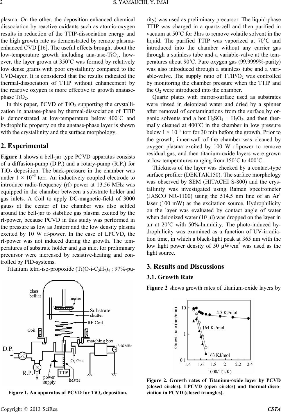 S. YAMAUCHI, Y. IMAI 2 plasma. On the other, the deposition enhanced chemical dissociation by reactive oxidants such as atomic-oxygen results in reduction of the TTIP-dissociation energy and the high growth rate as demonstrated by remote plasma- enhanced CVD [16]. The useful effects brought about the low-temperature growth including ana-tase-TiO2, how- ever, the layer grown at 350˚C was formed by relatively low dense grains with poor crystallinity compared to the CVD-layer. It is considered that the results indicated the thermal-dissociation of TTIP without enhancement by the reactive oxygen is more effective to growth anatase- phase TiO2. In this paper, PCVD of TiO2 supporting the crystalli- zation in anatase-phase by thermal-dissociation of TTIP is demonstrated at low-temperature below 400˚C and hydrophilic property on the anatase-phase layer is shown with the crystallinity and the surface morphology. 2. Experimental Figure 1 shows a bell-jar type PCVD apparatus consists of a diffusion-pump (D.P.) and a rotary-pump (R.P.) for TiO2 deposition. The back-pressure in the chamber was under 1 × 10⁻⁵ torr. An inductively coupled electrode to introduce radio-frequency (rf) power at 13.56 MHz was equipped in the chamber between a substrate holder and gas inlets. A Coil to apply DC-magnetic-field of 3000 gauss at the center of the chamber was also settled around the bell-jar to stabilize gas plasma excited by the rf-power, because PCVD in this study was performed in the pressure as low as 3mtorr and the low density plasma excited by 10 W rf-power. In the case of LPCVD, the rf-power was not induced during the growth. The tem- peratures of substrate holder and gas inlet for preliminary precursor were increased by resistive-heating and con- trolled by PID-systems. Titanium tetra-iso-propoxide (Ti(O-i-C3H7)4 : 97%-pu- Figure 1. An apparatus of PCVD for TiO2 deposition. rity) was used as preliminary precursor. The liquid-phase TTIP was charged in a quartz-cell and then purified in vacuum at 50˚C for 3hrs to remove volatile solvent in the liquid. The purified TTIP was vaporized at 70˚C and introduced into the chamber without any carrier gas through a stainless tube and a variable-valve at the tem- peratures about 90˚C. Pure oxygen gas (99.9999%-purity) was also introduced through a stainless tube and a vari- able-valve. The supply ratio of TTIP/O2 was controlled by monitoring the chamber pressure when the TTIP and the O2 were introduced into the chamber. Quartz plates with mirror-surface used as substrates were rinsed in deionized water and dried by a spinner after removal of contaminations from the surface by or- ganic solvents and a hot H2SO4 + H2O2, and then ther- mally cleaned at 400˚C in the chamber in low pressure below 1 × 10⁻⁵ torr for 30 min before the growth. Prior to the growth, inner-wall of the chamber was cleaned by oxygen plasma excited by 100 W rf-power to remove residual gas, and then titanium-oxide layers were grown at low temperatures ranging from 150˚C to 400˚C. Thickness of the layer was checked by a contact-type surface profiler (DEKTAK150). The surface morphology was observed by SEM (HITACHI S-800) and the crys- tallinity was investigated using Raman spectrometer (JASCO NR-1100) using the 514.5 nm line of an Ar+ laser (100 mW) as the excitation source. Hydrophilicity on the layer was evaluated by contact angle of water when deionized water (10 μl) was dropped on the layer in air at 20˚C with 50%-humidity. The photo-induced hy- drophilicity was examined as a function of UV-irradia- tion time, in which a black-light peak at 365 nm with the low light power density of 50 μW/cm2 was used as the light source. 3. Results and Discussions 3.1. Growth Rate Figure 2 shows growth rates of titanium-oxide layers by Figure 2. Growth rates of Titanium-oxide layer by PCVD (closed circles), LPCVD (open circles) and thermal-disso- ciation in PCVD (closed triangles). Copyright © 2013 SciRes. CSTA 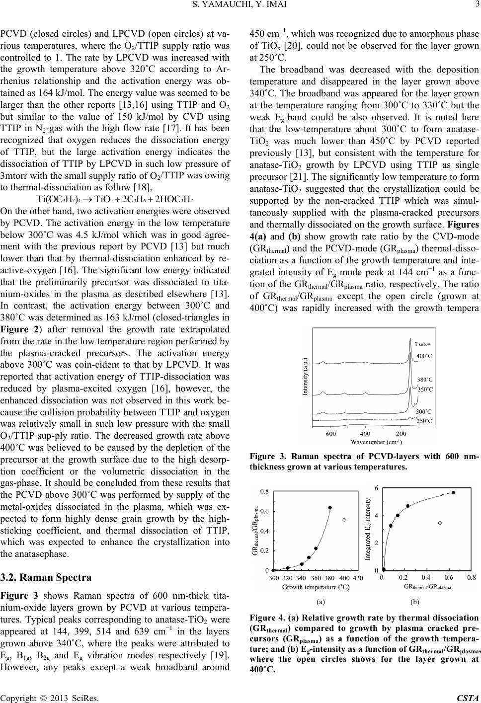 S. YAMAUCHI, Y. IMAI 3 PCVD (closed circles) and LPCVD (open circles) at va- rious temperatures, where the O2/TTIP supply ratio was controlled to 1. The rate by LPCVD was increased with the growth temperature above 320˚C according to Ar- rhenius relationship and the activation energy was ob- tained as 164 kJ/mol. The energy value was seemed to be larger than the other reports [13,16] using TTIP and O2 but similar to the value of 150 kJ/mol by CVD using TTIP in N2-gas with the high flow rate [17]. It has been recognized that oxygen reduces the dissociation energy of TTIP, but the large activation energy indicates the dissociation of TTIP by LPCVD in such low pressure of 3mtorr with the small supply ratio of O2/TTIP was owing to thermal-dissociation as follow [18], 3742 3637Ti(OC H )TiO2C H2HOC H On the other hand, two activation energies were observed by PCVD. The activation energy in the low temperature below 300˚C was 4.5 kJ/mol which was in good agree- ment with the previous report by PCVD [13] but much lower than that by thermal-dissociation enhanced by re- active-oxygen [16]. The significant low energy indicated that the preliminarily precursor was dissociated to tita- nium-oxides in the plasma as described elsewhere [13]. In contrast, the activation energy between 300˚C and 380˚C was determined as 163 kJ/mol (closed-triangles in Figure 2) after removal the growth rate extrapolated from the rate in the low temperature region performed by the plasma-cracked precursors. The activation energy above 300˚C was coin-cident to that by LPCVD. It was reported that activation energy of TTIP-dissociation was reduced by plasma-excited oxygen [16], however, the enhanced dissociation was not observed in this work be- cause the collision probability between TTIP and oxygen was relatively small in such low pressure with the small O2/TTIP sup-ply ratio. The decreased growth rate above 400˚C was believed to be caused by the depletion of the precursor at the growth surface due to the high desorp- tion coefficient or the volumetric dissociation in the gas-phase. It should be concluded from these results that the PCVD above 300˚C was performed by supply of the metal-oxides dissociated in the plasma, which was ex- pected to form highly dense grain growth by the high- sticking coefficient, and thermal dissociation of TTIP, which was expected to enhance the crystallization into the anatasephase. 3.2. Raman Spectra Figure 3 shows Raman spectra of 600 nm-thick tita- nium-oxide layers grown by PCVD at various tempera- tures. Typical peaks corresponding to anatase-TiO2 were appeared at 144, 399, 514 and 639 cm−1 in the layers grown above 340˚C, where the peaks were attributed to Eg, B1g, B2g and Eg vibration modes respectively [19]. However, any peaks except a weak broadband around 450 cm−1, which was recognized due to amorphous phase of TiOx [20], could not be observed for the layer grown at 250˚C. The broadband was decreased with the deposition temperature and disappeared in the layer grown above 340˚C. The broadband was appeared for the layer grown at the temperature ranging from 300˚C to 330˚C but the weak Eg-band could be also observed. It is noted here that the low-temperature about 300˚C to form anatase- TiO2 was much lower than 450˚C by PCVD reported previously [13], but consistent with the temperature for anatase-TiO2 growth by LPCVD using TTIP as single precursor [21]. The significantly low temperature to form anatase-TiO2 suggested that the crystallization could be supported by the non-cracked TTIP which was simul- taneously supplied with the plasma-cracked precursors and thermally dissociated on the growth surface. Figures 4(a) and (b) show growth rate ratio by the CVD-mode (GRthermal) and the PCVD-mode (GRplasma) thermal-disso- ciation as a function of the growth temperature and inte- grated intensity of Eg-mode peak at 144 cm−1 as a func- tion of the GRthermal/GRplasma ratio, respectively. The ratio of GRthermal/GRplasma except the open circle (grown at 400˚C) was rapidly increased with the growth tempera Figure 3. Raman spectra of PCVD-layers with 600 nm- thickness grown at various temperatures. (a) (b) Figure 4. (a) Rel ative growth rate by thermal dissociation (GRtherma l) compared to growth by plasma cracked pre- cursors (GRplasma) as a function of the growth tempera- ture; and (b) Eg-intensity as a function of GR thermal/GRplasma, where the open circles shows for the layer grown at 400˚C. Copyright © 2013 SciRes. CSTA 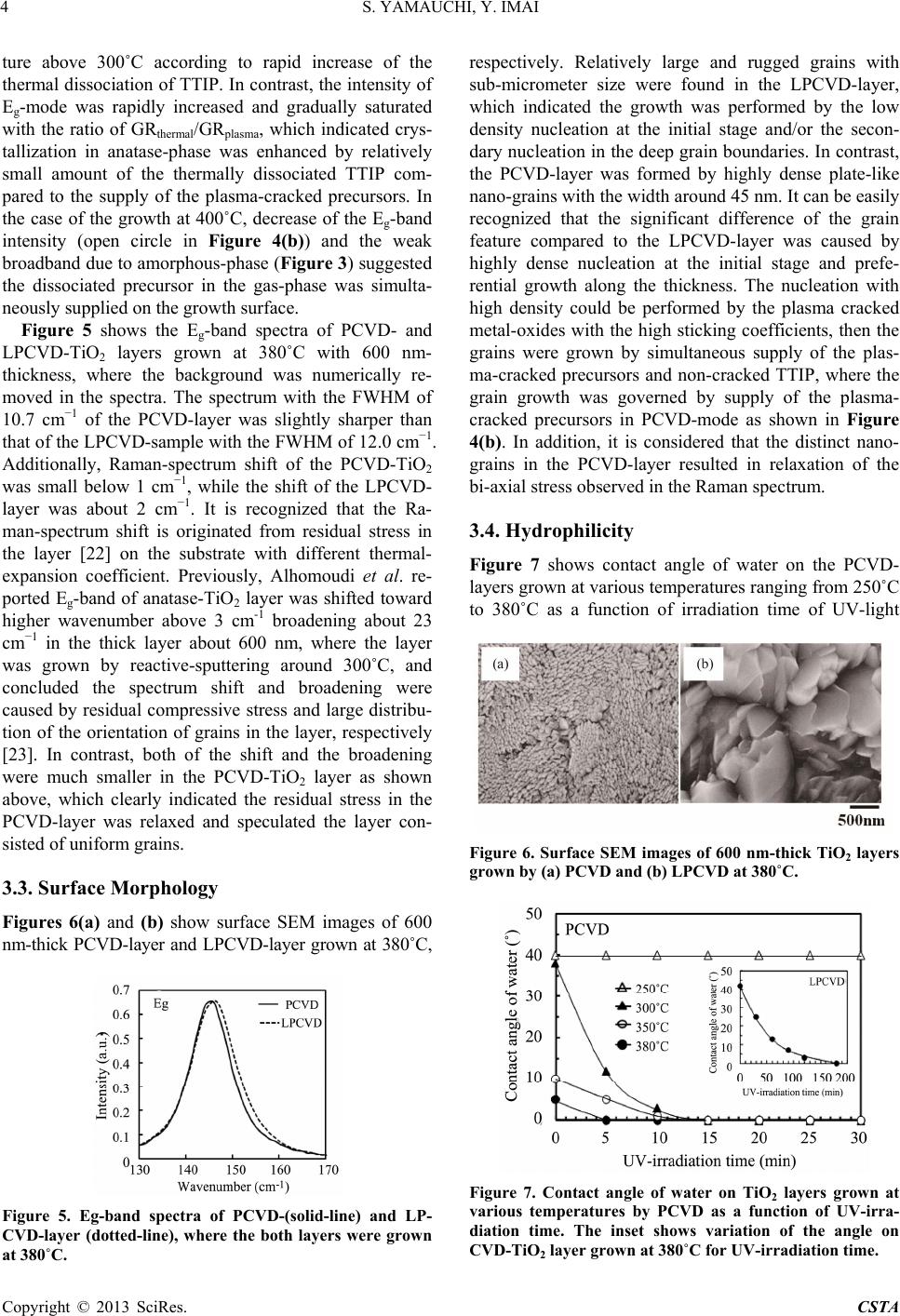 S. YAMAUCHI, Y. IMAI 4 ture above 300˚C according to rapid increase of the thermal dissociation of TTIP. In contrast, the intensity of Eg-mode was rapidly increased and gradually saturated with the ratio of GRthermal/GRplasma, which indicated crys- tallization in anatase-phase was enhanced by relatively small amount of the thermally dissociated TTIP com- pared to the supply of the plasma-cracked precursors. In the case of the growth at 400˚C, decrease of the Eg-band intensity (open circle in Figure 4(b)) and the weak broadband due to amorphous-phase (Figure 3) suggested the dissociated precursor in the gas-phase was simulta- neously supplied on the growth surface. Figure 5 shows the Eg-band spectra of PCVD- and LPCVD-TiO2 layers grown at 380˚C with 600 nm- thickness, where the background was numerically re- moved in the spectra. The spectrum with the FWHM of 10.7 cm−1 of the PCVD-layer was slightly sharper than that of the LPCVD-sample with the FWHM of 12.0 cm−1. Additionally, Raman-spectrum shift of the PCVD-TiO2 was small below 1 cm−1, while the shift of the LPCVD- layer was about 2 cm−1. It is recognized that the Ra- man-spectrum shift is originated from residual stress in the layer [22] on the substrate with different thermal- expansion coefficient. Previously, Alhomoudi et al. re- ported Eg-band of anatase-TiO2 layer was shifted toward higher wavenumber above 3 cm-1 broadening about 23 cm−1 in the thick layer about 600 nm, where the layer was grown by reactive-sputtering around 300˚C, and concluded the spectrum shift and broadening were caused by residual compressive stress and large distribu- tion of the orientation of grains in the layer, respectively [23]. In contrast, both of the shift and the broadening were much smaller in the PCVD-TiO2 layer as shown above, which clearly indicated the residual stress in the PCVD-layer was relaxed and speculated the layer con- sisted of uniform grains. 3.3. Surface Morphology Figures 6(a) an d (b) show surface SEM images of 600 nm-thick PCVD-layer and LPCVD-layer grown at 380˚C, Figure 5. Eg-band spectra of PCVD-(solid-line) and LP- CVD-layer (dotted-line), where the both layers were grown at 380˚C. respectively. Relatively large and rugged grains with sub-micrometer size were found in the LPCVD-layer, which indicated the growth was performed by the low density nucleation at the initial stage and/or the secon- dary nucleation in the deep grain boundaries. In contrast, the PCVD-layer was formed by highly dense plate-like nano-grains with the width around 45 nm. It can be easily recognized that the significant difference of the grain feature compared to the LPCVD-layer was caused by highly dense nucleation at the initial stage and prefe- rential growth along the thickness. The nucleation with high density could be performed by the plasma cracked metal-oxides with the high sticking coefficients, then the grains were grown by simultaneous supply of the plas- ma-cracked precursors and non-cracked TTIP, where the grain growth was governed by supply of the plasma- cracked precursors in PCVD-mode as shown in Figure 4(b). In addition, it is considered that the distinct nano- grains in the PCVD-layer resulted in relaxation of the bi-axial stress observed in the Raman spectrum. 3.4. Hydrophilicity Figure 7 shows contact angle of water on the PCVD- layers grown at various temperatures ranging from 250˚C to 380˚C as a function of irradiation time of UV-light Figure 6. Surface SEM images of 600 nm-thick TiO2 layers grown by (a) PCVD and (b) LPCVD at 380˚C. Figure 7. Contact angle of water on TiO2 layers grown at various temperatures by PCVD as a function of UV-irra- diation time. The inset shows variation of the angle on CVD-TiO2 layer grown at 380˚C for UV-irradiation time. Copyright © 2013 SciRes. CSTA 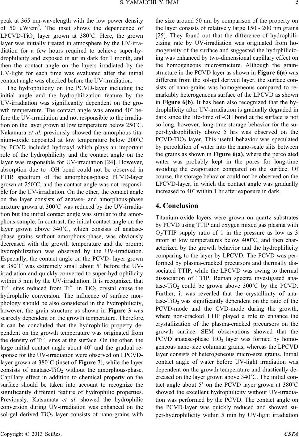 S. YAMAUCHI, Y. IMAI 5 peak at 365 nm-wavelength with the low power density of 50 μW/cm2. The inset shows the dependence of LPCVD-TiO2 layer grown at 380˚C. Here, the grown layer was initially treated in atmosphere by the UV-irra- diation for a few hours required to achieve super-hy- drophilicity and exposed in air in dark for 1 month, and then the contact angle on the layers irradiated by the UV-light for each time was evaluated after the initial contact angle was checked before the UV-irradiation. The hydrophilicity on the PCVD-layer including the initial angle and the hydrophilization feature by the UV-irradiation was significantly dependent on the gro- wth temperature. The contact angle was around 40˚ be- fore the UV-irradiation and not responsible to the irradia- tion on the layer grown at low temperature below 250˚C. Nakamura et al. previously showed the amorphous tita- nium-oxide deposited at low temperature below 200˚C by PCVD included hydroxyl which plays an important role of the hydrophilicity and the contact angle on the layer was responsible for UV-irradiation [24]. However, absorption due to -OH bond could not be observed in FTIR spectrum of the amorphous-phase PCVD-layer grown at 250˚C, and the contact angle was not responsi- ble for the UV-irradiation. On the other, the contact angle on the layer consists of anatase- and amorphous-phase mixture grown at 300˚C was reduced by the UV-irradia- tion but the initial contact angle was similar to the amor- phous-sample. In contrast, the initial contact angle on the layer grown above 340˚C, which consists of anatase- phase grains without amorphous-phase, was obviously decreased with the growth temperature and the prompt hydrophilization was observed by the UV-irradiation. Especially, the contact angle on the PCVD- layer grown at 380˚C was extremely small about 5˚ before the UV- irradiation and quickly converted to super-hydrophilicity within 5 min by the UV-irradiation. It is recognized that Ti3+ sites reduced from Ti4+ in TiO2 crystal cause the hydrophilic conversion. The influence of surface mor- phology should be also considered in the hydrophilicity, however, the grain structure as shown in Figure 3 was scarcely dependent on the growth temperature. Therefore, it can be concluded that the hydrophilic property de- pendent on the growth temperature was originated from the density of Ti3+ sites at the surface. On the other, the large initial contact angle about 40˚ and the gradual re- sponse for the UV-irradiation were observed on LPCVD- layer grown at 380˚C (inset of Figure 7), while the layer consists of anatase-TiO2 without the amorphous-phase. Capillary effect in addition to chemical property on the surface should be taken into account to recognize the significantly different feature of hydrophilic properties. Previously, Katsumata et al. showed the hydrophilic conversion during UV-irradiation was enhanced on the sol-gel derived TiO2 layer consists of nano-grains with the size around 50 nm by comparison of the property on the layer consists of relatively large 150 - 200 nm grains [25]. They found out that the difference of hydrophili- cizing rate by UV-irradiation was originated from ho- mogeneity of the surface and suggested the hydrphiliciz- ing was enhanced by two-dimensional capillary effect on the homogeneous microstructure. Although the grain- structure in the PCVD layer as shown in Figure 6(a) was different from the sol-gel derived layer, the surface con- sists of nano-grains was homogeneous compared to re- markably heterogeneous surface of the LPCVD as shown in Figure 6(b). It has been also recognized that the hy- drophilicity after UV-irradiation is gradually degraded in dark since the life-time of -OH bond at the surface is not so long, however, long-time storage behavior for the su- per-hydrophilicity above 5 hrs was observed on the PCVD-TiO2 layer. This useful behavior was speculated by percolation of water into the nano-scale slits between the grains as shown in Figure 6(a), where the percolated water was probably kept in the pores for long-time avoiding the evaporation compared on the surface. Of course, the storage behavior could not be observed on the LPCVD-layer, in which the contact angle was gradually increased to 40˚ within 1 hr after exposure in dark. 4. Conclusion Titanium-oxide layers were grown on quartz substrates by PCVD using TTIP and oxygen mixed gas plasma with O2/TTIP supply ratio of 1 in the pressure as low as 3 mtorr at low temperatures below 400˚C, and then char- acterized by the growth behavior and the hydrophilicity comparing to the layer by LPCVD. The PCVD was per- formed by plasma-cracked precursors and thermally dis- sociated TTIP, while the LPCVD was owing to thermal dissociation of TTIP. Raman spectra investigated ana- tase-TiO2 could be grown above 300˚C by the PCVD. Further, it was revealed that the crystallinity of ana- tase-TiO2 was significantly dependent on the ratio of the PCVD-mode and the CVD-mode during the growth, where non-cracked TTIP played a role to enhance the crystallization of the plasma-cracked precursors on the growth surface. SEM observations showed that the PCVD anatase-phase TiO2 layer was formed by homo- geneous nano-size columnar grains, whereas the LPCVD layer consists of heterogeneous micro-size grains. Initial contact angle of water before UV-light irradiation was dependent on the growth temperature and drastically de- creased on the layer grown above 340˚C. The initial con- tact angle about 5˚ on the PCVD layer grown at 380˚C showed the excellent hydrophilicity without UV-irradia- tion was performed by the PCVD. The contact angle on the PCVD-layer was quickly reduced and showed su- per-hydrophilicity within 5 min by UV-light irradiation Copyright © 2013 SciRes. CSTA 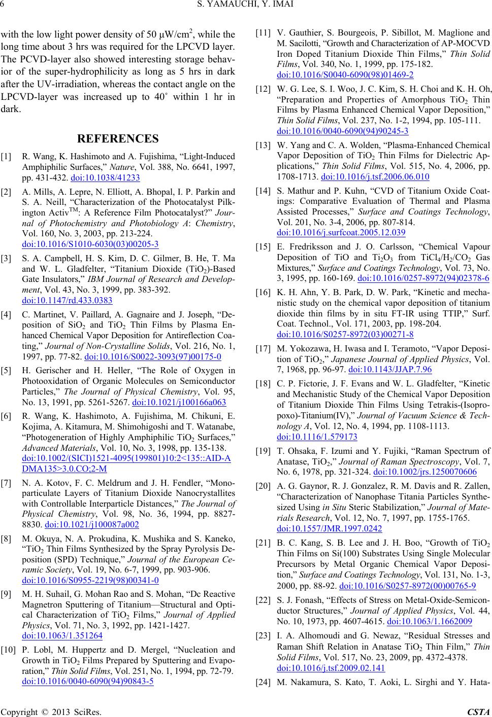 S. YAMAUCHI, Y. IMAI 6 with the low light power density of 50 μW/cm2, while the long time about 3 hrs was required for the LPCVD layer. The PCVD-layer also showed interesting storage behav- ior of the super-hydrophilicity as long as 5 hrs in dark after the UV-irradiation, whereas the contact angle on the LPCVD-layer was increased up to 40˚ within 1 hr in dark. REFERENCES [1] R. Wang, K. Hashimoto and A. Fujishima, “Light-Induced Amphiphilic Surfaces,” Nature, Vol. 388, No. 6641, 1997, pp. 431-432. doi:10.1038/41233 [2] A. Mills, A. Lepre, N. Elliott, A. Bhopal, I. P. Parkin and S. A. Neill, “Characterization of the Photocatalyst Pilk- ington ActivTM: A Reference Film Photocatalyst?” Jour- nal of Photochemistry and Photobiology A: Chemistry, Vol. 160, No. 3, 2003, pp. 213-224. doi:10.1016/S1010-6030(03)00205-3 [3] S. A. Campbell, H. S. Kim, D. C. Gilmer, B. He, T. Ma and W. L. Gladfelter, “Titanium Dioxide (TiO2)-Based Gate Insulators,” IBM Journal of Research and Develop- ment, Vol. 43, No. 3, 1999, pp. 383-392. doi:10.1147/rd.433.0383 [4] C. Martinet, V. Paillard, A. Gagnaire and J. Joseph, “De- position of SiO2 and TiO2 Thin Films by Plasma En- hanced Chemical Vapor Deposition for Antireflection Coa- ting,” Journal of Non-Crystalline Solids, Vol. 216, No. 1, 1997, pp. 77-82. doi:10.1016/S0022-3093(97)00175-0 [5] H. Gerischer and H. Heller, “The Role of Oxygen in Photooxidation of Organic Molecules on Semiconductor Particles,” The Journal of Physical Chemistry, Vol. 95, No. 13, 1991, pp. 5261-5267. doi:10.1021/j100166a063 [6] R. Wang, K. Hashimoto, A. Fujishima, M. Chikuni, E. Kojima, A. Kitamura, M. Shimohigoshi and T. Watanabe, “Photogeneration of Highly Amphiphilic TiO2 Surfaces,” Advanced Materials, Vol. 10, No. 3, 1998, pp. 135-138. doi:10.1002/(SICI)1521-4095(199801)10:2<135::AID-A DMA135>3.0.CO;2-M [7] N. A. Kotov, F. C. Meldrum and J. H. Fendler, “Mono- particulate Layers of Titanium Dioxide Nanocrystallites with Controllable Interparticle Distances,” The Journal of Physical Chemistry, Vol. 98, No. 36, 1994, pp. 8827- 8830. doi:10.1021/j100087a002 [8] M. Okuya, N. A. Prokudina, K. Mushika and S. Kaneko, “TiO2 Thin Films Synthesized by the Spray Pyrolysis De- position (SPD) Technique,” Journal of the European Ce- ramic Society, Vol. 19, No. 6-7, 1999, pp. 903-906. doi:10.1016/S0955-2219(98)00341-0 [9] M. H. Suhail, G. Mohan Rao and S. Mohan, “Dc Reactive Magnetron Sputtering of Titanium—Structural and Opti- cal Characterization of TiO2 Films,” Journal of Applied Physics, Vol. 71, No. 3, 1992, pp. 1421-1427. doi:10.1063/1.351264 [10] P. Lobl, M. Huppertz and D. Mergel, “Nucleation and Growth in TiO2 Films Prepared by Sputtering and Evapo- ration,” Thin Solid Films, Vol. 251, No. 1, 1994, pp. 72-79. doi:10.1016/0040-6090(94)90843-5 [11] V. Gauthier, S. Bourgeois, P. Sibillot, M. Maglione and M. Sacilotti, “Growth and Characterization of AP-MOCVD Iron Doped Titanium Dioxide Thin Films,” Thin Solid Films, Vol. 340, No. 1, 1999, pp. 175-182. doi:10.1016/S0040-6090(98)01469-2 [12] W. G. Lee, S. I. Woo, J. C. Kim, S. H. Choi and K. H. Oh, “Preparation and Properties of Amorphous TiO2 Thin Films by Plasma Enhanced Chemical Vapor Deposition,” Thin Solid Films, Vol. 237, No. 1-2, 1994, pp. 105-111. doi:10.1016/0040-6090(94)90245-3 [13] W. Yang and C. A. Wolden, “Plasma-Enhanced Chemical Vapor Deposition of TiO2 Thin Films for Dielectric Ap- plications,” Thin Solid Films, Vol. 515, No. 4, 2006, pp. 1708-1713. doi:10.1016/j.tsf.2006.06.010 [14] S. Mathur and P. Kuhn, “CVD of Titanium Oxide Coat- ings: Comparative Evaluation of Thermal and Plasma Assisted Processes,” Surface and Coatings Technology, Vol. 201, No. 3-4, 2006, pp. 807-814. doi:10.1016/j.surfcoat.2005.12.039 [15] E. Fredriksson and J. O. Carlsson, “Chemical Vapour Deposition of TiO and Ti2O3 from TiCl4/H2/CO2 Gas Mixtures,” Surface and Coatings Technology, Vol. 73, No. 3, 1995, pp. 160-169. doi:10.1016/0257-8972(94)02378-6 [16] K. H. Ahn, Y. B. Park, D. W. Park, “Kinetic and mecha- nistic study on the chemical vapor deposition of titanium dioxide thin films by in situ FT-IR using TTIP,” Surf. Coat. Technol., Vol. 171, 2003, pp. 198-204. doi:10.1016/S0257-8972(03)00271-8 [17] M. Yokozawa, H. Iwasa and I. Teramoto, “Vapor Deposi- tion of TiO2,” Japanese Journal of Applied Physics, Vol. 7, 1968, pp. 96-97. doi:10.1143/JJAP.7.96 [18] C. P. Fictorie, J. F. Evans and W. L. Gladfelter, “Kinetic and Mechanistic Study of the Chemical Vapor Deposition of Titanium Dioxide Thin Films Using Tetrakis-(Isopro- poxo)-Titanium(IV),” Journal of Vacuum Scie nce & Tec h- nology A, Vol. 12, No. 4, 1994, pp. 1108-1113. doi:10.1116/1.579173 [19] T. Ohsaka, F. Izumi and Y. Fujiki, “Raman Spectrum of Anatase, TiO2,” Journal of Raman Spectroscopy, Vol. 7, No. 6, 1978, pp. 321-324. doi:10.1002/jrs.1250070606 [20] A. G. Gaynor, R. J. Gonzalez, R. M. Davis and R. Zallen, “Characterization of Nanophase Titania Particles Synthe- sized Using in Situ Steric Stabilization,” Journal of Mate- rials Research, Vol. 12, No. 7, 1997, pp. 1755-1765. doi:10.1557/JMR.1997.0242 [21] B. C. Kang, S. B. Lee and J. H. Boo, “Growth of TiO2 Thin Films on Si(100) Substrates Using Single Molecular Precursors by Metal Organic Chemical Vapor Deposi- tion,” Surface and Coatings Technology, Vol. 131, No. 1-3, 2000, pp. 88-92. doi:10.1016/S0257-8972(00)00765-9 [22] S. J. Fonash, “Effects of Stress on Metal-Oxide-Semicon- ductor Structures,” Journal of Applied Physics, Vol. 44, No. 10, 1973, pp. 4607-4615. doi:10.1063/1.1662009 [23] I. A. Alhomoudi and G. Newaz, “Residual Stresses and Raman Shift Relation in Anatase TiO2 Thin Film,” Thin Solid Films, Vol. 517, No. 23, 2009, pp. 4372-4378. doi:10.1016/j.tsf.2009.02.141 [24] M. Nakamura, S. Kato, T. Aoki, L. Sirghi and Y. Hata- Copyright © 2013 SciRes. CSTA  S. YAMAUCHI, Y. IMAI Copyright © 2013 SciRes. CSTA 7 naka, “Role of Terminal OH Groups on the Electrical and Hydrophilic Properties of Hydro-Oxygenated Amorphous TiOx: OH Thin Films,” Journal of Applied Physics, Vol. 90, No. 7, 2001, pp. 3391-3395. doi:10.1063/1.1398599 [25] K. Katsumata, A. Nakajima, H. Yoshikawa, T. Shiota, N. Yoshida, T. Watanabe, Y. Kameshima and K. Okada, “Ef- fect of Microstructure on Photoinduced Hydrophilicity of Transparent Anatase Thin Films,” Surface Science, Vol. 579, No. 2-3, 2005, pp. 123-130. doi:10.1016/j.susc.2005.01.035 |