

Paper Menu >>
Journal Menu >>
 Vol.2, No.10, 1061-1065 (2010) Natural Science http://dx.doi.org/10.4236/ns.2010.210131 Copyright © 2010 SciRes. OPEN ACCESS Preparation and characterization of genipin-cross-linked chitosan microparticles by water-in-oil emulsion solvent diffusion method Jesada Karnchanajindanun, Mangkorn Srisa-ard, Prasong Srihanam, Yodthong Baimark* Department of Chemistry and Center of Excellence for Innovation in Chemistry, Faculty of Science, Mahasarakham University, Mahasarakham, Thailand; *Corresponding Author: [email protected]. Received 27 July 2010; revised 30 August 2010; accepted 5 September 2010. ABSTRACT Chitosan (CS) microparticles with and without cross-linking were prepared by a water-in-oil emulsion solvent diffusion method without any surfactants. Aqueous CS solution and ethyl ace- tate were used as water and oil phases, respec- tively. Genipin was used as a cross-linker. Influ- ences of genipin ratios and cross-linking times on CS microparticle characteristics were inves- tigated. Non-cross-linked and cross-linked CS microparticles were spherical in shape and rough in surface. Microparticle matrices showed po- rous structures. Surface roughness, mean par- ticle sizes and bulk density of CS microparticles increased and their dissolutions in acetic acid solution decreased when genipin ratio and cross- linking time increased. Keywords: Chitosan microparticles; Porous structure; Genipin; Cross-linking; Morphology 1. INTRODUCTION Chitosan is a copolymer of 2-glucosamine and N- acetyl-2-glucosamine prepared by alkaline deacetylation of chitin that has received great attention for its possible uses in medical, pharmaceutical and metal ion treatment applications because of its biodegradability, biocompati- bility and high concentration of amine functional groups [1-4]. Chitosan microparticles have usually been fabri- cated by precipitation, spray drying and water-in-oil (W / O) emulsification-cross-linking methods [5]. The current study describes an alternative method for preparation of cross-linked and non-cross-linked CS microparticles by the W / O emulsion solvent diffusion method. Genipin was selected as a biocompatible cross-linker. Genipin is a natural water-soluble bi-functional cross- linker. It is obtained from geniposide, a component of traditional Chinese medicine and is isolated from the fruits of the plant, Gardenial jasminoides Ellis [6]. Genipin is a fully biocompatible reagent about 10,000 times less cytotoxic than glutaraldehyde [7]. The CS device matrices were successfully cross-linked with ge- nipin [8-10]. In this work, a novel approach to the preparation of genipin-cross-linked chitosan microparticles by the sim- ple W / O emulsion solvent diffusion method is reported. Cross-linked chitosan microparticles were solidified and formed after diffusion out of water from emulsion drop- lets of chiosan solution to external continuous phase, ethyl acetate. The influences of cross-linker ratio and cross-linking time on CS microparticle characteristics including morphology, particle size, dissolution and bulk density were investigated and discussed. 2. EXPERIMENTAL 2.1. Materials Chitosan with degree of de-acetylation and average molecular weight of 90% and 100 kDa, respectively was purchased from Seafresh Chitosan Lab Co., Ltd. (Thai- land). Genipin (Challenge Bioproducts Co. Ltd., Taiwan) and analytical grade ethyl acetate (Lab Scan) were used without further purification. 2.2. Preparation of Chitosan Microparticles Chitosan solution with 1% w / v was prepared by us- ing a 2% (v / v) acetic acid aqueous solution as a solvent. Chitosan microparticles were prepared by the water- in-oil emulsion solvent diffusion method. The 0.5 mL of 0.5% w / v chitosan solution was added drop-wise to 200 mL of ethyl acetate with a stirring speed of 900 rpm for 1 h. The beaker was tightly sealed with aluminum foil during the emulsification-diffusion process to prevent ethyl acetate evaporation. The chitosan microparticles suspended in ethyl acetate were collected by centrifuga-  J. Karnchanajindanun et al. / Natural Science 2 (2010) 1061-1065 Copyright © 2010 SciRes. OPEN ACCESS 1062 tion before drying in a vacuum oven at room tempera- ture for 6 h. Genipin-cross-linked chitosan microparticles were pro- duced by the same method. The chitosan and genipin solutions were mixed together under constant stirring at room temperature for cross-linking before microparticle preparation. The final chitosan concentration was 0.5% w / v after cross-linking. The different genipin concen- trations (5%, 10% and 20% w / w) and cross-linking times (1.5, 3 and 6 h) were investigated. 2.3. Characterization of Chitosan Microparticles Morphology of the chitosan microparticles was de- termined by scanning electron microscopy (SEM) using a JEOL JSM-6460LV SEM. The microparticles were coated with gold for enhancing conductivity before scan. Mean microparicle size and the coefficient of variation (CV) were calculated for each on SEM images by count- ing a minimum of 100 particles using smile view soft- ware (version 1.02). The CV value was calculated from the following equation: CV = p D 100 (1) where is a standard deviation, and Dp is a mean micropar- ticle diameter measured from SEM images. Lower CV val- ues indicate high microparticle monodispersity in size. Percentage of dissolution of the chitosan microparticles was investigated by shaking 50 mg of the chitosan micro- particles in 1.5 mL of 2% w / v acetic acid aqueous solu- tion at room temperature for 24 h. The residue microparti- cles were recovered by centrifugation before drying in a vacuum oven at 50C until its weight remained constant. The percentage of dissolution of the chitosan microparti- cles was then calculated by following the equation. Each average percentage of dissolution value was calculated from a mean of three measurements (see Eq. 2 ). where initial and remaining CS microparticles are the weights of chitosan microparticles before and after dis- solution test, respectively. Bulk density of the chitosan microparticles was meas- ured by gas displacement method using an ultrapyc- nometer 1000 (Quantachrom, USA) under helium gas. Each average density value was calculated from a mean of five determinations. 3. RESULTS AND DISCUSSIONS Here, the water-in-oil (W / O) emulsion solvent diffu- sion method without any surfactant was used to prepare chitosan (CS) microparticles. The maximum water solu- bility in ethyl acetate is 3.30% (CAS No. 141-78-6). Then the polymer particles should form, if the less than 3.3 mL of aqueous polymer solution (W phase) was added drop-wise into 100 mL of ethyl acetate (O phase) with stirring. The water in dispersed emulsion droplets of CS solution diffused out to the continuous phase, ethyl ace- tate. It was found in our cases that the CS microparticles were successfully prepared using this method. In our preliminary test, the magnetic stirring speed of 900 rpm is found the most appropriate for microparticle prepara- tion. The almost aqueous CS solution could not be bro- ken to form uniform droplets when the stirring speed was lower than 900 rpm. Meanwhile, almost all particles were stuck at the wall of glassware during emulsifica- tion-diffusion process when higher stirring speed than 900 rpm was applied. The chitosan microparticles could not be completely solidified when the stirring time was shorter than 1 h. The CS aggregates were found when higher 0.5 mL of CS solution was used due to its high viscosity of CS solution. 3.1. Morphology and Sizes of CS Microparticles Figure 1 shows SEM images of non-cross-linked and cross-linked CS microparticles prepared with different genipin ratios. They were nearly spherical in shape sug- gested that the genipin ratios did not effect on the parti- cle shape. Surfaces of these CS microparticles are illus- trated in Figure 2. The non-cross-linked CS microparti- cles in Figure 2(a) showed rough surfaces. This may occur from diffusion out of water from dispersed drop- lets of CS solution to continuous ethyl acetate phase during particle solidification. The surface roughness increased with the genipin ratio as shown in Figures 2(b) -2(d). The results may be explained that the cross- link- ing can increase viscosity of CS solution. The diffusion out of water from cross-linked CS solution droplets was difficult. The cross-linked CS microparticles prepared with dif- ferent cross-linking times also showed the spherical-like shape, as illustrated in Figures 3(b)-3(d). This indicates that cross-linking times did not effect on the particle shape. The CS microparticles prepared with longer cross- linking time showed rougher surface than the shorter cross-linking time, as shown in Figure 4. This may be due to increasing of the viscosity of CS solution when the cross-linking time increased. % dissolution = 100 (mg) clesmicroparti CS initial ](mg) clesmicroparti CS remaining(mg) clesmicroparti CS [initial (2) 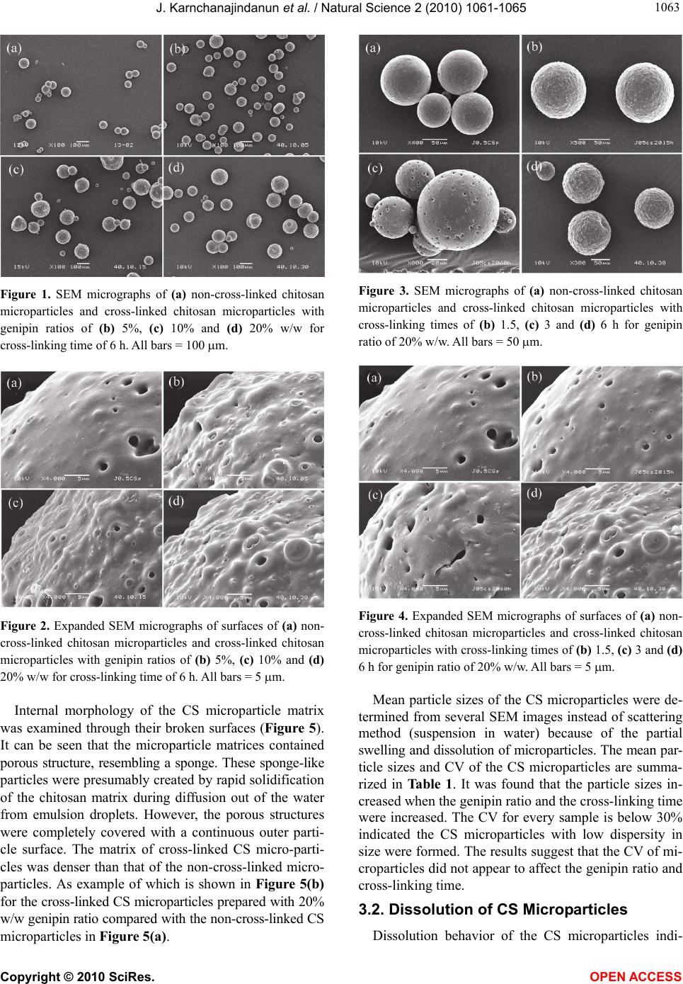 J. Karnchanajindanun et al. / Natural Science 2 (2010) 1061-1065 Copyright © 2010 SciRes. OPEN ACCESS 106 1063 Figure 1. SEM micrographs of (a) non-cross-linked chitosan microparticles and cross-linked chitosan microparticles with genipin ratios of (b) 5%, (c) 10% and (d) 20% w/w for cross-linking time of 6 h. All bars = 100 m. Figure 2. Expanded SEM micrographs of surfaces of (a) non- cross-linked chitosan microparticles and cross-linked chitosan microparticles with genipin ratios of (b) 5%, (c) 10% and (d) 20% w/w for cross-linking time of 6 h. All bars = 5 m. Internal morphology of the CS microparticle matrix was examined through their broken surfaces (Figure 5). It can be seen that the microparticle matrices contained porous structure, resembling a sponge. These sponge-like particles were presumably created by rapid solidification of the chitosan matrix during diffusion out of the water from emulsion droplets. However, the porous structures were completely covered with a continuous outer parti- cle surface. The matrix of cross-linked CS micro-parti- cles was denser than that of the non-cross-linked micro- particles. As example of which is shown in Figure 5(b) for the cross-linked CS microparticles prepared with 20% w/w genipin ratio compared with the non-cross-linked CS microparticles in Figure 5(a). Figure 3. SEM micrographs of (a) non-cross-linked chitosan microparticles and cross-linked chitosan microparticles with cross-linking times of (b) 1.5, (c) 3 and (d) 6 h for genipin ratio of 20% w/w. All bars = 50 m. Figure 4. Expanded SEM micrographs of surfaces of (a) non- cross-linked chitosan microparticles and cross-linked chitosan microparticles with cross-linking times of (b) 1.5, (c) 3 and (d) 6 h for genipin ratio of 20% w/w. All bars = 5 m. Mean particle sizes of the CS microparticles were de- termined from several SEM images instead of scattering method (suspension in water) because of the partial swelling and dissolution of microparticles. The mean par- ticle sizes and CV of the CS microparticles are summa- rized in Table 1. It was found that the particle sizes in- creased when the genipin ratio and the cross-linking time were increased. The CV for every sample is below 30% indicated the CS microparticles with low dispersity in size were formed. The results suggest that the CV of mi- croparticles did not appear to affect the genipin ratio and cross-linking time. 3.2. Dissolution of CS Microparticles Dissolution behavior of the CS microparticles indi- 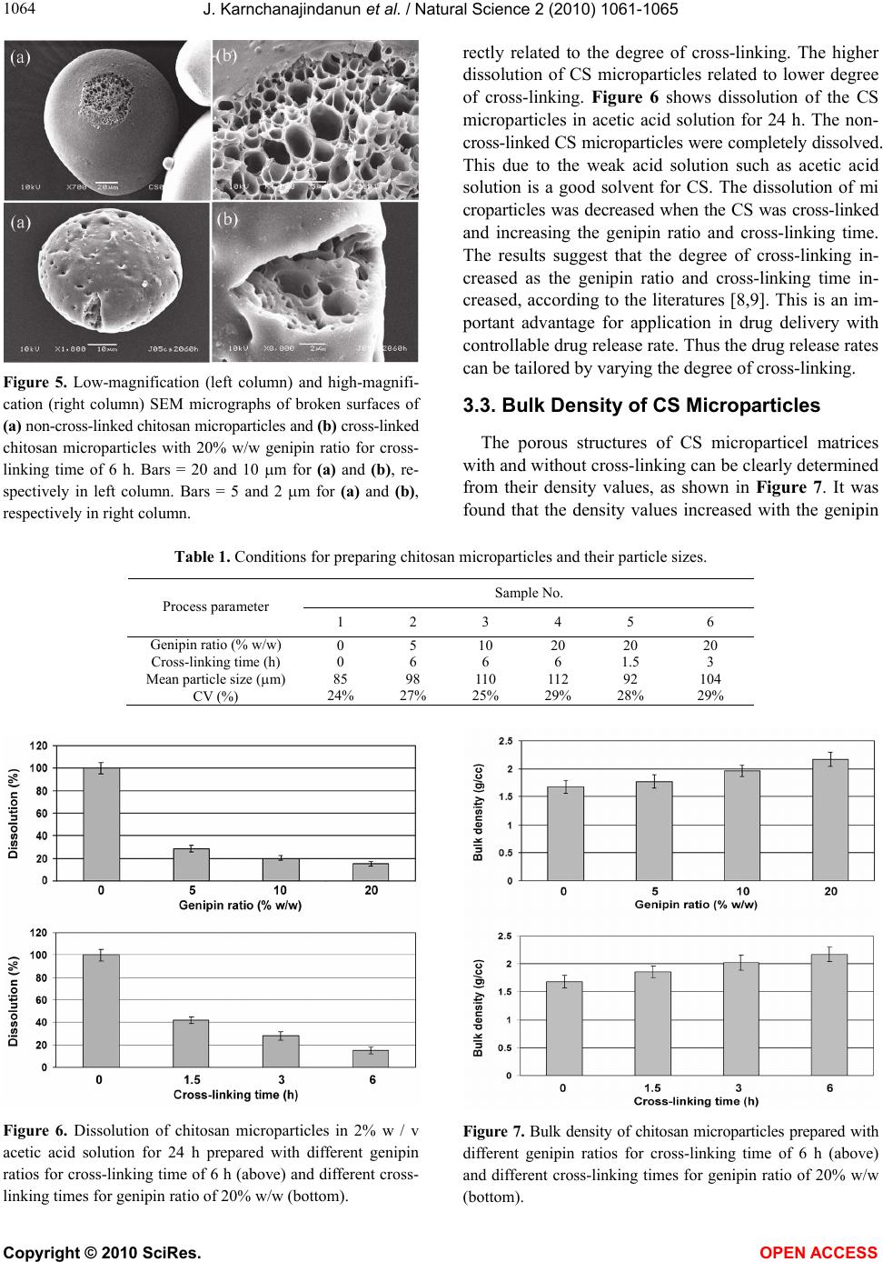 J. Karnchanajindanun et al. / Natural Science 2 (2010) 1061-1065 Copyright © 2010 SciRes. OPEN ACCESS 1064 Figure 5. Low-magnification (left column) and high-magnifi- cation (right column) SEM micrographs of broken surfaces of (a) non-cross-linked chitosan microparticles and (b) cross-linked chitosan microparticles with 20% w/w genipin ratio for cross- linking time of 6 h. Bars = 20 and 10 m for (a) and (b), re- spectively in left column. Bars = 5 and 2 m for (a) and (b), respectively in right column. rectly related to the degree of cross-linking. The higher dissolution of CS microparticles related to lower degree of cross-linking. Figure 6 shows dissolution of the CS microparticles in acetic acid solution for 24 h. The non- cross-linked CS microparticles were completely dissolved. This due to the weak acid solution such as acetic acid solution is a good solvent for CS. The dissolution of mi croparticles was decreased when the CS was cross-linked and increasing the genipin ratio and cross-linking time. The results suggest that the degree of cross-linking in- creased as the genipin ratio and cross-linking time in- creased, according to the literatures [8,9]. This is an im- portant advantage for application in drug delivery with controllable drug release rate. Thus the drug release rates can be tailored by varying the degree of cross-linking. 3.3. Bulk Density of CS Microparticles The porous structures of CS microparticel matrices with and without cross-linking can be clearly determined from their density values, as shown in Figure 7. It was found that the density values increased with the genipin Table 1. Conditions for preparing chitosan microparticles and their particle sizes. Sample No. Process parameter 1 2 3 4 5 6 Genipin ratio (% w/w) Cross-linking time (h) Mean particle size (m) CV (%) 0 0 85 24% 5 6 98 27% 10 6 110 25% 20 6 112 29% 20 1.5 92 28% 20 3 104 29% Figure 6. Dissolution of chitosan microparticles in 2% w / v acetic acid solution for 24 h prepared with different genipin ratios for cross-linking time of 6 h (above) and different cross- linking times for genipin ratio of 20% w/w (bottom). Figure 7. Bulk density of chitosan microparticles prepared with different genipin ratios for cross-linking time of 6 h (above) and different cross-linking times for genipin ratio of 20% w/w (bottom).  J. Karnchanajindanun et al. / Natural Science 2 (2010) 1061-1065 Copyright © 2010 SciRes. OPEN ACCESS 106 1065 ratio and cross-linking time. This can be explained that the CS molecules were closer together when the higher genipin ratio and cross-linking time were used. There- fore, the denser microparticles were obtained. The bulk density change after cross-linking corresponded to the microparticle matrices of the broken CS microparticles from the SEM images in Figure 5. 4. CONCLUSIONS Non-cross-linked and genipin-cross-linked chitosan microparticles with spherical-like shapes have been suc- cessfully prepared using the simple and rapid W / O emu- lsion solvent diffusion method. The surface roughness of microparticles increased with genipin ratio and cross- linking time but the particle shape did not change. All chitosan microparticle matricess contained porous struc- tures. The cross-linked microparticles showed denser ma- trices than that of non-cross-linked microparticles. The mean particle sizes and bulk density of microparticles slightly increased as increasing the genipin ratio and the cross-linking time. This simple W / O emulsion solvent diffusion method is promising for the preparation of drug-loaded chitosan microparticles with and without cross-linking, especially water-soluble drugs. Drug release rates from microparti- cles might be controlled by adjusting the genipin ratio and/or cross-linking time. 5. ACKNOWLEDGEMENTS This work was supported by Mahasarakham University (fiscal year 2011), the National Metal and Materials Technology Center (MTEC), National Science and Technology Development Agency (NSTDA), Ministry of Science and Technology, Thailand (MT-B-52-BMD- 68-180-G) and the Center of Excellence for Innovation in Chemistry (PERCH-CIC), Commission on Higher Education, Ministry of Educa- tion, Thailand. REFERENCES [1] Kumar, M.N.V.R., Muzzarelli, R.A.A., Muzzarelli, C., Sashiwa, H. and Domb, A.J. (2004) Chitosan chemistry and pharmaceutical perspectives. Chemical Review, 104, 6017-6084. [2] Muzzarelli, R.A.A. and Muzzarelli, C. (2005) Chitosan chemistry: Relevance to the biomedical sciences. Advan- ces in Polymer Science, 186, 151-209. [3] Crini, G. (2005) Recent developments in polysaccharide- based materials used as adsorbents in wastewater treat- ment. Progress in Polymer Science, 30, 38-70. [4] Learoyd, T.P., Burrows, J.L., French, E. and Seville, P.C. (2008) Modified release of beclometasone dipropionate from chitosan-based spray-dried respirable powders. Pow- der Technology, 187, 231-238. [5] Agnihotri, S.A., Mallikarjuna, N.N. and Aminabhavi, T. M. (2004) Recent advances on chitosan-based micro- and nanoparticles in drug delivery. Journal of Controlled Re- lease, 100, 5-28. [6] Mi, F.L., Shyu, S.S. and Peng, C.K. (2005) Characteriza- tion of ring-opening Polymerization of genipin and pH- dependent cross-linking reactions between chitosan and genipin, Journal of Polymer Science, Part A: Polymer Chemistry, 43, 1985-2000. [7] Nishi, C., Nakajima, N. and Ikada, Y. (1995) In vitro evaluation of cytotoxicity of diepoxy compounds used for biomaterial modification. Journal of Biomedical Ma- terial Research, 29, 829-834. [8] Yuan, Y., Chesnutt, B.M., Utturkarr, G., Haggard, W.O., Yang, Y., Ong, J.L. and Bumgardner, J.D. (2007) The ef- fect of cross-linking of chitosan microspheres with geni- pin on protein release. Carbohydrate Polymers, 68, 561- 567. [9] Silva, S.S., Motta, A., Rodrigues, M.T., Pinheiro, A.F.M., Gomes, M.E., Mano, J.F., Reis, R.L. and Migliaresi, C. (2008) Novel genipin-cross-linked chitosan/silk fibroin sponges for cartilage engineering strategies. Biomacromo- lecules, 9, 2764-2774. [10] Muzzarelli, R.A.A. (2009) Genipin-crosslinked chitosan hydrogels as biomedical and pharmaceutical aids. Carbo- hydrate Polymers, 77, 1-9. |