

Paper Menu >>
Journal Menu >>
 Advances in Infectious Diseases, 2013, 3, 55-59 http://dx.doi.org/10.4236/aid.2013.31007 Published Online March 2013 (http://www.scirp.org/journal/aid) 55 Estimates of Genetic Variability of Mycobacterium tuberculosis Complex and Its Association with Drug Resistance in Cameroon Larissa Kamgue Sidze1,2, Emmanuel Mouafo Tekwu1,2, Christopher Kuaban3, Jean-Paul Assam Assam1, Jean-Claude Tedom1, Stefan Niemann4, Matthias Frank2, Véronique N. Penlap Beng1* 1Laboratory for Tuberculosis Research, Biotechnology Centre of Nkolbisson (BTC), University of Yaoundé I, Yaoundé, Cameroon; 2Institute for Tropical Medicine, University of Tübingen, Tübingen, Germany; 3Pneumology Unit, Jamot Hospital, Yaoundé, Cam- eroon; 4Research Centre Borstel, Molecular Mycobacteriology, Borstel, Germany. Email: *[email protected] Received November 5th, 2012; revised December 8th, 2012; accepted January 1st, 2013 ABSTRACT The present study investigates the genetic diversity among Mycobacterium tuberculosis complex circulating in the Cen- tre region of Cameroon and analyzes the relationship between genotypes and drug resistance patterns. Spoligotyping was performed by PCR-amplification followed by the reverse hybridization of 298 cultured specimens. Spoligotypes patterns were identified by comparison to reference strains in SPolDB4 database via the MIRU VNTR plus web appli- cation. About 97.65% of all tuberculosis (TB) cases were attributed to M. tuberculosis. A total of 65 different profiles were identified. Of these, 40 were represented as Shared Types (ST) while the others were orphans. LAM10_CAM and Haarlem families were the most prevalent genetic families with 51.01% and 14.09% respectively. ST 61, a member of the LAM10_ CAM family formed the largest cluster with 128 (42.95%) isolates. No association was found between genotypes with regard to drug resistance and HIV sero-status. However, there was a significant association between genotypes and age groups. Patients belonging to 15 - 24 and 35 - 44 age groups were more likely infected by LAM10_CAM strains compared to others. The population structure of Mycobacterium tuberculosis complex strains from the Centre region was found to be diverse and the spoligotype 61 of the LAM10_CAM family was highly pre- dominant. Isolates of the LAM10_CAM seem to be not associated with drug resistance. Keywords: M. tuberculosis; Spoligotyping; LAM10_CAM 1. Introduction Tuberculosis (TB) is a cause of great mortality and suf- fering, especially in poor and less-developed countries. Its association with the HIV/AIDS pandemic forms a lethal combination. In addition, multidrug resistant (MDR) TB and extensively drug resistant (XDR) TB severely complicate the management and control of the disease worldwide [1]. More recently, the discovery of totally drug resistant (TDR) TB, a deadly form of the disease highlighted a crisis of mismanagement of the disease [2]. Elimination of TB by 2050 is a long-term goal of the World Health Organization (WHO) and their strategy is heavily based on the improvements in the current diag- nostics, treatment and vaccination, as well as on the development of new strategies to control and fight the epidemic [3]. Any strategy for combating the epidemic should be based on a thorough appreciation of the pro- blem. Interventions driven by a poor understanding of the pathogen in a specific geographical context will neces- sarily entail a high risk of failure [4]. Our understanding of the transmission of tuberculosis (TB) has been greatly enhanced since the introduction of DNA fingerprinting techniques for Mycobacterium tu- berculosis [5]. Spoligotyping is a very practical and re- producible PCR-based method, which assays the pre- sence or the absence of a set of target sequences in the direct repeat (DR) locus [6]. The resulting genotype has a simple binary format, which has recently leaded to the construction of large databases, intended to facilitate recognition of the origin of a particular clinical isolate [7]. Another advantage of spoligotyping is that it can be used simultaneously for the detection and typing of the M. tuberculosis complex bacteria in one assay. As in most resource poor countries, TB epidemiology in Cameroon has so far largely consisted of reporting the *Corresponding author. Copyright © 2013 SciRes. AID 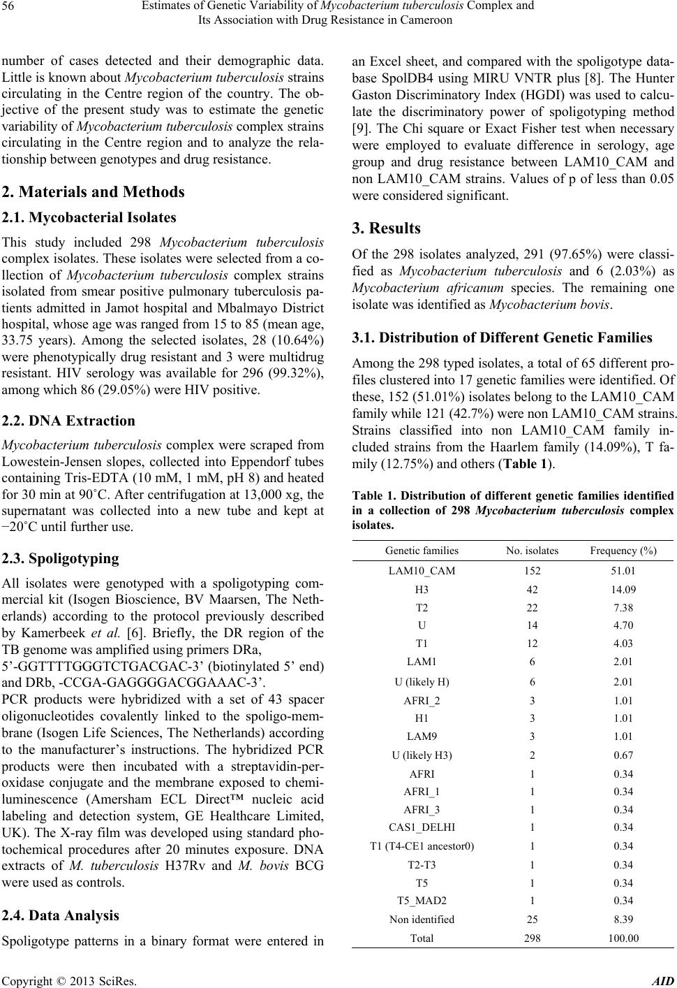 Estimates of Genetic Variability of Mycobacterium tuberculosis Complex and Its Association with Drug Resistance in Cameroon 56 number of cases detected and their demographic data. Little is known about Mycobacterium tuberculosis strains circulating in the Centre region of the country. The ob- jective of the present study was to estimate the genetic variability of Mycobacterium tuberculosis complex strains circulating in the Centre region and to analyze the rela- tionship between genotypes and drug resistance. 2. Materials and Methods 2.1. Mycobacterial Isolates This study included 298 Mycobacterium tuberculosis complex isolates. These isolates were selected from a co- llection of Mycobacterium tuberculosis complex strains isolated from smear positive pulmonary tuberculosis pa- tients admitted in Jamot hospital and Mbalmayo District hospital, whose age was ranged from 15 to 85 (mean age, 33.75 years). Among the selected isolates, 28 (10.64%) were phenotypically drug resistant and 3 were multidrug resistant. HIV serology was available for 296 (99.32%), among which 86 (29.05%) were HIV positive. 2.2. DNA Extraction Mycobacterium tuberculosis complex were scraped from Lowestein-Jensen slopes, collected into Eppendorf tubes containing Tris-EDTA (10 mM, 1 mM, pH 8) and heated for 30 min at 90˚C. After centrifugation at 13,000 xg, the supernatant was collected into a new tube and kept at −20˚C until further use. 2.3. Spoligotyping All isolates were genotyped with a spoligotyping com- mercial kit (Isogen Bioscience, BV Maarsen, The Neth- erlands) according to the protocol previously described by Kamerbeek et al. [6]. Briefly, the DR region of the TB genome was amplified using primers DRa, 5’-GGTTTTGGGTCTGACGAC-3’ (biotinylated 5’ end) and DRb, -CCGA-GAGGGGACGGAAAC-3’. PCR products were hybridized with a set of 43 spacer oligonucleotides covalently linked to the spoligo-mem- brane (Isogen Life Sciences, The Netherlands) according to the manufacturer’s instructions. The hybridized PCR products were then incubated with a streptavidin-per- oxidase conjugate and the membrane exposed to chemi- luminescence (Amersham ECL Direct™ nucleic acid labeling and detection system, GE Healthcare Limited, UK). The X-ray film was developed using standard pho- tochemical procedures after 20 minutes exposure. DNA extracts of M. tuberculosis H37Rv and M. bovis BCG were used as controls. 2.4. Data Analysis Spoligotype patterns in a binary format were entered in an Excel sheet, and compared with the spoligotype data- base SpolDB4 using MIRU VNTR plus [8]. The Hunter Gaston Discriminatory Index (HGDI) was used to calcu- late the discriminatory power of spoligotyping method [9]. The Chi square or Exact Fisher test when necessary were employed to evaluate difference in serology, age group and drug resistance between LAM10_CAM and non LAM10_CAM strains. Values of p of less than 0.05 were considered significant. 3. Results Of the 298 isolates analyzed, 291 (97.65%) were classi- fied as Mycobacterium tuberculosis and 6 (2.03%) as Mycobacterium africanum species. The remaining one isolate was identified as Mycobacterium bovis. 3.1. Distribution of Different Genetic Families Among the 298 typed isolates, a total of 65 different pro- files clustered into 17 genetic families were identified. Of these, 152 (51.01%) isolates belong to the LAM10_CAM family while 121 (42.7%) were non LAM10_CAM strains. Strains classified into non LAM10_CAM family in- cluded strains from the Haarlem family (14.09%), T fa- mily (12.75%) and others (Table 1). Table 1. Distribution of different genetic families identified in a collection of 298 Mycobacterium tuberculosis complex isolates. Genetic families No. isolates Frequency (%) LAM10_CAM 152 51.01 H3 42 14.09 T2 22 7.38 U 14 4.70 T1 12 4.03 LAM1 6 2.01 U (likely H) 6 2.01 AFRI_2 3 1.01 H1 3 1.01 LAM9 3 1.01 U (likely H3) 2 0.67 AFRI 1 0.34 AFRI_1 1 0.34 AFRI_3 1 0.34 CAS1_DELHI 1 0.34 T1 (T4-CE1 ancestor0) 1 0.34 T2-T3 1 0.34 T5 1 0.34 T5_MAD2 1 0.34 Non identified 25 8.39 Total 298 100.00 Copyright © 2013 SciRes. AID 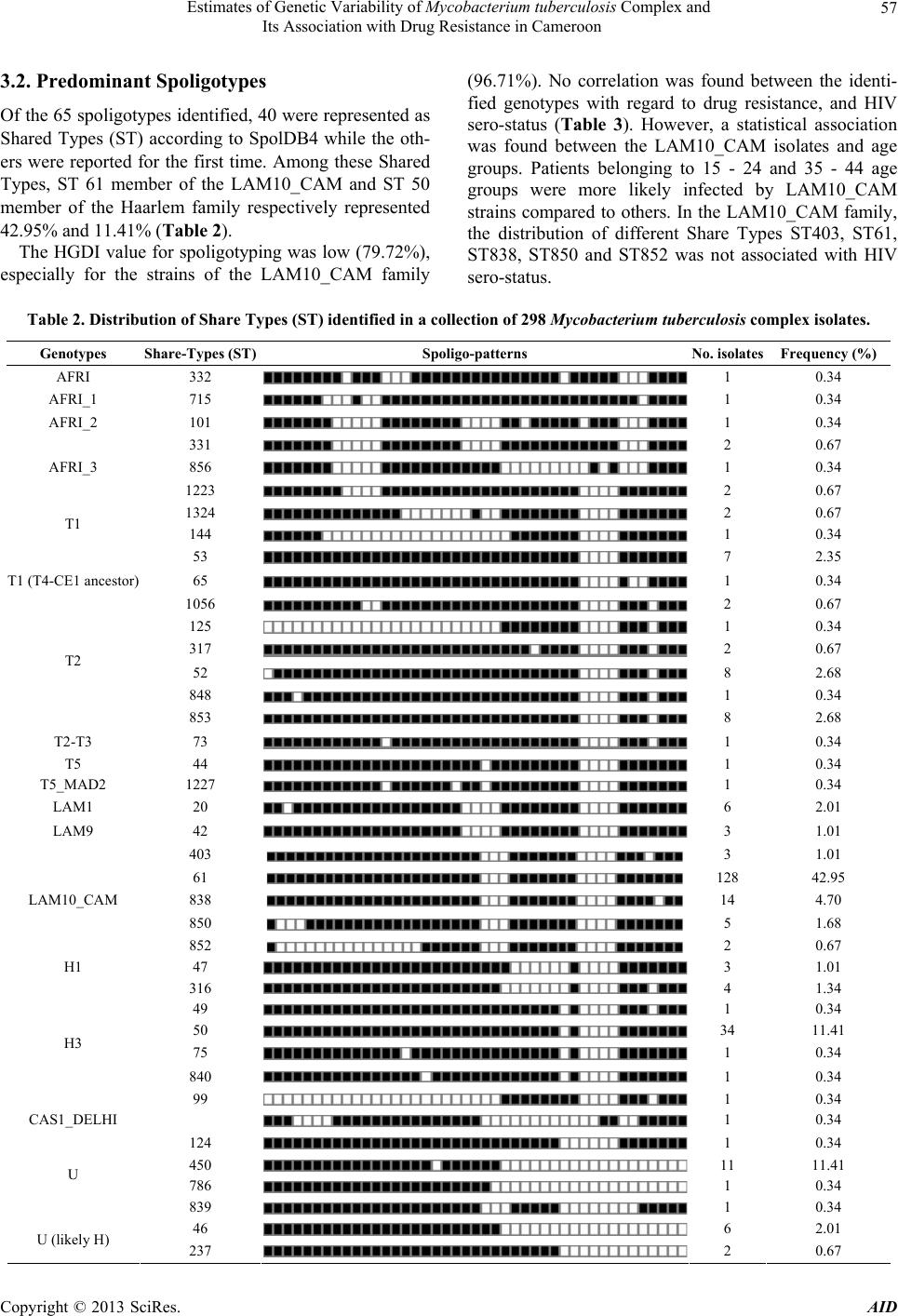 Estimates of Genetic Variability of Mycobacterium tuberculosis Complex and Its Association with Drug Resistance in Cameroon Copyright © 2013 SciRes. AID 57 3.2. Predominant Spoligotypes Of the 65 spoligotypes identified, 40 were represented as Shared Types (ST) according to SpolDB4 while the oth- ers were reported for the first time. Among these Shared Types, ST 61 member of the LAM10_CAM and ST 50 member of the Haarlem family respectively represented 42.95% and 11.41% (Table 2). The HGDI value for spoligotyping was low (79.72%), especially for the strains of the LAM10_CAM family (96.71%). No correlation was found between the identi- fied genotypes with regard to drug resistance, and HIV sero-status (Table 3). However, a statistical association was found between the LAM10_CAM isolates and age groups. Patients belonging to 15 - 24 and 35 - 44 age groups were more likely infected by LAM10_CAM strains compared to others. In the LAM10_CAM family, the distribution of different Share Types ST403, ST61, ST838, ST850 and ST852 was not associated with HIV sero-status. Table 2. Distribution of Share Types (ST) identified in a collection of 298 Mycobacterium tuberculosis complex isolates. Genotypes Share-Types (ST) Spoligo-patterns No. isolates Frequency (%) AFRI 332 1 0.34 AFRI_1 715 1 0.34 AFRI_2 101 1 0.34 331 2 0.67 AFRI_3 856 1 0.34 1223 2 0.67 1324 2 0.67 144 1 0.34 T1 53 7 2.35 T1 (T4-CE1 ancestor) 65 1 0.34 1056 2 0.67 125 1 0.34 317 2 0.67 52 8 2.68 848 1 0.34 T2 853 8 2.68 T2-T3 73 1 0.34 T5 44 1 0.34 T5_MAD2 1227 1 0.34 LAM1 20 6 2.01 LAM9 42 3 1.01 403 3 1.01 61 128 42.95 838 14 4.70 850 5 1.68 LAM10_CAM 852 2 0.67 H1 47 3 1.01 316 4 1.34 49 1 0.34 50 34 11.41 75 1 0.34 840 1 0.34 H3 99 1 0.34 CAS1_DELHI 1 0.34 124 1 0.34 450 11 11.41 786 1 0.34 U 839 1 0.34 46 6 2.01 U (likely H) 237 2 0.67 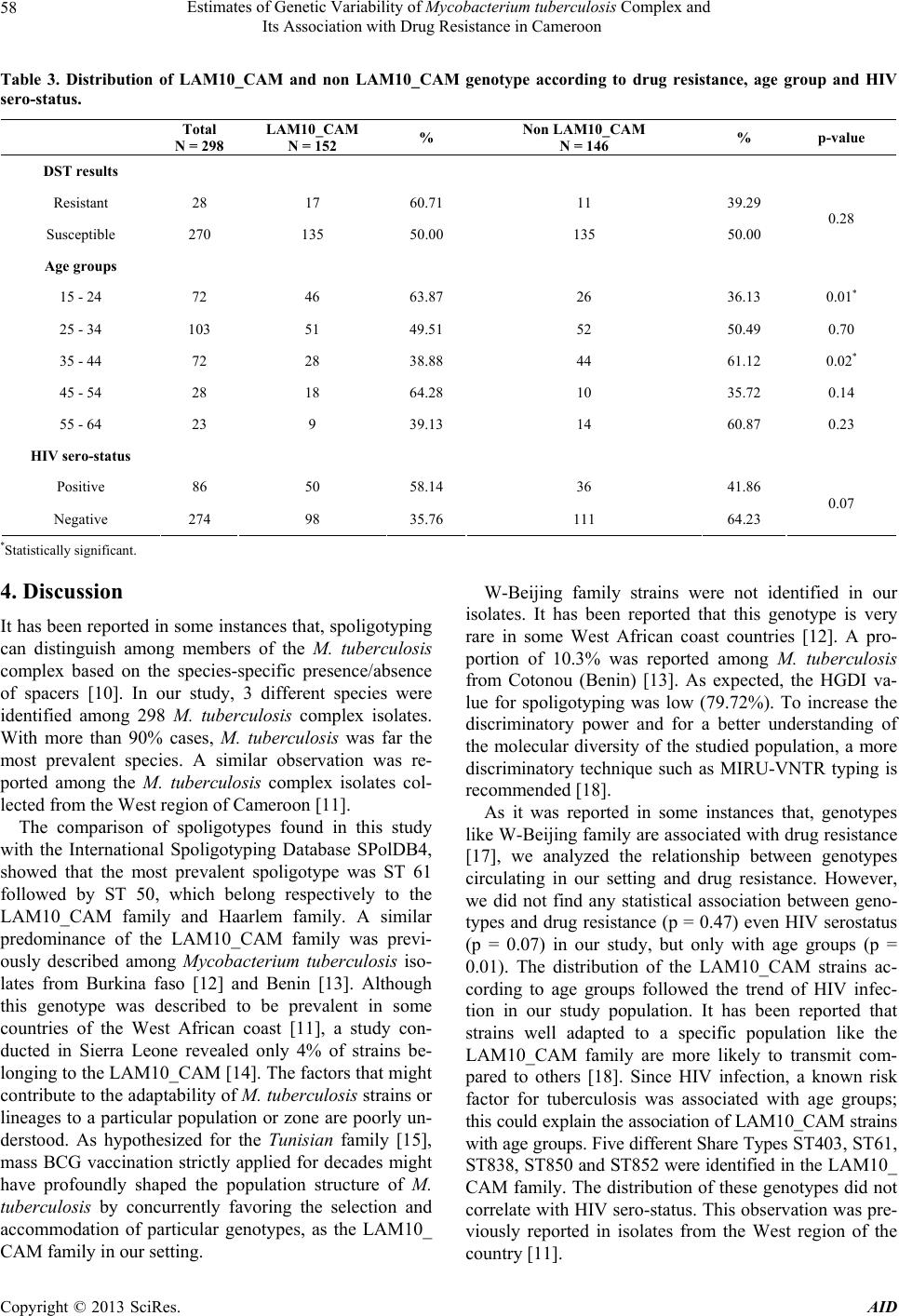 Estimates of Genetic Variability of Mycobacterium tuberculosis Complex and Its Association with Drug Resistance in Cameroon 58 Table 3. Distribution of LAM10_CAM and non LAM10_CAM genotype according to drug resistance, age group and HIV sero-status. Total N = 298LAM10_CAM N = 152 % Non LAM10_CAM N = 146 % p-value DST results Resistant 28 17 60.71 11 39.29 Susceptible 270 135 50.00 135 50.00 0.28 Age groups 15 - 24 72 46 63.87 26 36.13 0.01* 25 - 34 103 51 49.51 52 50.49 0.70 35 - 44 72 28 38.88 44 61.12 0.02* 45 - 54 28 18 64.28 10 35.72 0.14 55 - 64 23 9 39.13 14 60.87 0.23 HIV sero-status Positive 86 50 58.14 36 41.86 Negative 274 98 35.76 111 64.23 0.07 *Statistically significant. 4. Discussion It has been reported in some instances that, spoligotyping can distinguish among members of the M. tuberculosis complex based on the species-specific presence/absence of spacers [10]. In our study, 3 different species were identified among 298 M. tuberculosis complex isolates. With more than 90% cases, M. tuberculosis was far the most prevalent species. A similar observation was re- ported among the M. tuberculosis complex isolates col- lected from the West region of Cameroon [11]. The comparison of spoligotypes found in this study with the International Spoligotyping Database SPolDB4, showed that the most prevalent spoligotype was ST 61 followed by ST 50, which belong respectively to the LAM10_CAM family and Haarlem family. A similar predominance of the LAM10_CAM family was previ- ously described among Mycobacterium tuberculosis iso- lates from Burkina faso [12] and Benin [13]. Although this genotype was described to be prevalent in some countries of the West African coast [11], a study con- ducted in Sierra Leone revealed only 4% of strains be- longing to the LAM10_CAM [14]. The factors that might contribute to the adaptability of M. tuberculosis strains or lineages to a particular population or zone are poorly un- derstood. As hypothesized for the Tunisian family [15], mass BCG vaccination strictly applied for decades might have profoundly shaped the population structure of M. tuberculosis by concurrently favoring the selection and accommodation of particular genotypes, as the LAM10_ CAM family in our setting. W-Beijing family strains were not identified in our isolates. It has been reported that this genotype is very rare in some West African coast countries [12]. A pro- portion of 10.3% was reported among M. tuberculosis from Cotonou (Benin) [13]. As expected, the HGDI va- lue for spoligotyping was low (79.72%). To increase the discriminatory power and for a better understanding of the molecular diversity of the studied population, a more discriminatory technique such as MIRU-VNTR typing is recommended [18]. As it was reported in some instances that, genotypes like W-Beijing family are associated with drug resistance [17], we analyzed the relationship between genotypes circulating in our setting and drug resistance. However, we did not find any statistical association between geno- types and drug resistance (p = 0.47) even HIV serostatus (p = 0.07) in our study, but only with age groups (p = 0.01). The distribution of the LAM10_CAM strains ac- cording to age groups followed the trend of HIV infec- tion in our study population. It has been reported that strains well adapted to a specific population like the LAM10_CAM family are more likely to transmit com- pared to others [18]. Since HIV infection, a known risk factor for tuberculosis was associated with age groups; this could explain the association of LAM10_CAM strains with age groups. Five different Share Types ST403, ST61, ST838, ST850 and ST852 were identified in the LAM10_ CAM family. The distribution of these genotypes did not correlate with HIV sero-status. This observation was pre- viously reported in isolates from the West region of the country [11]. Copyright © 2013 SciRes. AID  Estimates of Genetic Variability of Mycobacterium tuberculosis Complex and Its Association with Drug Resistance in Cameroon 59 The population structure of Mycobacterium tuberculo- sis complex strains from the Centre region was diverse and included 65 different genotypes. The majority of strains belonged to the LAM10_CAM which can be sub- divided in 5 spoligotypes. The consequence of this diver- sity for the TB epidemic are not yet clear and need to be addressed in further studies. 5. Acknowledgements This study was financially supported by EDCTP grant through the CANTAM-TB “Central Africa Network for Tuberculosis, AIDS/HIV and Malaria” project. Larissa Kamgue Sidze and Emmanuel Mouafo Tekwu were re- search fellow students at the Institute for Tropical Medi- cine in Tübingen (Germany). We thank Mrs. Augusta Tsasse (Centre Pasteur of Cameroon) and Tanja Ubben (Research Centre Borstel, Germany) for their technical assistance. REFERENCES [1] C. Dye, “Global Epidezmiology of Tuberculosis,” Lancet, Vol. 367, No. 9514, 2006, pp. 938-940. doi:10.1016/S0140-6736(06)68384-0 [2] K. Rowland, “Totally Drug-Resistant TB Emerges in India,” Nature News and Comment, 13 January 2012. [3] World Health Organization, “Global Tuberculosis Control: WHO Report 2011,” Geneva, 2011. [4] G. D. Van der Spuy, R. M. Warren and P. D. Van Helden, “The Role of Molecular Epidemiology in Low-Income, High-Burden Countries,” International Journal of Tuber- cle and Lung Diseases, Vol. 13, No. 4, 2009, pp. 419- 420. [5] D. Van Soolingen, P. W. Hermans, P. E. De Haas, D. R. Soll and J. D. Van Embden, “Occurrence and Stability of Insertion Sequences in Mycobacterium tuberculosis Com- plex Strains: Evaluation of an Insertion Sequence-De- pendent DNA Polymorphism as a Tool in the Epidemiol- ogy of Tuberculosis,” Journal of Clinical Microbiology, Vol. 29, No. 11, 1991, pp. 2578-2586. [6] J. Kamerbeek, L. Schouls, A. Kolk, M. Van Agterveld, D. Van Soolingen, S. Kuijper, A. Bunschoten, H. Molhuizen, R. Shaw, M. Goyal, et al., “Simultaneous Detection and Strain Differentiation of Mycobacterium tuberculosis for Diagnosis and Epidemiology,” Journal of Clinical Mi- crobiology, Vol. 35, No. 4, 1997, pp. 907-914. doi:10.3201/eid0811.020125 [7] I. Filliol, J. R. Driscoll, D. Van Soolingen, B. N. Kreis- wirth, K. Kremer, G. Valetudie, D. D. Anh, R. Barlow, D. Banerjee, P. J. Bifani, et al., “Global Distribution of My- cobacterium tuberculosis Spoligotypes,” Emerging Infec- tious Diseases, Vol. 8, No. 11, 2002, pp. 1347-1349. [8] T. Weniger, J. Krawczyk, P. Supply, S. Niemann and D. Harmsen, “MIRU-VNTR plus: A Web Tool for Polypha- sic Genotyping of Mycobacterium tuberculosis Complex Bacteria,” Nucleic Acids Research, Vol. 38, 2010, pp. W326-W331. [9] P. R. Hunter and M. A. Gaston, “Numerical Index of the Discriminatory Ability of Typing Sysems: An Applica- tion of Simpson’s Index of Diversity,” Journal of Clinical Microbiology, Vol. 26, No. 1, 1988, pp. 2465-2466. [10] B. Mathema, N. E. Kurepina, P. J. Bifani and B. N. Krei- swirth, “Molecular Epidemiology of Tuberculosis: Cur- rent Insights,” Clinical Microbiology Review, Vol. 19, No. 4, 2006, pp. 658-685. doi:10.1128/CMR.00061-05 [11] S. N. Niobe-Eyangoh, C. Kuaban, P. Sorlin, P. Cunin, J. Thonnon, C. Sola, et al., “Genetic Biodiversity of Myco- bacterium tuberculosis Complex Strains from Patients with Pulmonary Tuberculosis in Cameroon,” Journal of Clinical Microbiology, Vol. 41, No. 6, 2003, pp. 2547- 2553. doi:10.1128/JCM.41.6.2547-2553.2003 [12] S. Godreuil, G. Torrea, D. Terru, F. Chevenet, S. Diag- bouga, P. Supply, et al., “First Molecular Epidemiology Study of Mycobacterium tuberculosis in Burkina Faso,” Journal of Clinical Microbiology, Vol. 45, No. 3, 2007, pp. 921-927. doi:10.1128/JCM.01918-06 [13] D. Affolabi, G. Anyo, F. Faihun, N. Sanoussi, I. C. Sham- puta, L. Rigouts, et al., “First Molecular Epidemiological Study of Tuberculosis in Benin,” International Journal of Tubercle and Lung Diseases, Vol. 13, No. 13, 2009, pp. 317-322. [14] S. Homolka, E. Post, B. Oberhauser, A. G. George, L. Westman, F. Dafae, S. Rusch-Gerdes and S. Niemann, “High Genetic Diversity among Mycobacterium tubercu- losis Complex Strains from Sierra Leone,” BMC Micro- biology, Vol. 8, No. 103, 2008, pp. 489-495. [15] A. Namouchi, A. Karboul, B. Mhenni, N. Khabouchi, R. Haltiti, R. Ben Hassine, et al., “Genetic Profiling of My- cobacterium tuberculosis in Tunisia: Predominance and Evidence for the Establishment of a Few Genotypes,” Journal of Medical Microbiology, Vol. 57, No. 7, 2008, pp. 864-872. doi:10.1099/jmm.0.47483-0 [16] P. Supply, E. Mazars, S. Lesjean, V. Vincent, B. Gicquel and C. Locht, “Variable Human Minisatellite-Like Re- gions in the Mycobacterium tuberculosis Genome,” Mo- lecular Microbiology, Vol. 36, No. 3, 2000, pp. 762-771. doi:10.1046/j.1365-2958.2000.01905.x [17] W. A. Githui, A. M. Jordaan, E. S. Juma, P. Kinyanjui, F. G. Karimi, J. Kimwomi, et al., “Identification of MDR- TB Beijing/W and Other Mycobacterium tuberculosis Genotypes in Nairobi, Kenya,” International Journal of Tubercle and Lung Diseases, Vol. 8, No. 3, 2004, pp. 352-360. [18] S. Gagneux and P. M. Small, “Global Phylogeography of Mycobacterium tuberculosis and Implications for Tuber- culosis Product Development,” Lancet Infectious Dis- eases, Vol. 7, No. 5, 2007, pp. 328-337. doi:10.1016/S1473-3099(07)70108-1 Copyright © 2013 SciRes. AID |