

Paper Menu >>
Journal Menu >>
 Food and Nutrition Sciences, 2013, 4, 299-304 http://dx.doi.org/10.4236/fns.2013.43040 Published Online March 2013 (http://www.scirp.org/journal/fns) Quantitative Adhesion of Staphylococcus aureus on Stainless Steel Coated with Milk Fatima Hamadi1,2, Hassan Latrache2*, Fatima Asserne2, Soumya Elabed3,4, Ha f i da Zahir2, Ibnsouda Koraichi Saad3,4, Hafida Hanine2, Jamaa Bengourram5 1Laboratory of Biotechnology and Vaorization of Natural Resources, Department of Biology, Faculty of Science, University Ibn Zohr, Agadir, Marocco; 2Laboratory of Valorization and Safety of Food Products, Faculty of Sciences and Technics, University Sul- tan Moulay Slimane, Beni Mellal, Morocco; 3Laboratoy of Microbial Biotechnology, Faculty of Science and Technics, University Sidi Mohamed Ben Abdellah, Fés-Sáis, Marocco; 4Regional University Center of Interface, University Sidi Mohamed Ben Abdellah, Fés, Marocco; 5Laboratory of industrial Engineering, Food and Environment, Faculty of Sciences and Technics, University Sultan Moulay Slimane, Beni Mellal, Marocco. Email: *[email protected] Received September 14th, 2011; revised December 9th, 2011; accepted December 16th, 2011 ABSTRACT The surface energy characteristics of uncoated (clean) and coated stainless steel with UHT milk at various contact time (5 min, 30 min, 1 hours, 3 hours, 6 hours, 24 hours) were determined using contact angle measurement. Whatever the contact time, the clean stainless steel coupons became more hydrophobic and more electron acceptor when they are coated by milk. Inversely, the electron donor character seems to decreasing in this condition. The calculated surface energy component of coated stainless steel was found to vary with contact time. Its hydrophobicity and its electron ac- ceptor were minimal after 3 hours of contact, but its electron donor was minimal after 1 hours of contact. Adhesion ex- periments of Staphylococcus aureus were carried out on uncoated and coated stainless steels at various contact times. For all contact times, the adhesion results show that milk reduce S. aureus adhesion, and the level of this reduction de- pend on contact time. This reduction was lower and higher after 1 hour, 5 min and 30 min of contact respectively. Keywords: Surface Energy Characteristics; S. aureus; Adhesion; Milk; Stainless Steel 1. Introduction The formation of biofilm creates major problems in the food industry since it may represent an important source of contamination for materials or foodstuffs coming into contact with them, so leading to food spoilage or trans- mission of diseases. Biofilms are of interest in the dairy industry, as bacteria within biofilms are more difficult to eliminate than plank tonic cells, and bacteria detached from biofilms can contaminate milk and milk products [1]. This biotransfer may affect hygiene and the com- mercial value of the product. To control these problems, it has been recognized that a greater understanding of the interactions between microorganisms and food—process- ing surface is required [2-4]. The adhesion of bacteria to surface is the first and es- sential stage in the formation of biofilm. This adhesion depends on both physicochemical properties of cell sur- face and solid surface, and also on characteristics of the surrounding medium. Stainless steel is the most frequently used material for food processing equipment because of its high impor- tance related to food safety reasons. There are many cir- cumstances in dairies where substratum surface is either continuously or periodically in contact with liquids that contain microorganisms. These conditions could affect the substratum surface properties and consequently the adhesion process. Staphylococcus aureus is a gram positive bacterium, which is an important food-borne pathogen [5-7]. In food industry this organism could be able to attach and to form biofilms on the food-processing surface [5,7,8]. S. aureus was studied here because little information [5-7] is available of its adhesion behaviour in dairy industry in comparison with other organisms such as Listeria mono- cytognes and bacillus [4,9-12]. The aim of this study was therefore to investigate the surface properties of stainless steel at various times of contact with milk. The adhesion of S. aureus to stainless steel was also examined and discussed in terms of phys- icochemical properties of cell surface and substratum surface. *Corresponding author. Copyright © 2013 SciRes. FNS 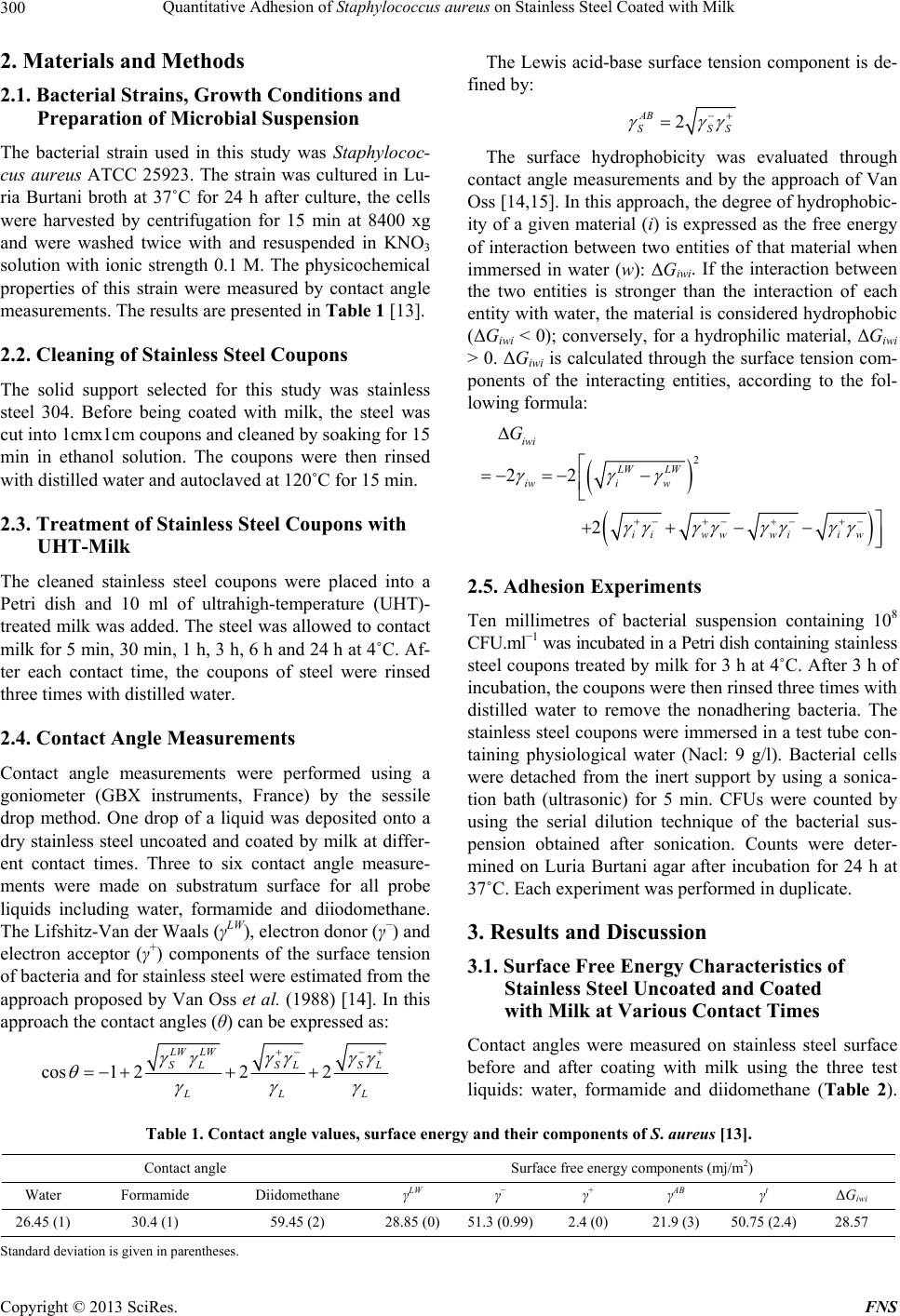 Quantitative Adhesion of Staphylococcus aureus on Stainless Steel Coated with Milk 300 2. Materials and Methods 2.1. Bacterial Strains, Growth Conditions and Preparation of Microbial Suspension The bacterial strain used in this study was Staphylococ- cus aureus ATCC 25923. The strain was cultured in Lu- ria Burtani broth at 37˚C for 24 h after culture, the cells were harvested by centrifugation for 15 min at 8400 xg and were washed twice with and resuspended in KNO3 solution with ionic strength 0.1 M. The physicochemical properties of this strain were measured by contact angle measurements. The results are presented in Table 1 [13]. 2.2. Cleaning of Stainless Steel Coupons The solid support selected for this study was stainless steel 304. Before being coated with milk, the steel was cut into 1cmx1cm coupons and cleaned by soaking for 15 min in ethanol solution. The coupons were then rinsed with distilled water and autoclaved at 120˚C for 15 min. 2.3. Treatment of Stainless Steel Coupons with UHT-Milk The cleaned stainless steel coupons were placed into a Petri dish and 10 ml of ultrahigh-temperature (UHT)- treated milk was added. The steel was allowed to contact milk for 5 min, 30 min, 1 h, 3 h, 6 h and 24 h at 4˚C. Af- ter each contact time, the coupons of steel were rinsed three times with distilled water. 2.4. Contact Angle Measurements Contact angle measurements were performed using a goniometer (GBX instruments, France) by the sessile drop method. One drop of a liquid was deposited onto a dry stainless steel uncoated and coated by milk at differ- ent contact times. Three to six contact angle measure- ments were made on substratum surface for all probe liquids including water, formamide and diiodomethane. The Lifshitz-Van der Waals (γLW), electron donor (γ−) and electron acceptor (γ+) components of the surface tension of bacteria and for stainless steel were estimated from the approach proposed by Van Oss et al. (1988) [14]. In this approach the contact angles (θ) can be expressed as: cos1 222 LW LW SLSL SL LL The Lewis acid-base surface tension component is de- fined by: 2 AB SSS The surface hydrophobicity was evaluated through contact angle measurements and by the approach of Van Oss [14,15]. In this approach, the degree of hydrophobic- ity of a given material (i) is expressed as the free energy of interaction between two entities of that material when immersed in water (w): ΔGiwi. If the interaction between the two entities is stronger than the interaction of each entity with water, the material is considered hydrophobic (ΔGiwi < 0); conversely, for a hydrophilic material, ΔGiwi > 0. ΔGiwi is calculated through the surface tension com- ponents of the interacting entities, according to the fol- lowing formula: 2 22 2 iwi LW LW iwi w iiww wiiw G 2.5. Adhesion Experiments Ten millimetres of bacterial suspension containing 108 CFU.ml−1 was incubated in a Petri dish containing stainless steel coupons treated by milk for 3 h at 4˚C. After 3 h of incubation, the coupons were then rinsed three times with distilled water to remove the nonadhering bacteria. The stainless steel coupons were immersed in a test tube con- taining physiological water (Nacl: 9 g/l). Bacterial cells were detached from the inert support by using a sonica- tion bath (ultrasonic) for 5 min. CFUs were counted by using the serial dilution technique of the bacterial sus- pension obtained after sonication. Counts were deter- mined on Luria Burtani agar after incubation for 24 h at 37˚C. Each experiment was performed in duplicate. 3. Results and Discussion 3.1. Surface Free Energy Characteristics of Stainless Steel Uncoated and Coated with Milk at Various Contact Times Contact angles were measured on stainless steel surface before and after coating with milk using the three test liquids: water, formamide and diidomethane (Table 2). L Table 1. Contact angle values, surface energy and their components of S. aureus [13]. Contact angle Surface free energy components (mj/m2) Water Formamide Diidomethane γLW γ− γ+ γAB γt ΔGiwi 26.45 (1) 30.4 (1) 59.45 (2) 28.85 (0) 51.3 (0.99)2.4 (0) 21.9 (3) 50.75 (2.4) 28.57 Standard deviation is given in parentheses. Copyright © 2013 SciRes. FNS 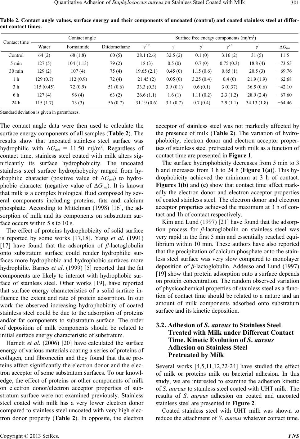 Quantitative Adhesion of Staphylococcus aureus on Stainless Steel Coated with Milk 301 Table 2. Contact angle values, surface energy and their components of uncoated (control) and coated stainless steel at differ- ent contact times. Contact angle Surface free energy components (mj/m2) Contact time Water Formamide DiidomethaneγLW γ− γ+ γAB γt ΔGiwi Control 64 (2) 68 (1.8) 60 (5) 28.1 (2.6)32.5 (2) 0.1 (0) 3.16 (2) 31 (5) 11.5 5 min 127 (5) 104 (1.13) 79 (2) 18 (3) 0.5 (0) 0.7 (0) 0.75 (0.3) 18.8 (4) −73.53 30 min 129 (2) 107 (4) 75 (4) 19.65 (2.1)0.45 (0) 1.15 (0.6)0.85 (1) 20.5 (3) −69.76 1 h 129 (0.7) 112 (0.9) 72 (4) 21.45 (2)0.05 (0) 3.25 (0.4)0.4 (0) 21.9 (1.9) −62.68 3 h 115 (0.45) 72 (0.9) 51 (0.6) 33.3 (0.3)3.9 (0.1) 0.6 (0.1) 3 (0.37) 36.5 (0.6) −42.10 6 h 127 (4) 96 (4) 63 (2) 26.6 (1.1)1.6 (1) 1.11 (0.2)2.3 (1.2) 28.9 (2.4) −67.60 24 h 115 (1.7) 73 (3) 56 (0.7) 31.19 (0.6)3.1 (0.7) 0.7 (0.4) 2.9 (1.1) 34.13 (1.8) −64.46 Standard deviation is given in parentheses. The contact angle data were then used to calculate the surface energy components of all samples (Table 2). The results show that uncoated stainless steel surface was hydrophilic with ΔGiwi = 11.50 mj/m2. Regardless of contact time, stainless steel coated with milk alters sig- nificantly its surface hydrophobicity. The uncoated stainless steel surface hydrophobycity ranged from hy- drophilic character (positive value of ΔGiwi) to hydro- phobic character (negative value of ΔGiwi). It is known that milk is a complex biological fluid composed by sev- eral components including proteins, fats and calcium phosphate. According to Mittelman (1998) [16], the ad- sorption of milk and its components on substratum sur- face occurs within 5 s to 10 s. The effect of proteins hydrophobicity of solid surface is reported by some works [17,18]. Yang et al. (1991) [17] have found that the adsorption of β-lactoglobulin onto substratum surface could render hydrophilic sur- faces more hydrophobic and hydrophobic surfaces more hydrophilic. Barnes et al. (1999) [5] reported that the fat components are likely to interact with hydrophobic sur- face of stainless steel. Other works [19], have reported that surface energy characteristics of a solid surface in- fluence the extent and rate of protein adsorption. In our work the observed increasing hydrophobicity of coated stainless steel could be due to the adsorption of proteins and/or fat components to substratum surface. The order of deposition of milk components should be related to initial surface energy characteristic of substratum. Harnett et al. (2006) [20] have calculated the surface energy of various materials coating a series of proteins of collagen, and fibroncetin and they found that these pro- teins affect significantly the electron donor and the elec- tron acceptor of some substratum surfaces. To our knowl- edge, the effect of proteins or other components of milk on electron donor/electron acceptor properties of sub- stratum surface were not examined previously. Stainless steel coated with milk has a very lower electron donor compared to stainless steel uncoated with very high elec- tron donor property (Table 2). In opposite, the electron acceptor of stainless steel was not markedly affected by the presence of milk (Table 2). The variation of hydro- phobicity, electron donor and electron acceptor proper- ties of stainless steel pretreated with milk as a function of contact time are presented in Figure 1. The surface hydrophobicity decreases from 5 min to 3 h and increases from 3 h to 24 h (Figure 1(a)). This hy- drophobicity achieved the minimum at 3 h of contact. Figures 1(b) and (c) show that contact time affect mark- edly the electron donor and electron acceptor properties of coated stainless steel. The electron donor and electron acceptor properties achieved the maximum at 3 h of con- tact and 1h of contact respectively. Kim and Lund (1997) [21] have found that the adsorp- tion process for β-lactoglobulin on stainless steel was very rapid in the first 5 min and essentially reached equi- librium within 10 min. These authors have also reported that the precipitation of calcium phosphate onto the stain- less steel surface was very slow compared to monolayer deposition of β-lactoglobulin. Addesso and Lund (1997) [19] show that protein adsorption onto a surface depends on protein concentration. The random observed variation of physicochemical properties of stainless steel as a func- tion of contact time should be related to a nature and an amount of milk components adsorbed onto substratum surface and its kinetic deposition. 3.2. Adhesion of S. aureus to Stainless Steel Treated with Milk under Different Contact Time. Kinetic Evolution of S. aureus Adhesion on Stainless Steel Pretreated by Milk Several works [4,5,11,12,22-24] have studied the effect of milk or proteins milk on bacterial adhesion. In this study, we are interested to examine the adhesion kinetic of S. aureus to stainless steel coated with UHT milk. The results of S. aureus adhesion on coated and uncoated stainless steel are presented in Figure 2. Coated stainless steel with UHT milk was shown to reduce the attachment of S. aureus whatever contact time. Copyright © 2013 SciRes. FNS 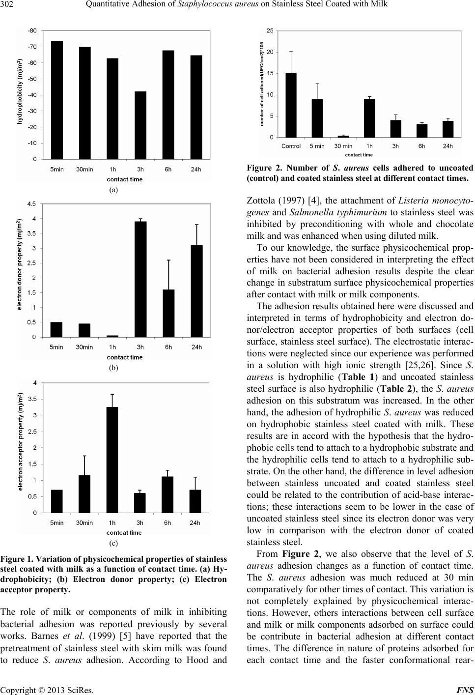 Quantitative Adhesion of Staphylococcus aureus on Stainless Steel Coated with Milk 302 (a) (b) (c) Figure 1. Variation of physicochemical properties of stai nl e ss steel coated with milk as a function of contact time. (a) Hy- drophobicity; (b) Electron donor property; (c) Electron acceptor property. The role of milk or components of milk in inhibiting bacterial adhesion was reported previously by several works. Barnes et al. (1999) [5] have reported that the pretreatment of stainless steel with skim milk was found to reduce S. aureus adhesion. According to Hood and Figure 2. Number of S. aureus cells adhered to uncoated (control) and coated stainless steel at different contact times. Zottola (1997) [4], the attachment of Listeria monocyto- genes and Salmonella typhimurium to stainless steel was inhibited by preconditioning with whole and chocolate milk and was enhanced when using diluted milk. To our knowledge, the surface physicochemical prop- erties have not been considered in interpreting the effect of milk on bacterial adhesion results despite the clear change in substratum surface physicochemical properties after contact with milk or milk components. The adhesion results obtained here were discussed and interpreted in terms of hydrophobicity and electron do- nor/electron acceptor properties of both surfaces (cell surface, stainless steel surface). The electrostatic interac- tions were neglected since our experience was performed in a solution with high ionic strength [25,26]. Since S. aureus is hydrophilic (Table 1) and uncoated stainless steel surface is also hydrophilic (Table 2), the S. aureus adhesion on this substratum was increased. In the other hand, the adhesion of hydrophilic S. aureus was reduced on hydrophobic stainless steel coated with milk. These results are in accord with the hypothesis that the hydro- phobic cells tend to attach to a hydrophobic substrate and the hydrophilic cells tend to attach to a hydrophilic sub- strate. On the other hand, the difference in level adhesion between stainless uncoated and coated stainless steel could be related to the contribution of acid-base interac- tions; these interactions seem to be lower in the case of uncoated stainless steel since its electron donor was very low in comparison with the electron donor of coated stainless steel. From Figure 2, we also observe that the level of S. aureus adhesion changes as a function of contact time. The S. aureus adhesion was much reduced at 30 min comparatively for other times of contact. This variation is not completely explained by physicochemical interac- tions. However, others interactions between cell surface and milk or milk components adsorbed on surface could be contribute in bacterial adhesion at different contact times. The difference in nature of proteins adsorbed for each contact time and the faster conformational rear- Copyright © 2013 SciRes. FNS  Quantitative Adhesion of Staphylococcus aureus on Stainless Steel Coated with Milk 303 rangement undergone by one protein at surface relative to that of other proteins could be the origin of the varia- tion observed in adhesion results. Barnes et al. (1999) [5] have found that the pre-treatment of stainless steel with the individual milk proteins α-, β- and casein and α-lactalbumin at equal concentration reduce attachment of S. aureus and this reduction was marked with β casein. McEldowney and Fletcher (1987) [27] observed that hy- drated layers of polymers and proteins that form on inert surfaces can either facilitate or reduce bacterial adhesion. Al Makhlafi et al. (1994) [22] examined the effect of competitive adsorption of bovine serum albumin (BSA) and β-lactoglobulin on Listeria monocytogenes adhesion to silica, and they found that the film formed by the ad- sorption of β-lactoglobulin followed by BSA encouraged adhesion more than the film formed by the adsorption of BSA followed by β-lactoglobulin. 4. Conclusion The results obtained here show that the physicochemical properties including hydrophobicity and electron donor- electron acceptor properties of stainless steel surface were markedly affected by treatment by milk. The adhesion results show that whatever the contact time, the pre- treatment of substratum by milk reduce the adhesion level. This reduction is random with the contact time. This research suggests that it is very important to take into account the contact time between the substratum and milk in the cleaning and sanitizing process. REFERENCES [1] S. Flint, J. Plamer, K. Bloemen, J. Brooks and R. Craw- ford, “The Growth of Bacillus strearothermophilus on Stainless Steel,” Journal of Applied Microbiology, Vol. 90, No. 2, 2001, pp. 151-157. doi:10.1046/j.1365-2672.2001.01215.x [2] B. Carpentier and O. Cerf, “Biofilms and Their Conse- quences with Particular Reference to Hygiene in the Food Industry,” Journal of Applied Microbiology, Vol. 75, No. 6, 1993, pp. 499-511. [3] J. D. Bryers, “Biofilms and the Technological Implica- tions of Microbial Cell Adhesion,” Colloids and Surfaces B: Biointerfaces, Vol. 2, No. 1-3, 1994, pp. 9-23. doi:10.1016/0927-7765(94)80013-8 [4] S. K. Hood and E. A. Zottola, “Adherence to Stainless Steel by Foodborne Microorganisms during Growth in Model Food Systems,” International Journal of Food Microbiology, Vol. 37, No. 9, 1997, pp. 145-153. doi:10.1016/S0168-1605(97)00071-8 [5] L. M. Barnes, M. F. Lo, M. R. Adams and A. H. L. Chamberlain, “Effect of Milk Proteins on Adhesion of Bacteria to Stainless Steel Surfaces,” Applied Environ- mental Microbiology, Vol. 65, No. 10, 1999, pp. 4543- 4548. [6] D. M. C. Pompermayer and C. C. Gaylarde, “The Influ- ence of Temperature on the Adhesion of Mixed Cultures of Staphylococcus aureus and Escherichia coli to Poly- propylene,” Food Microbiology, Vol. 17, No. 5, 2000, pp. 361-365. doi:10.1006/fmic.1999.0291 [7] N. Oulahal, W. Brice, A. Martial and P. Degraeve, “Quantitative Analysis of Survival of Staphylococcus aureus or Listeria innocua on Two Types of Surfaces: Polypropylene and Stainless Steel in Contact with Three Different Dairy Products,” Food Control, Vol. 19, No. 21, 2008, pp. 78-185. [8] S. C. Marques, J. D. G. O. S. Rezende, L. A. F. Alves, B. C. Silva, E. Alves, L. Ronaldo de Abreu and R. H. Piccoli, “Formation of biofilm by Staphylococcus aureus on Stainless Steel and Glass Surface and Its Resistance to Some Selected Chemical Sanitizers,” Brazilian Journal of Microbiology, Vol. 38, No. 3, 2007, pp. 539-543. doi:10.1590/S1517-83822007000300029 [9] H. Al-Makhlafi, A. Nasir, J. Mcguire and M. Daeschel, “Adhesion of Listeria monocytogenes to Silica Surfaces after Sequential and Competitive Adsorption of Bovine Serum Albumin and β-Lactoglobulin,” Applied and En- vironmental Microbiology, Vol. 61, No. 5, 1995, pp. 2013-2015 [10] S. H. Flint, P. J. Bremer and J. D. Brooks, “Biofilms in Dairy Manufacturing Plant-Description, Current Concerns and Methods of Control,” Biofouling: The Journal of Bio- adhesion and Biofilm Research, Vol. 11, No. 1, 1996, pp. 81-97. doi:10.1080/08927019709378321 [11] S. G. Parkar, S. H. Flint, J. S. Palmer and J. D. Brooks, “Factors Influencing Attachment of Thermophilic Bacilli to Stainless Steel,” Journal of Applied Microbiology, Vol. 90, No. 6, 2001, pp. 901-908. doi:10.1046/j.1365-2672.2001.01323.x [12] R. Rosmaninho, O. Santos, T. Nylander, M. Paulsson, M. Beuf, T. Benezech, S. Yiantsios, N. Andritsos, A. Karabe- las, G. Rizzo, H. Muller-Steinhagen and L. F. Melo, “Modified Stainless Steel Surfaces Targeted to Reduce Fouling—Evaluation of Fouling by Milk Components,” Journal of Food Engineering, Vol. 80, No. 4, 2007, pp. 1176-1187. doi:10.1016/j.jfoodeng.2006.09.008 [13] F. Hamadi and H. Latrache, “Comparison of Contact Angle Measurement and Microbial Adhesion to Solvents for Assaying Electron Donor-Electron Acceptor (Acid- Base) Properties of Bacterial Surface,” Colloids and Sur- faces B: Biointerfaces, Vol. 65, No. 1, 2008 pp. 134-139. doi:10.1016/j.colsurfb.2008.03.010 [14] C. J. Van Oss, R. J. Good and M. K. Chaudhury, “Addi- tive and Nonadditive Surface Tension Components and the Interpretation of Contact Angles,” Langmuir, Vol. 4, No. 4, 1988, pp. 884-891. doi:10.1021/la00082a018 [15] C. J. Van Oss, “Hydrophobicity of Biosurfaces—Origin, Quantitative-Determination and Interaction Energies,” Colloids and Surfaces B: Biointerfaces, Vol. 5, No. 3-4, 1995, pp. 91-110. doi:10.1016/0927-7765(95)01217-7 [16] M. M. Mittelman, “Structure and Functional Characteris- tics of Bacterial Biofilms in Fluid Processing Operations,” Journal of Dairy Science, Vol. 81, No. 10, 1998, pp. 2760-2764. doi:10.3168/jds.S0022-0302(98)75833-3 [17] J. Yang, J. McGuire and E. R. Kolbe, “Use of Equilib- Copyright © 2013 SciRes. FNS  Quantitative Adhesion of Staphylococcus aureus on Stainless Steel Coated with Milk Copyright © 2013 SciRes. FNS 304 rium Contact Angle as an Index of Contact Surface Cleanliness,” Journal of Food Protection, Vol. 54, No. 1991, pp. 879-884. [18] Y. Tamada and Y. Ikada, “Effect of Preadsorbed Proteins on Cell Adhesion to Polymer Surface,” Journal of Colloid and Interface Science, Vol. 155, No. 2, 1993, pp. 334-339. doi:10.1006/jcis.1993.1044 [19] A. Addesso and D. B. Lund, “Influence of Solid Surface Energy on Protein Adsorption,” Journal of Food Proc- essing and Preservation, Vol. 21, No. 4, 1997, pp. 319- 333. doi:10.1111/j.1745-4549.1997.tb00786.x [20] E. M. Harnett, J. Alderman and T. Wood, “The Surface Energy of Various Coated with Adhesion Molecules Used in Cell Culture,” Colloids and Surfaces B: Biointerfaces, Vol. 55, No. 1, 2006, pp. 90-97. doi:10.1016/j.colsurfb.2006.11.021 [21] J. C. Kim and D. B. Lund, “Adsorption Behavior of β-Lactoglobulin onto Stainless Steel Surfaces,” Journal of Food Processing and Preservation, Vol. 21, No. 4, 1997, pp. 303-317. doi:10.1111/j.1745-4549.1997.tb00785.x [22] H. Al-Makhlafi, J. Mcguire and M. Daeschel, “Influence of Preadsorbed Milk Proteins on Adhesion of Listeria monocytogenes to Hydrophobic and Hydrophilic Silica Surfaces,” Applied and Environmental Microbiology, Vol. 60, No. 10, 1994, pp. 3560-3565. [23] S. K. Hood and E. A. Zottola, “Biofilms in Food Proc- essing,” Food Control, Vol. 6, No. 1, 1995, pp. 9-18. doi:10.1016/0956-7135(95)91449-U [24] J. S. Peng, W. C. Tsai and C. C. Chou, “Surface Charac- teristics of Bacillus cereus and Its Adhesion to Stainless Steel,” International Journal of Food Microbiology, Vol. 65, No. 1, 2001, pp. 105-111. doi:10.1016/S0168-1605(00)00517-1 [25] G. Lerebour, S. Cupferman and M. N. Bellon-Fontaine, “Adhesion of Staphylococcus aureus and Staphylococcus epidermidis to the Episkin Reconstructed Epidermis Model and to an Inert 304 Stainless steel Substrate,” Journal of Applied Microbiology, Vol. 97, No. 1, 2004, pp. 7-16. doi:10.1111/j.1365-2672.2004.02181.x [26] A. M. Gallardo-Moreno, M. L. Gonzalez-Martin, C. Perez- Giraldo, J. M. Bruque and A. C. Gomez-Garcia, “The Measurment Temperature: An Important Factor Relating Physicochemical and Adhesive Properties of Yeast Cells to Biomaterials,” Journal of Colloid and Interface Sci- ence, Vol. 272, No. 2, 2004, pp. 351-358. doi:10.1016/j.jcis.2003.12.008 [27] S. McEldowney and M. Fletcher, “Adhesion of Bacteria from Mixed Cell Suspension to Solid Surfaces,” Archives of Microbiology, Vol. 148, No. 1, 1987, pp. 57-62. doi:10.1007/BF00429648 |