

Paper Menu >>
Journal Menu >>
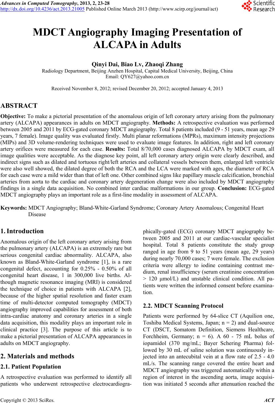 Advances in Computed Tomography, 2013, 2, 23-28 http://dx.doi.org/10.4236/act.2013.21005 Published Online March 2013 (http://www.scirp.org/journal/act) MDCT Angiography Imaging Presentation of ALCAPA in Adults Qinyi Dai, Biao Lv, Zhaoqi Zhang Radiology Department, Beijing Anzhen Hospital, Capital Medical University, Beijing, China Email: [email protected] Received November 8, 2012; revised December 20, 2012; accepted January 4, 2013 ABSTRACT Objective: To make a pictorial presentation of the anomalous origin of left coronary artery arising from the pulmonary artery (ALCAPA) appearances in adults on MDCT angiography. Methods: A retrospective evaluation was performed between 2005 and 2011 by ECG-gated coronary MDCT angiography. Total 8 patients included (9 - 51 years, mean age 29 years, 7 female). Image quality was evaluated firstly. Multi planar reformations (MPRs), maximum intensity projections (MIPs) and 3D volume-rendering techniques were used to evaluate image features. In addition, right and left coronary artery orifices were measured for each case. Results: Total 8/70,000 cases diagnosed ALCAPA by MDCT exam, all image qualities were acceptable. As the diagnose key point, all left coronary artery origin were clearly described, and indirect signs such as dilated and tortuous right/left arteries and collateral vessels between them, enlarged left ventricle were also well showed, the dilated degree of both the RCA and the LCA were marked with ages, the diameter of RCA for each case were a mild wider than that of left one. Other combined signs like papillary muscle calcification, bronchial arteries from aorta to the cardiac and coronary artery degeneration change were also included by MDCT angiography findings in a single data acquisition. No combined inter cardiac malformations in our group. Conclusion: ECG-gated MDCT angiography plays an important role as a first-line modality in assessment of ALCAPA. Keywords: MDCT Angiography; Bland-White-Garland Syndrome; Coronary Artery Anomalous; Congenital Heart Disease 1. Introduction Anomalous origin of the left coronary artery arising from the pulmonary artery (ALCAPA) is an extremely rare but serious congenital cardiac abnormality. ALCAPA, also known as Bland-White-Garland syndrome [1], is a rare congenital defect, accounting for 0.25% - 0.50% of all congenital heart disease, 1 in 300,000 live births. Al- though magnetic resonance imaging (MRI) is considered the technique of choice in patients with ALCAPA [2], because of the higher spatial resolution and faster exam time of multi-detector computed tomography (MDCT) angiography improved capabilities for assessment of both intra-cardiac anatomy and coronary arteries in a single data acquisition, this modality plays an important role in clinical practice [3]. The purpose of this article is to make a pictorial presentation of ALCAPA appearances in adults on MDCT angiography. 2. Materials and methods 2.1. Patient Population A retrospective evaluation was performed to identify all patients who underwent retrospective electrocardiogra- phically-gated (ECG) coronary MDCT angiography be- tween 2005 and 2011 at our cardiac-vascular specialist hospital. Total 8 patients constitute the study group ranged in age from 9 to 51 years (mean age, 29 years) during nearly 70,000 cases; 7 were female. The exclusion criteria were allergy to iodine containing contrast me- dium, renal insufficiency (serum creatinine concentration > 120 μmol/L) and unstable clinical condition. All pa- tients were written the informed consent before examina- tion. 2.2. MDCT Scanning Protocol Patients were performed by 64-slice CT (Aquilion one, Toshiba Medical Systems, Japan; n = 2) and dual-source CT (DSCT, Somatom Definition, Siemens Healthcare, Forchheim, Germany; n = 6). A 60 - 75 mL bolus of iopamidol (370 mg/mL; Bayer Schering Pharma) fol- lowed by 30 mL of saline solution was continuously in- jected into an antecubital vein at a flow rate of 2.5 - 4.0 mL/s. The scanning range covered the entire heart and MDCT angiography was triggered automatically within a region of interest in the ascending aorta, image acquisi- tion was initiated 5 seconds after attenuation reached the C opyright © 2013 SciRes. ACT 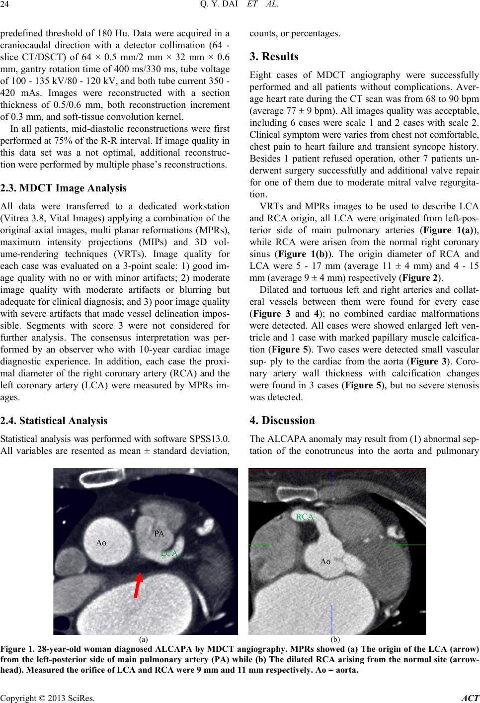 Q. Y. DAI ET AL. 24 predefined threshold of 180 Hu. Data were acquired in a craniocaudal direction with a detector collimation (64 - slice CT/DSCT) of 64 × 0.5 mm/2 mm × 32 mm × 0.6 mm, gantry rotation time of 400 ms/330 ms, tube voltage of 100 - 135 kV/80 - 120 kV, and both tube current 350 - 420 mAs. Images were reconstructed with a section thickness of 0.5/0.6 mm, both reconstruction increment of 0.3 mm, and soft-tissue convolution kernel. In all patients, mid-diastolic reconstructions were first performed at 75% of the R-R interval. If image quality in this data set was a not optimal, additional reconstruc- tion were performed by multiple phase’s reconstructions. 2.3. MDCT Image Analysis All data were transferred to a dedicated workstation (Vitrea 3.8, Vital Images) applying a combination of the original axial images, multi planar reformations (MPRs), maximum intensity projections (MIPs) and 3D vol- ume-rendering techniques (VRTs). Image quality for each case was evaluated on a 3-point scale: 1) good im- age quality with no or with minor artifacts; 2) moderate image quality with moderate artifacts or blurring but adequate for clinical diagnosis; and 3) poor image quality with severe artifacts that made vessel delineation impos- sible. Segments with score 3 were not considered for further analysis. The consensus interpretation was per- formed by an observer who with 10-year cardiac image diagnostic experience. In addition, each case the proxi- mal diameter of the right coronary artery (RCA) and the left coronary artery (LCA) were measured by MPRs im- ages. 2.4. Statistical Analysis Statistical analysis was performed with software SPSS13.0. All variables are resented as mean ± standard deviation, counts, or percentages. 3. Results Eight cases of MDCT angiography were successfully performed and all patients without complications. Aver- age heart rate during the CT scan was from 68 to 90 bpm (average 77 ± 9 bpm). All images quality was acceptable, including 6 cases were scale 1 and 2 cases with scale 2. Clinical symptom were varies from chest not comfortable, chest pain to heart failure and transient syncope history. Besides 1 patient refused operation, other 7 patients un- derwent surgery successfully and additional valve repair for one of them due to moderate mitral valve regurgita- tion. VRTs and MPRs images to be used to describe LCA and RCA origin, all LCA were originated from left-pos- terior side of main pulmonary arteries (Figure 1(a)), while RCA were arisen from the normal right coronary sinus (Figure 1(b)). The origin diameter of RCA and LCA were 5 - 17 mm (average 11 ± 4 mm) and 4 - 15 mm (average 9 ± 4 mm) respectively (Figure 2). Dilated and tortuous left and right arteries and collat- eral vessels between them were found for every case (Figure 3 and 4); no combined cardiac malformations were detected. All cases were showed enlarged left ven- tricle and 1 case with marked papillary muscle calcifica- tion (Figure 5). Two cases were detected small vascular sup- ply to the cardiac from the aorta (Figure 3). Coro- nary artery wall thickness with calcification changes were found in 3 cases (Figure 5), but no severe stenosis was detected. 4. Discussion The ALCAPA anomaly may result from (1) abnormal sep- tation of the conotruncus into the aorta and pulmonary PA Ao LCA Ao RCA (a) (b) Figure 1. 28-year-old woman diagnosed ALCAPA by MDCT angiography. MPRs showed (a) The origin of the LCA (arrow) from the left-posterior side of main pulmonary artery (PA) while (b) The dilated RCA arising from the normal site (arrow- head). Measured the orifice of LCA and RCA were 9 mm and 11 mm respectively. Ao = aorta. Copyright © 2013 SciRes. ACT 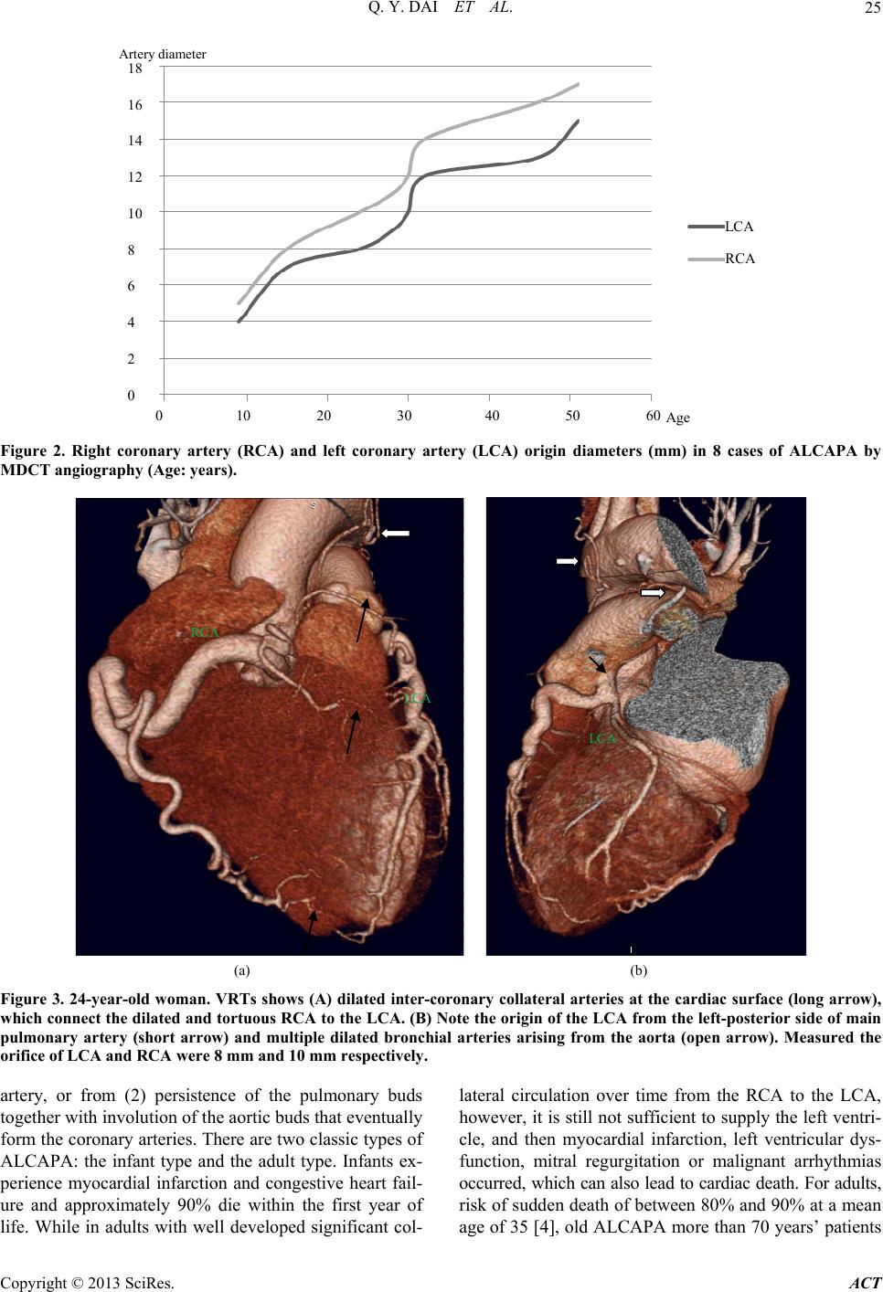 Q. Y. DAI ET AL. 25 0 10 20 30 40 50 60 18 16 14 12 10 8 6 4 2 0 RCA LCA Age A rtery di ameter Figure 2. Right coronary artery (RCA) and left coronary artery (LCA) origin diameters (mm) in 8 cases of ALCAPA by MDCT angiography (Age: years). RCA LCA LCA (a) (b) Figure 3. 24-year-old woman. VRTs shows (A) dilated inter-coronary collateral arteries at the cardiac surface (long arrow), which connect the dilated and tortuous RCA to the LCA. (B) Note the origin of the LCA from the left-posterior side of main pulmonary artery (short arrow) and multiple dilated bronchial arteries arising from the aorta (open arrow). Measured the orifice of LCA and RCA were 8 mm and 10 mm respectively. artery, or from (2) persistence of the pulmonary buds together with involution of the aortic buds that eventually form the coronary arteries. There are two classic types of ALCAPA: the infant type and the adult type. Infants ex- perience myocardial infarction and congestive heart fail- ure and approximately 90% die within the first year of life. While in adults with well developed significant col- lateral circulation over time from the RCA to the LCA, however, it is still not sufficient to supply the left ventri- cle, and then myocardial infarction, left ventricular dys- function, mitral regurgitation or malignant arrhythmias occurred, which can also lead to cardiac death. For adults, risk of sudden death of between 80% and 90% at a mean age of 35 [4], old ALCAPA ore than 70 years’ patients m Copyright © 2013 SciRes. ACT 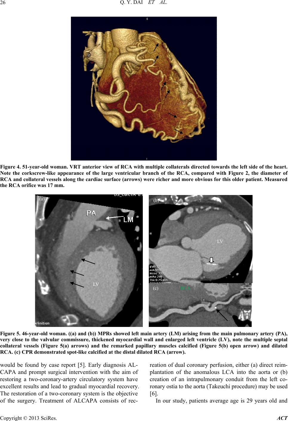 Q. Y. DAI ET AL. 26 Figure 4. 51-year-old woman. VRT anterior view of RCA with mult iple collaterals directed towards the left side of the heart. Note the corkscrew-like appearance of the large ventricular branch of the RCA, compared with Figure 2, the diameter of RCA and collateral vessels along the cardiac surface (arrows) were richer and more obvious for this older patient. Measured the RCA orifice was 17 mm. LV (a) LV (c) RCA ( b ) Figure 5. 46-year-old woman. (( a) and (b)) MPRs showed left main artery (LM) arisi ng from the main pulmonary artery (PA), very close to the valvular commissure, thickened myocardial wall and enlarged left ventricle (LV), note the multiple septal collateral vessels (Figure 5(a) arrows) and the remarked papillary muscles calcified (Figure 5(b) open arrow) and dilated RCA. (c) CPR demonstrated spot-like calcified at the distal dilated RCA (arrow). would be found by case report [5]. Early diagnosis AL- CAPA and prompt surgical intervention with the aim of restoring a two-coronary-artery circulatory system have excellent results and lead to gradual myocardial recovery. The restoration of a two-coronary system is the objective of the surgery. Treatment of ALCAPA consists of rec- reation of dual coronary perfusion, either (a) direct reim- plantation of the anomalous LCA into the aorta or (b) creation of an intrapulmonary conduit from the left co- ronary ostia to the aorta (Takeuchi procedure) may be used [6]. In our study, patients average age is 29 years old and Copyright © 2013 SciRes. ACT  Q. Y. DAI ET AL. 27 symptoms were varies from mild chest not comfortable to syncope or heart failure, the lack of serious symptoms besides well-developed collateral vessels, may also been due in part to systemic hypertension, which preserved antegrade blood flow in the RCA [4]. Occurrence is gen- erally no predilection in sex or race, and is not consid- ered an inheritable congenital heart defect. In our study, all 8 patients are Chinese, female predominant and with- out combined with intra-cardiac disease. ECG-gated MDCT angiography findings of ALCAPA in adults include direct visualization of the LCA original from the main pulmonary artery, which is the diagnostic key point viewed by MIPs and VRTs, the LCA typically arises from the left-posterior aspect of the main pulmo- nary artery beyond the pulmonary valve (Figure 1(a)), which were the most common position, however, its po- sition within the sinus is extremely variable, particularly it may be very close to a valvular commissure (Figure 5(a)). Rarely, the anomalous ostium originates from the right posterior sinus or even one of the main pulmonary arteries. To know the exact LCA origin position may help the surgical doctor to determine the optional opera- tion method. In our study, 7 of 8 cases allowed operation successfully, including direct re-implantation (n = 3) and Takeuchi (n = 4) surgery. Such images give the surgeons a better understanding of the complex anatomy before re- pair. The remarkable secondary imaging finding is the RCA and the LCA dilated and tortuous, and dilated intracoro- nary collateral arteries are seen along the epicardial sur- face of the heart by VRTs (Figure 3(a)) or within the interventricular septum by MIPs or MPRs (Figure 5(a)). All cases represent the well-developed collateral path- ways between the RCA and the LCA, MDCT images offers excellent spatial resolution, which is required to assess small vessels such as the coronary arteries. In our study, the dilated degree of both the RCA and the LCA marked with ages, the diameter of RCA for each case were a mild wider than that of left one (Table 2, Figure 1 and 3), and collateral vessels along the cardiac surface were richer for older patients (Figure 4). Moreover, the dilated bronchial arteries from the aorta (Figure 3), which act as systemic collateral vessels to the LCA area’s blood supply, are best depicted at ECG-gated MDCT angiography because of their small size. Besides, left ventricular hypertrophy and dilatation result from chronic myocardial ischemia also be seen in MDCT images (Figure 5), may be used to assess left ventricular function. If ischemia of the papillary muscles happened and adjacent myocardium may cause mitral insufficiency, as a finding, papillary muscles calcified can easily be seen at MPRs besides left ventricle en- larged (Figure 5(b)). Due to the marked dilatation develops over time as blood is rapidly shunted from the RCA into the LCA and then into the low-pressure pulmonary circulation, the de- generation change of the coronary artery could be found in our mean age 29 years ALCAPA group (Figure 5(c)). 2D echocardiography with Doppler color flow map- ping in some situations replaces angiography [7], echo is sensitive to determine the mitral valve and left ventricle function, which is also a convenient method used for fol- low-up. Another technique used to non-invasively image the coronary arteries is MR angiography, MR could ac- curately depict the origin and proximal course of anoma- lous coronary arteries [8] and no radiation exposure, even more crucial for MR is the ability to assess myocardial viability, which can be used as an important prognostic factor to consider by the surgical doctor. Conventional coronary angiography still remains as a gold standard for the diagnosis of anomalous coronary arteries; however, it is invasive and limited to a projection two-dimensional view of the complex coronary artery system [9], especial- ly for the retrograde ostium. ALCAPA should be different with other disease which may cause coronary artery dilated, such as Kawasaki di- sease, coronary artery fistula or secondary to the athe- rosclerosis change, the essential feature is to show and determine the LCA origin area. ECG-gated MDCT angiography plays an important role as a first-line modality in assessment of ALCAPA, and MDCT angiography also would be a valuable evalu- ated post-operation condition [10]. But our study is lim- ited both by its retrospective nature and a relatively small number of patients. Because of potential drawbacks of MDCT such as radiation exposure and contrast medium- related complications, performing MDCT should be limi- ted to patients presenting symptoms suggesting the pres- ence of anomalous coronary arteries. REFERENCES [1] H. Brooks, “Two Cases of an Abnormal Coronary Artery of the Heart Arising from the Pulmonary Artery: With Some Remarks upon the Effect of This Anomaly in Pro- ducing Circoid Dilation of the Vessels,” Journal of Anat- omy and Physiology, Vol. 20, No. 1, 1885, pp. 26-29. [2] J. C. Post, A. C. van Rossum, J. G. Bronzwaer, C. C. de Cock, M. B. Hofman and J. Valk, “Magnetic Resonance Angiography of Anomalous Coronary Arteries. A New Gold Standard for Delineating the Proximal Course?” Circulation, Vol. 92, No. 11, 1995, pp. 3163-3171. doi:10.1161/01.CIR.92.11.3163 [3] T. Horisaki, T. Yamashita, H. Yokoyama, K. Urasawa and A. Kitabatake, “Three-Dimensional Reconstruction of Computed Tomographic Images of Anomalous Origin of the Left Main Coronary Artery from the Pulmonary Trunk in an Adult,” American Journ al of Cardiology, Vol. 92, No. 7, 2003, pp. 898-899. doi:10.1016/S0002-9149(03)00913-5 Copyright © 2013 SciRes. ACT  Q. Y. DAI ET AL. Copyright © 2013 SciRes. ACT 28 [4] C. Frescura, C. Basso, G. Thiene, D. Corrado, T. Pennelli and A. Angelini, “Anomalous Origin of Coronary Arter- ies and Risk of Sudden Death: A Study Based on an Au- topsy Population of Congenital Heart Disease,” Human Pathology, Vol. 29, No. 7, 1998, pp. 689-695. doi:10.1016/S0046-8177(98)90277-5 [5] A. Khanna, D. A. Torgian, V. A. Ferrari, R. J. Bross and M. A. Rose, “Anomalous Origin of the Left Coronary Artery from the Pulmonary Artery in Adulthood CT and MRI,” American Journal of Roentgenology, Vol. 185, No. 2, 2005, pp. 326-329. [6] J. Caspi, T. W. Pettitt, C. Sperrazza, T. Mulder and A. Stopa, “Reimplantation of Anomalous Left Coronary Ar- tery from the Pulmonary Artery without Mitral Valve Re- pair,” Annals of Thoracic Surgery, Vol. 84, No. 2, 2007, pp. 619- 623. doi:10.1016/j.athoracsur.2007.03.036 [7] Y. L. Yang, N. C. Nanda, X. F. Wang, M. X. Xie, Lu Q., L. He and X. F. Lu, “Echocardiographic Diagnosis of Anomalous Origin of the Left Coronary Artery from the Pulmonary Artery,” Echocardiography, Vol. 24, No. 4, 2007, pp. 405-411. doi:10.1111/j.1540-8175.2006.00406.x [8] N. H. Bunce, C. H. Lorenz, J. Keegan, J. Lesser, E. M. Reyes, D. N. Firmin and D. J. Pennell, “Coronary Artery Anomalies: Assessment with Free-Breathing Three-Di- mensional Coronary MR Angiography,” Radiology, Vol. 227, No. 1, 2003, pp. 201-208. doi:10.1148/radiol.2271020316 [9] I. A. Williams, W. M. Gersony, W. E. Hellenbrand, “Ano- malous Right Coronary Artery Arising from the Pulmo- nary Artery: A Report of 7 Cases and a Review of the Literature,” American Heart Journal, Vol. 152, No. 5, 2006, pp. 9-17. [10] E. Peρa, E. Nguyen, N. Merchant and G. Dennie, “AL- CAPA Syndrome: Not Just a Pediatric Disease,” Radio- graphics, Vol. 29, No. 2, 2009, pp. 553-565. doi:10.1148/rg.292085059 |