

Paper Menu >>
Journal Menu >>
 Advances in Computed Tomography, 2013, 2, 20-22 http://dx.doi.org/10.4236/act.2013.21004 Published Online March 2013 (http://www.scirp.org/journal/act) Giant Calcified Renal Artery Aneurysm: Traditional RX versus Thr ee-Dimensional Computed Tomography Mauro Gacci1, Omar Saleh1, Annalisa Mantella1, Leonidas Azas2, Paola Romagnani3, Andrea Minervini1, Sergio Serni1, Marco Carini1 1Department of Urology, University of Florence, Florence, Italy 2Department of Vascular Surgery, University of Florence, Florence, Italy 3Excellence Center for Research, Transfer and High Education (DENOTHE), University of Florence, Florence, Italy Email: [email protected] Received November 7, 2012; revised December 16, 2012; accepted December 26, 2012 ABSTRACT A 65-year-old woman with no history of previous flank trauma, renal stone or upper urinary tract infections, presented for flank pain and left hydro-uretero-nephrosis seven days after hysterectomy. Percutaneous pielography revealed nar- rowing of the distal ureter, without endoureteral mass. The plain abdomen film incidentally showed a 3-cm calcified ring on the left renal shadow, who resulted external to the collecting system at pielography. A 3-dimensional-CT scan with angiographic reconstruction revealed a 3-cm calcified renal artery aneurysm. The vascular surgeon suggested a watchful waiting. The patient underwent ur eteral reimplantation with ureteral stenting, allowing a co mplete recovery of iatrogenic stenosis two months postoperatively. Keywords: Renal Artery Aneurysm; 3D-CT; Angiographic Reconstruction 1. Introduction Renal artery aneurysms have been encountered with increasing frequency over the past decade. It is slightly more common in women than men and in the right than left renal artery [1]. Many cases are asymptomatic and found incidentally, and their occurrence has been recen- tly increasing with the advancement of imaging techni- ques. Angiography is the gold standard in the diagnosis of renovascular injuries, and it has the additional advan- tage to possesses the poten tial of therapeutic intervention [2]. Therefore, after any invasive urological procedure, CT angiography can be considered as the first choice for renal artery injury [3,4]. In the present case, we inciden- tally discovered a lesion of the left renal artery and we completed the diagnostic work-up with a 3D-CT scan. 2. Case Report A 67-year-old woman was referred to the Department of Urology for left flank pain developed seven days after hysterectomy for uterine leiomyomatosis. The patient had no history of previous flank trauma, renal stone or upper urinary tract infections. Physical examination was unremarkable, with only minimal flank pain at Giordano manoeuvre; body temperature was 37˚C, blood pressure was 120/75 mmHg. White cell count was within the limits (9 × 10⁹ white blood cells in a litre of blood); ren al and liver functions were normal (creatinine 0.9 mg/dL, total bilirubin 0.8 mg/dL). A severe left hydronephrosis was detected at ultrasound, with no sign of uretheral stones or masses. The plain abdomen film showed a 3-cm calcified ring on the left renal shadow (Figure 1(a)). Percutaneous pielography confirmed hydronephrosis, and showed no relationship of the calcified mass with th e col- lecting system (Figure 1(b)). A 3-dimensional-CT scan revealed a 3-cm round hol- low mass with calcified walls located in front of anterior renal surface. The lesion had no connection with renal pelvis (Figure 2(a)) but was firmly related with renal artery, and it was compatible with calcified renal artery aneurysm (Figure 2(b)). The patient was treated with an open access ureteral reimplantation performed on the suture of the previous surgical approach. A direct ure- theral reimplantation on the upper bladder wall was per- formed, with psoas hitch and double J uretheral stenting. At the 1 month follow up visit there was a complete recovery of the obstruction. The vascular surgeon sugge- sted a watchful waiting by monitoring blood pressure, renal function, and imaging ever y 6 months. 3. Discussion Renal artery aneurysms (RAA) are rare, with an estima- ted incidence below 1%. Hypertension and fibro-mus- cular disease of the renal artery are the leading classes of C opyright © 2013 SciRes. ACT 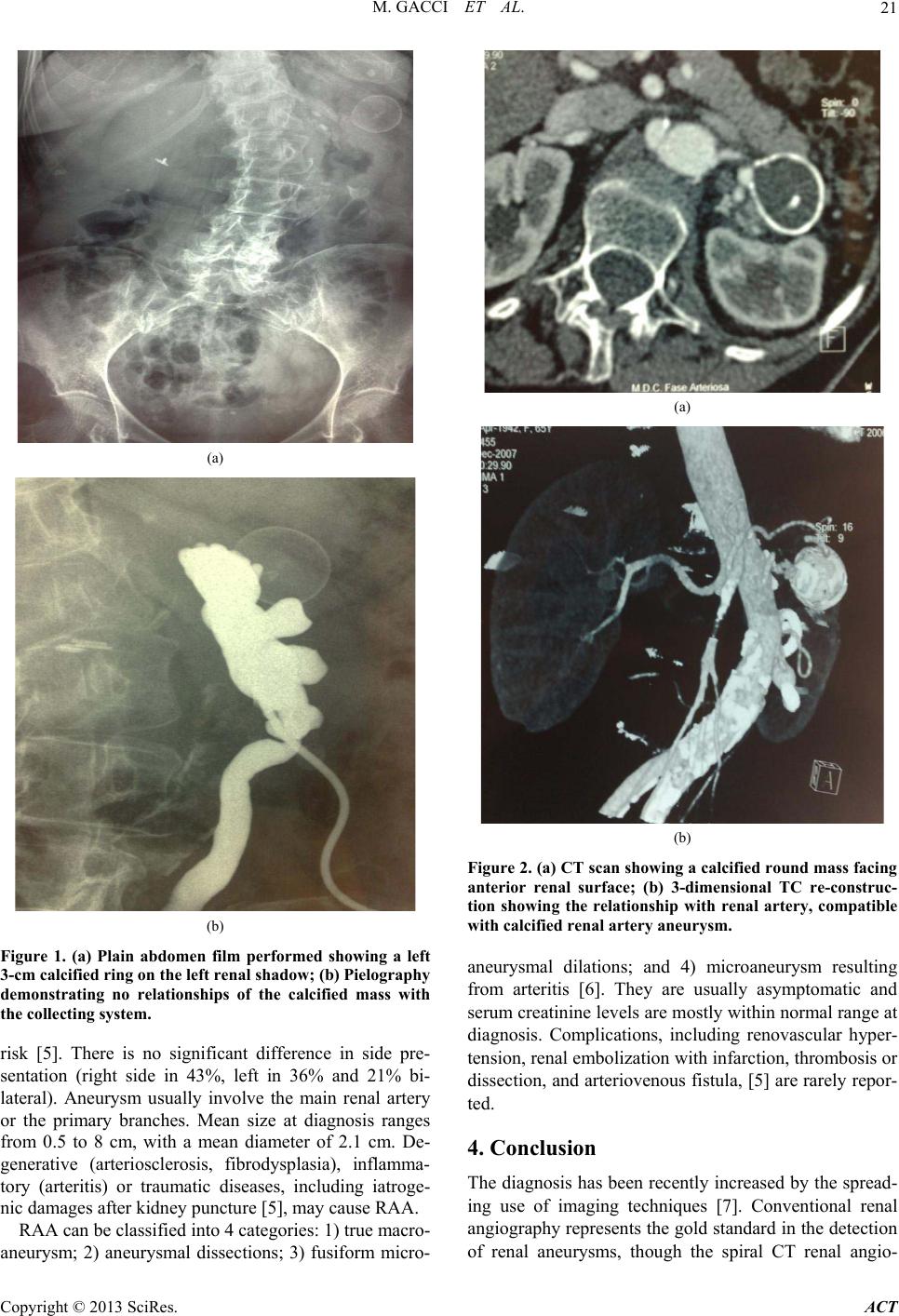 M. GACCI ET AL. 21 (a) (b) Figure 1. (a) Plain abdomen film performed showing a left 3-cm calcified ring on the left renal shadow; (b) Pielography demonstrating no relationships of the calcified mass with the collecting system. risk [5]. There is no significant difference in side pre- sentation (right side in 43%, left in 36% and 21% bi- lateral). Aneurysm usually involve the main renal artery or the primary branches. Mean size at diagnosis ranges from 0.5 to 8 cm, with a mean diameter of 2.1 cm. De- generative (arteriosclerosis, fibrodysplasia), inflamma- tory (arteritis) or traumatic diseases, including iatroge- nic damages after kidney puncture [5], may cause RAA. RAA can be classified into 4 categories: 1) true macro- aneurysm; 2) aneurysmal dissections; 3) fusiform micro- (a) (b) Figure 2. (a) CT scan show ing a calcified round mass faci ng anterior renal surface; (b) 3-dimensional TC re-construc- tion showing the relationship with renal artery, compatible with calcified renal artery aneurysm. aneurysmal dilations; and 4) microaneurysm resulting from arteritis [6]. They are usually asymptomatic and serum creatinine levels are mostly within normal range at diagnosis. Complications, including renovascular hyper- tension, renal embolization with in farction , thro mbosis or dissection, and arteriovenous fistula, [5] are rarely repor- ted. 4. Conclusion The diagnosis has been recently increased by the spread- ing use of imaging techniques [7]. Conventional renal angiography represents the gold standard in the detection of renal aneurysms, though the spiral CT renal angio- Copyright © 2013 SciRes. ACT  M. GACCI ET AL. Copyright © 2013 SciRes. ACT 22 graphy with three dimensional reconstruction (3D-CT) adds the benefits of being quicker, more costeffective, and non-invasive. Moreover, the use of 3D-CT worksta- tion allows accurate treatment planning, especially for endovascular proce d ur es [8]. Management options include observation, trans-cathe- ter-occlusion or surgical interven tion. The indications for surgery include: symptomatic or enlarging aneurysms, renal embolization, aneurysms in pregnant females or those considering pregnancy, renovascular hypertension, aneurysms >2.5 cm [9]. REFERENCES [1] J. C. Stanley, L. M. Messina and G. B. Zelenock, “Splan- ckmc and Renal Artery Aneurysms,” In: W. S. Moore, Ed., Vascular Surgery: A Comprehensive Review, WB Saunders, Philadelphia, 1491, pp. 335-349. [2] Y. H. Rokni and Y. Moharamzad, “Endovascular Treat- ment of Renal Arteriovenous Fistula Following a Stab Wound,” Urology Journal, Vol. 5, No. 2, 2008, pp. 129- 131. [3] L. J. Zhang, G. F. Yang, J. Qi and W. Shen, “Renal Ar- tery Aneurysm: Diagnosis and Surveillance with Multi- Detector Row Computed Tomography,” Acta Radiolo- gica, Vol. 48, No. 3, 2007, pp. 274-279. doi:10.1080/02841850601161521 [4] F. Y. Dönmez, M. Cokun, A. Uyuur, et al., “Non-Inva- sive Imaging Findings of Idiopathic Renal Arteriovenous Fistula,” Diagnostic and Interventional Radiology, Vol. 14, No. 2, 2008, pp. 103-105. [5] A. Lumsden, T. Salam and K. Walton, “Renal Artery Aneurysm: A Report of 28 Cases,” Cardiovascular Sur- gery, Vol. 4, No. 2, 1996, pp. 185-189. doi:10.1016/0967-2109(96)82312-X [6] J. C. Stanley, E. L. Rhodes, B. L. Cewertz, C. Y. Chang, J. F. Walter and W. J. Fry, “Renal Artery Aneurysms: Sig- nificance of Macroaneurysms Exclusive of Dissections and Fibrodysplastic Mural Dilations,” Archives of Sur- gery, 1975; Vol. 110, No. 11, 1975, pp. 1327-1333. doi:10.1001/archsurg.1975.01360170067009 [7] S. P. Stanl and E. Fishman, “Three Dimensional Ct Angi- ography: Renal Applications,” Semin Ultrasound CT MR, Vol. 19, No. 5, 1998, pp. 413-424. doi:10.1016/S0887-2171(98)90018-X [8] W. Higashiura, S. Sakaguchi, N. Tabayashi, S. Taniguchi and K. Kichikawa, “Impact of 3-Dimensional-Computed Tomography Workstation for Precise Planning of Endo- vascular Aneurysm Repair,” Circulation Journal, Vol. 72, No. 12, 2008, pp. 2028-2034. doi:10.1253/circj.CJ-08-0331 [9] C. Dzsinich, P. Gloviczki, M. A. McKusick, P. C. Pai- rolero, T. C. Bower, J. W. Hallett Jr. and K. J. Cherry Jr., “Surgical Management of Renal Artery Aneurysm,” Car- diovascular Surgery, Vol. 1, No. 3, 1993, pp. 243-247. Abbreviation and Acronym Renal artery aneurysms (RAA) Units |