

Paper Menu >>
Journal Menu >>
 Advances in Computed Tomography, 2013, 2, 13-19 http://dx.doi.org/10.4236/act.2013.21003 Published Online March 2013 (http://www.scirp.org/journal/act) How to Reduce Unnecessary Invasive Angiograms When Patients Are Initially Evaluated by Coronary Computed Tomography? Edouard Cheneau1, Bruno Vahdat1, Noémie Resseguier2, Laurent Bernard1, Annamaria Molon1, Roch Giorgi2, Dimitri Panagides1 1Service de Cardiologie, Clinique Bouchard, Marseille, France 2Service de Santé Publique et d’Information Médicale, Hôpital de la Timone, Marseille, France Email: edouard.c[email protected] Received November 5, 2012; revised December 16, 2012; accepted December 26, 2012 ABSTRACT Purpose: When Coronary Computed Tomography (CCT) detects coronary obstruction, patients are regularly referred to invasive angiogram. With higher sensitivity than specificity, CCT might induce unnecessary angiograms (“false posi- tive CCT”). We sought to determine the patients and CCT findings associated with false positive CCT. Methods: Pa- tients were prospectively referred to CCT with a 64-slice CT scan for suspected CAD. Inclusion criteria were: 1) clini- cally suspected angina pectoris; or 2) suspected silent ischemia on resting EKG. Exclusion criteria were acute coronary syndrome and non sinus rhythm. Invasive coronary angiogram (ICA) was performed on the basis of CCT findings (stenosis >50%). Analysis was performed on a per patient basis. Results: Out of 702 patients, 228 had suspected sig- nificant stenosis by CCT and ICA was performed in 176 patients. Coronary stenosis >50% was not confirmed by ICA in 44 (25%). In multivariate analysis, we observed that atypical angina (OR 3.63, CI 1.43 - 9.66), silent ischemia (OR 5.11, CI 1.89 - 14.6) and number of suspected stenosed arteries by CCT (OR 1.81, CI 1.15 - 2.94) were independently predictive of false positive CCT (p < 0.05). Lesion location and coronary plaque characteristics did not impact on CCT accuracy. Conclusions: Performing CCT for atypical angina or silent ischemia is associated with higher rate of unnec- essary invasive coronary angiograms. We failed to identify lesions characteristics prone to be “false positive” of CCT. Keywords: Angina; Coronary Disease; Diagnosis; CCT 1. Introduction Coronary computed tomography (CCT) is a rapidly de- veloping technique and plays a rising role in angina di- agnosis [1,2]. CCT has shown high sensibility for de- tecting obstructive coronary disease in a non-invasive manner [3,4]. Despite CCT can adequately rule out ob- structive coronary artery disease (CAD), its high sensi- tivity is associated with a lower positive predictive value and a number of coronary stenosis on CCT are not con- firmed on invasive coronary angiograms. Since CCT has a growing role as first line work-up for angina pectoris diagnosis, referring patients with a positive CCT to inva- sive coronary angiography might induce numerous un- necessary invasive angiograms [5]. From retrospective analysis, CCT usefulness appears to be dependent on pre-test probability for having CAD and might be most advantageous in subsets of patients, especially those with an intermediate risk [6]. Consequently, patient selection prior to CCT enables to increase CCT accuracy. A complementary approach would be to individualize subset of patients or CCT findings that are more likely to be false positive of CCT after they were screened by this technique. In these patients, a positive CCT would not justify to directly undergo cardiac catheterization and al- ternative work-up might be developed. In this study, we sought to determine the patient characteristics and CCT findings associated with false positive CCT. 2. Material and Method 2.1. Patients We included all patients referred to our center for CT scan as initial work up test to evaluate possible obstruc- tive coronary heart disease. Patients presenting with ty- pical or atypical angina pectoris or suspected silent ischemia on resting EKG were referred for CCT and prospectively included into the study. CCT is the front- line test routinely used in our institution for CAD diag- nosis. Inclusion criteria were patients referred for CCT without previous myocardial stress imaging or treadmill C opyright © 2013 SciRes. ACT  E. CHENEAU ET AL. 14 test and patients with preceding equivocal stress test dur- ing a six month period. A dedicated information sheet and database was used for data collection. Inclusions were opened from January 2009 to December 2010. Typical angina was defined as having three characteristics: 1) substernal discomfort; 2) that is precipitated by physical exertion or emotion; and 3) relieved with rest or sublingual nitroglycerin. Atypical angina pectoris was defined as having 2 of 3 of the defi- nition characteristics. Suspected silent ischemia was de- fined as Q wave or negative T wave on resting EKG in at least in two derivations in patients with at least one risk factor. Inclusion criteria were patients referred for CCT as initial work up test and patients with preceding equi- vocal stress test. Exclusion criteria were non sinus rhy- thm, acute coronary syndromes, impaired renal function (serum creatinine >120 µmol/l) and known allergy to io- dinated contrast material. The estimated pretest prob- ability for obstructive CAD was estimated using the Duke Clinical Score, which includes type of chest dis- comfort, age, gender, and traditional risk factors [7]. The protocol was reviewed and accepted by the ethic com- mittee of our Board of Cardiology. 2.2. Scan Protocol Patients with a heart rate exceeding 70 beats/min re- ceived IV beta-blockers (Atenolol 5 to 15 mg) before CT examination. All scans were performed with a 64-slice CT scanner (Lightspeed VCT, General Electric Health- care) that features a gantry rotation time of 350 ms, a temporal resolution of 175 ms, and a spatial resolution of 0.54 mm3. Tube voltage was 120 kV( 100 kV for patients weight <70 kg), X-ray tube current was prospectively modulated on EKG (250 to 650 mA). CT was acquired in a breath-hold and ECG-gated. Rotation speed and pitch were adjusted to the acquisition protocols and the heart rate. Calcium scoring was not performed. A bolus of con- trast media (Ioxaglate 320 mG I/mL, Guerbet) was in- fused in an antebrachial vein with use of a dual-barrel injector (70 cc of contrast media at 5 cc/s washed out by 30 cc of isotonic solution at 3.5 cc/s). CT data were ana- lyzed by the use of an offline Advantage Workstation (General Electric) using (curved) multiplanar reconstruc- tion. Phases from 0 to 90% every 10% were systemati- cally reconstructed to allow for imaging coronary arteries. Segments were scored positive for significant CAD if there was ≥50% diameter reduction of the lumen (at least 2 mm) by visual assessment. Three experienced (more than 500 CT coronary angiograms) observers blinded about previous medical history and symptoms partici- pated in the study. Optimal quality CT scan was defined as lumen visual assessment available in all >2 mm di- ameter coronary arteries. Patients with non-optimal qual- ity CT (including heavy calcification without lumen as- sessment) were scored positive for significant CAD. All patients with significant stenosis by CCT were suggested to undergo to ICA. 2.3. Angiographic Analysis Baseline quantitative angiography was performed using the contrast-filled injection catheter for image calibration. Cine Angiographic stenosis was defined as stenosis grea- ter than 50%. Quantitative coronary angiographic (QCA) analysis was performed using the Integris H5000C soft- ware (Philips, The Netherlands). Patients were classified as positive for significant CAD if at least one (>2 mm diameter) coronary artery showed a significant stenosis (>50%). Patients were classified as true positive CCT if they were classified as positive for CAD by both CCT and ICA and false positive CCT if significant stenosis by CCT was not confirmed by ICA. 2.4. Statistics All statistical analyses were realized using the R software, version 2.12.0 (R Foundation for Statistical Computing, Vienna, Austria). First, a descriptive analysis of all re- corded data was performed among the overall study po- pulation. Bivariate analysis was then performed by com- paring the patients and lesions characteristics of the two groups, i.e. true positive CCT and false positive CCT. Continuous variables were expressed as mean ± standard deviation, and were compared using t-test after graphi- cally checking of the conditions of the test. Categorical variables were expressed as number of patients and per- centages, and were compared using chi-square test or Fisher test, when appropriate. Variables were retained for the multivariate analysis when their effect had a p-value less than 0.25 for the bivariate analysis. A multivariate logistic regression model was built. A backward stepwise selection procedure was applied to retain the significant (p < 0.05) independent variables and their interactions in the final model. Significance was determined using likelihood ratio test. The accuracy of the final model was assessed by Hosmer-Lemeshow test (HLgof.test R package). 3. Results We evaluated 702 patients (male 66%) by CCT. Age was 61 ± 11 years. CCT image quality was inadequate in 74 patients (10.5%). Significant obstruction was suspected in 228 patients (32%) by CCT and invasive coronary angiogram was performed in 176 patients. Coronary stenosis was >50% in 132 patients (75%) by ICA. False positive CCT were observed in 44 patients (25%). Patients’ characteristics were compared between true Copyright © 2013 SciRes. ACT 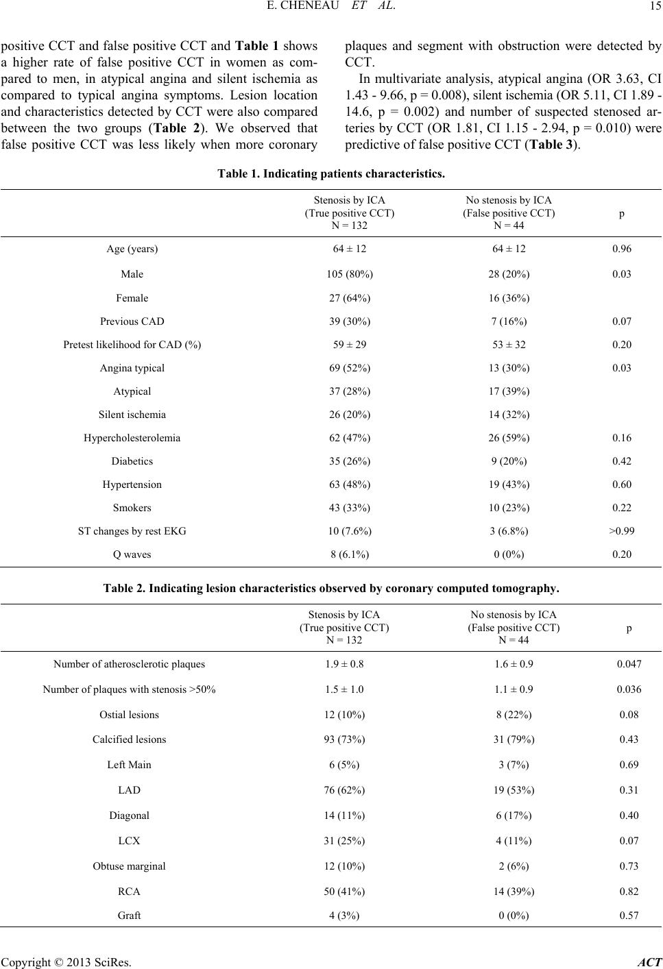 E. CHENEAU ET AL. Copyright © 2013 SciRes. ACT 15 positive CCT and false positive CCT and Table 1 shows a higher rate of false positive CCT in women as com- pared to men, in atypical angina and silent ischemia as compared to typical angina symptoms. Lesion location and characteristics detected by CCT were also compared between the two groups (Table 2). We observed that false positive CCT was less likely when more coronary plaques and segment with obstruction were detected by CCT. In multivariate analysis, atypical angina (OR 3.63, CI 1.43 - 9.66, p = 0.008), silent ischemia (OR 5.11, CI 1.89 - 14.6, p = 0.002) and number of suspected stenosed ar- teries by CCT (OR 1.81, CI 1.15 - 2.94, p = 0.010) were predictive of false positive CCT (Table 3). Table 1. Indicating patients characteristics. Stenosis by ICA (True positive CCT) N = 132 No stenosis by ICA (False positive CCT) N = 44 p Age (years) 64 ± 12 64 ± 12 0.96 Male 105 (80%) 28 (20%) 0.03 Female 27 (64%) 16 (36%) Previous CAD 39 (30%) 7 (16%) 0.07 Pretest likelihood for CAD (%) 59 ± 29 53 ± 32 0.20 Angina typical 69 (52%) 13 (30%) 0.03 Atypical 37 (28%) 17 (39%) Silent ischemia 26 (20%) 14 (32%) Hypercholesterolemia 62 (47%) 26 (59%) 0.16 Diabetics 35 (26%) 9 (20%) 0.42 Hypertension 63 (48%) 19 (43%) 0.60 Smokers 43 (33%) 10 (23%) 0.22 ST changes by rest EKG 10 (7.6%) 3 (6.8%) >0.99 Q waves 8 (6.1%) 0 (0%) 0.20 Table 2. Indicating lesion characteristics observed by coronary computed tomography. Stenosis by ICA (True positive CCT) N = 132 No stenosis by ICA (False positive CCT) N = 44 p Number of atherosclerotic plaques 1.9 ± 0.8 1.6 ± 0.9 0.047 Number of plaques with stenosis >50% 1.5 ± 1.0 1.1 ± 0.9 0.036 Ostial lesions 12 (10%) 8 (22%) 0.08 Calcified lesions 93 (73%) 31 (79%) 0.43 Left Main 6 (5%) 3 (7%) 0.69 LAD 76 (62%) 19 (53%) 0.31 Diagonal 14 (11%) 6 (17%) 0.40 LCX 31 (25%) 4 (11%) 0.07 Obtuse marginal 12 (10%) 2 (6%) 0.73 RCA 50 (41%) 14 (39%) 0.82 Graft 4 (3%) 0 (0%) 0.57 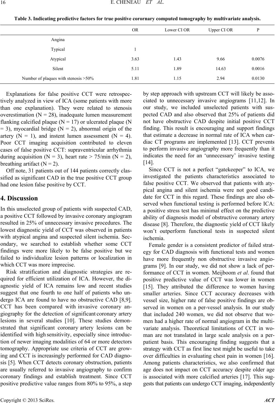 E. CHENEAU ET AL. 16 Table 3. Indicating predictive factors for true positive corornary computed tomography by multivariate analysis. OR Lower CI OR Upper CI OR P Angina Typical 1 Atypical 3.63 1.43 9.66 0.0076 Silent 5.11 1.89 14.63 0.0016 Number of plaques with stenosis >50% 1.81 1.15 2.94 0.0130 Explanations for false positive CCT were retrospec- tively analyzed in view of ICA (some patients with more than one explanation). They were related to stenosis overestimation (N = 28), inadequate lumen measurement flanking calcified plaque (N = 17) or ulcerated plaque (N = 3), myocardial bridge (N = 2), abnormal origin of the artery (N = 1), and instent lumen assessment (N = 4). Poor CCT imaging acquisition contributed to eleven cases of false positive CCT: supraventricular arrhythmia during acquisition (N = 3), heart rate > 75/min (N = 2), breathing artifact (N = 2). Off note, 31 patients out of 144 patients correctly clas- sified as significant CAD in the true positive CCT group had one lesion false positive by CCT. 4. Discussion In this unselected group of patients with suspected CAD, a positive CCT followed by invasive coronary angiogram resulted in 25% of unnecessary invasive procedures. The lowest diagnostic yield of CCT was observed in patients with atypical angina and suspected silent ischemia. Sec- ondary, we searched to establish whether some CCT findings were more likely to be false positive but we failed to individualize lesion patterns or localization in which CCT was more imprecise. Risk stratification and diagnostic strategies are re- quired for efficient utilization of ICA. However, the di- agnostic yield of ICA remains low and recent studies suggest that one fourth to one half of patients who un- dergo ICA are found to have no obstructive CAD [8,9]. CCT has been compared with invasive coronary an- giography for the detection of significant coronary artery lesions in several studies [10]. These studies demon- strated that significant coronary artery lesions can be identified with high sensitivity, especially since introduc- tion of newer imaging modalities of 64 or more detectors tomography. Appropriate use criteria of CCT are grow- ing and CCT is increasingly performed for CAD diagno- sis [5]. When CCT detects coronary obstruction, patients are usually referred to invasive angiography to confirm coronary findings and establish treatment. Since CCT positive predictive value ranges from 80% to 95%, a step by step approach with upstream CCT will likely be asso- ciated to unnecessary invasive angiograms [11,12]. In our study, we included unselected patients with sus- pected CAD and also observed that 25% of patients did not have obstructive CAD despite initial positive CCT finding. This result is encouraging and support findings that estimate a decrease in normal rate of ICA when car- diac CT programs are implemented [13]. CCT prevents to perform invasive angiography more frequently than it indicates the need for an ‘unnecessary’ invasive testing [14]. Since CCT is not a perfect “gatekeeper” to ICA, we investigated the patients characteristics associated to false positive CCT. We observed that patients with aty- pical angina and silent ischemia were not good candi- date for CCT in this regard. These findings are also ob- served when functional testing is performed before ICA: a positive stress test has minimal effect on the predictive ability of diagnosis model of obstructive coronary artery disease [8]. Therefore, the diagnostic yield of CCT likely won’t outperform functional tests in suspected silent ischemia. Female gender is a consistent predictor of failed strat- egy for CAD diagnosis with functional tests and women have more frequently non obstructive invasive angio- grams [9]. In our study, we did not show a lack of per- formance of CCT in women. Meijboom et al. found that positive predictive value of CCT was lower in women [15]. They attributed the difference to women having smaller arteries. Since CCT accuracy decreases with vessel size, higher rate of false positive findings are ob- served in women on a per-vessel analysis. In our study that included 240 women, we did not observe that wo- men had a higher rate of normal angiogram in the multi- variate analysis. Theoretical limitations of CCT in wo- man are not translated in large scale analysis on a per- patient basis. This encouraging finding suggests that a strategy with CCT as first line test might be useful to take over difficulties in evaluating chest pain in women [16]. Among patients characteristics, we also confirmed that age does not impact on CCT accuracy despite older age is associated with more calcified arteries [17]. This sug- gests that patients can undergo CCT imaging, independently Copyright © 2013 SciRes. ACT  E. CHENEAU ET AL. 17 from their age. Once CCT detects coronary obstruction in epicardial coronary vessel, patients are commonly referred for ca- theterization. The goal is to confirm CCT findings and decision-making for potential revascularization. However, a greater focus should be placed on CCT findings and evaluate the likelihood of false positive CCT. That strat- egy might avoid unnecessary catheterization. In studies evaluating 64-slice CT to invasive coronary angiogra- phy, positive predictive value differed between vessels: it was lower in left circumflex and right coronary arteries as compared to left descending and left main arteries as well as in stent and distal segments as compared to native and proximal segments [18]. However, positive predic- tive value of CCT remained constantly high in a narrow range (85% to 93%). As a result, lesion location im- pacted so slightly on CCT accuracy that anatomical fea- tures could not be predictive of false positive CCT. When we compared patients with or without confirmed coronary obstruction by ICA, we did not see difference in lesion characteristics or location. No anatomical sub- group can be identified as “prone to be false positive CCT”. Though, we logically found that risk for false positive decreased when more plaques and more signifi- cant lesions were detected by CCT. Our per-patient ana- lysis emphasizes the impact of the number of diseased vessels. Thus, patients with one vessel disease justify con- sideration before referring to catheterization although patients with more than one stenosed vessel by CCT can be more evidently directed to ICA. Performance of CCT was equivalent in calcified versus non-calcified plaques and lesion location did not impact on CCT accuracy. At a patient level, the CORE—64 trials did not find calcium score as a predictor of diagnostic accuracy of CCT [12]. Even if calcified lesions remain challenging in a per le- sion analysis, this suggests that calcium does not de- crease diagnosis accuracy on a per patient basis since the introduction of 64 detectors scans [18,19]. Overall, we observed that image quality had the great- est impact on false positive CCT. Our data sustain the previously described influence of heart rate and rhythm on image reconstruction [19-21]. In addition, we have faced overestimation in stenosis measurement. This oc- currence of stenosis overestimation is likely the most common mechanism leading to perform non obstructive invasive angiograms, especially in intermediate stenosis and complex lesions [22,23]. The definition of positive CCT we utilized was “50% diameter reduction of the lumen by visual assessment” to determine significant stenosis by CCT. Even if this definition is commonly accepted, it might be insufficient to quantify CAD ob- struction. New tools are developed to better evaluate lu- men stenosis with automated quantification [24]. Thus, predictive value of CCT might increase with computed quantification, especially in high-quality image recon- struction [25]. Overall, we utilized best acquisition and reconstruction methods available at the time of the study inclusion. However, we didn’t anticipate false positive CCT and did not implement our strategy in order to de- crease those occurrences throughout the study. We can- not state that a higher lumen percent stenosis threshold might have impacted on “false positive” CCT rate. This study had limited exclusion criteria. Therefore, it reflects a “real life” study evaluating coronary artery disease in a wide spectrum of patients. No functional testing was requested in the inclusion criteria and the impact of such test on CCT accuracy has not been evalu- ated. Secondly, the negative predictive value of CCT has not been established in this study. Definition for positive CCT might have been too restrictive and increase posi- tive predictive value and lower negative predictive value of CCT. Nevertheless, we observed a negative predictive value of 99% in a previous study with same inclusion criteria, CCT methods and analysis criteria [26]. This suggests that definitions we utilized in the current study were adequately set in order to preserve high negative predictive value of CCT. 5. Conclusion Performing CCT for atypical angina or silent ischemia is associated with higher rate of unnecessary invasive coronary angiograms. We failed to identify lesions char- acteristics prone to be “false positive” of CCT. Improv- ing CCT imaging and lumen stenosis measurement ap- pear the best means to reduce the rate of unnecessary an- giogram following CCT. REFERENCES [1] M. Henneman, J. D. Schuijf, J. M. van Werkhoven, G. Pundziute, E. E. van der Wall, J. W. Jukema and J. J. Bax “Multi-Slice Computed Tomography Coronary Angio- graphy for Ruling Out Suspected Coronary Artery Dis- ease: What Is the Prevalence of a Normal Study in a Ge- neral Clinical Population?” European Heart Journal, Vol. 29, No. 16, 2008, pp. 2006-2013. doi:10.1093/eurheartj/ehn284 [2] L. H. Piers, R. Dikkers, T. P. Willems, B. de Smet, M. Oudkerk, F. Zijlstra and R. A. Tio, “Computed Tomogra- phic Angiography or Conventional Coronary Angiogra- phy in Therapeutic Decision-Making,” European Heart Journal, Vol. 29, No. 23, 2008, pp. 2902-2907. doi:10.1093/eurheartj/ehn454 [3] W. B. Meijboom, M. F. Meijs, J. D. Schuijf, M. J. Cramer, N. R. Mollet, C. A. van Mieghem, K. Nieman, J. M. van Werkhoven, G. Pundziute, A. C. Weustink, A. M. de Vos, F. Pugliese, B. Rensing, a J. W. Jukem, J. J. Bax, M. Pro- kop, P. A. Doevendans, M. G. Hunink, G. P. Krestin and P. J. de Feyter, “Diagnostic Accuracy of 64-Slice Com- puted Tomography Coronary Angiography: A Prospec- Copyright © 2013 SciRes. ACT  E. CHENEAU ET AL. 18 tive, Multicenter, Multivendor Study,” Journal of the American College of Cardiology, Vol. 52, No. 25, 2008, pp. 2135-2144. doi:10.1016/j.jacc.2008.08.058 [4] M. W. von Ballmoos, B. Haring, P. Juillerat and H. Al- kadhi, “Meta-Analysis: Diagnostic Performance of Low- Radiation-Dose Coronary Computed Tomography Angio- graphy,” Annals of International Medicine, Vol. 154, No. 6, 2011, pp. 413-420. [5] A. J. Taylor, M. Cerqueira, J. M. Hodgson, D. Mark, J. Min, P. O’Gara and G. D. Rubin, “Appropriate Use Crite- ria for Cardiac Computed Tomography. A report of the American College of Cardiology Foundation Appropriate Use Criteria Task Force, the Society of Cardiovascular Computed Tomography, the American College of Radi- ology, the American Heart Association, the American Society of Echocardiography, the American Society of Nuclear Cardiology, the North American Society for Car- diovascular Imaging, the Society for Cardiovascular An- giography and Interventions, and the Society for Cardio- vascular Magnetic Resonance,” Circulation, Vol. 122, 2010, pp. e525-e555. doi:10.1161/CIR.0b013e3181fcae66 [6] W. B. Meijboom, C. van Mieghem, N. R. Mollet, F. Pug- liese, A. C. Weustink, N. van Pelt, F. Cademartiri, K. Nie- man, E. Boersma, P. de Jaegere, G. P. Krestin and P. J. de Feyter, “64-Slice Computed Tomography Coronary An- giography in Patients with High, Intermediate, or Low Pretest Probability of Significant Coronary Artery Dis- ease,” Journal of the American College of Cardiology, Vol. 50, No. 15, 2007, pp. 1469-1475. doi:10.1016/j.jacc.2007.07.007 [7] D. B. Pryor, L. Shaw, C. B. McCants, K. L. Lee, D. B. Mark, F. E. Harrell Jr., L. H. Muhlbaier and R. M. Califf, “Value of the History and Physical in Identifying Patients at Increased Risk for Coronary Artery Disease,” Annals of International Medicine, Vol. 118, No. 2, 1993, pp. 81-90. [8] M. R. Patel, E. D. Peterson, D. Dai, J. M. Brennan, R. F. Redberg, H. V. Anderson, R. G. Brindis and P. S. Dou- glas, “Low Diagnostic Yield of Elective Coronary Angio- graphy,”New England Journal of Medicine, Vol. 362, No. 5, 2010, pp. 886-895. doi:10.1056/NEJMoa0907272 [9] C. M. Taylor, K. H. Humphries, A. Pu, W. Ghali, M. Gao, M. Knudtson, U. Hoffmann, R. G. Carere, “A Proposed Clinical Model for Efficient Utilization of Invasive Co- ronary Angiography,”American Journal of Cardiology, Vol. 106, No. 4, 2010, pp. 457-462. doi:10.1016/j.amjcard.2010.03.061 [10] M. Hamon, R. Morello, J. W. Riddell and M. Hamon, “Coronary Arteries: Diagnostic Performance of 16-ver- sus 64-Section Spiral CT Compared with Invasive Coro- nary Angiography-Meta-Analysis,” Radiology, Vol. 245, No. 3, 2007, pp. 720-731. doi:10.1148/radiol.2453061899 [11] M. J. Budoff, D. Dowe, J. G. Jollis, M. Gitter, J. Su- therland, E. Halamert, M. Scherer, R. Bellinger, A. Mar- tin, R. Benton, A. Delago, J. K. Min, “Diagnostic Perfor- mance of 64-Multidetector Row Coronary Computed To- mographic Angiography for Evaluation of Coronary Ar- tery Stenosis in Individuals without Known Coronary Ar- tery Disease: Results from the Prospective Multicenter Accuracy (Assessment by Coronary Computed Tomo- graphic Angiography of Individuals Undergoing Invasive Coronary Angiography) Trial,”Journal of the American College of Cardiology, Vol. 52, No. 21, 2008, pp. 1724- 1732. doi:10.1016/j.jacc.2008.07.031 [12] J. M. Miller, C. E. Rochitte, M. Dewey, A. Arbab-Zadeh, H. Niinuma, I. Gottlieb, N. Paul, M. E. Clouse, E. P. Shapiro, J. Hoe, A. C. Lardo, D. E. Bush, A. de Roos, C. Cox, J. Brinker and J. A. Lima, “Diagnostic Performance of Coronary Angiography by 64-Row CT,” New England Journal of Medicine, Vol. 359, No. 22, 2008, pp. 2324- 2336. doi:10.1056/NEJMoa0806576 [13] B. J. Chow, A. Abraham, G. A. Wells, L. Chen, T. D. Ru- ddy, Y. Yam, N. Govas, P. D. Galbraith, C. Dennie and R. S. Beanlands, “Diagnostic Accuracy and Impact of Com- puted Tomographic Coronary Angiography on Utilization of Invasive Coronary Angiography,” Circ Cardiovasc Imaging, Vol. 2, No. 1, 2009, pp. 16-23. doi:10.1161/CIRCIMAGING.108.792572 [14] J. Hausleiter, T. Meyer, M. Hadamitzky, M. Zankl, P. Ge- rein, K. Dörrler, A. Kastrati, S. Martinoff and A. Schö- mig, “Non-Invasive Coronary Computed Tomographic Angiography for Patients with Suspected Coronary Ar- tery Disease: The Coronary Angiography by Computed Tomography with the Use of a Submillimeter Resolution (Cactus) Trial,” European Heart Journal, Vol. 28, No. 24, 2007, pp. 3034-3041 [15] W. B. Meijboom, A. C. Weustink, F. Pugliese, C. A. van Mieghem, N. R. Mollet, N. van Pelt, F. Cademartiri, K. Nieman, E. Vourvouri, E. Regar, G. P. Krestin, P. J. de Feyter, “Comparison of Diagnostic Accuracy of 64-Slice Computed Tomography Coronary Angiography in Wo- men versus Men with Angina Pectoris,” American Jour- nal of Cardiology, Vol. 100, No. 10, 2007, pp. 1532-1537. doi:10.1016/j.amjcard.2007.06.061 [16] J. H. Mieres, L. J. Shaw, A. Arai, M. J. Budoff, S. D. Flamm, W. G. Hundley, T. H. Marwick, L. Mosca, A. R. Patel, M. A. Quinones, R. F. Redberg, K. A. Taubert, A. J. Taylor, G. S. Thomas and N. K. Wenger, “Role of Non- invasive Testing in the Clinical Evaluation of Women with Suspected Coronary Artery Disease: Consensus State- ment from the Cardiac Imaging Committee, Council on Clinical Cardiology, and the Cardiovascular Imaging and Intervention Committee, Council on Cardiovascular Ra- diology and Intervention, American Heart Association,” Circulation, Vol. 111, No. 5, 2005, pp. 682-696. doi:10.1161/01.CIR.0000155233.67287.60 [17] M. Dewey, A. L. Vavere, A. Arbab-Zadeh, J. M. Miller, L. Sara, C. Cox, I. Gottlieb, K. Yoshioka, N. Paul, J. Hoe, A. de Roos, A. C. Lardo, J. A. Lima, M. E. Clouse, “Pa- tient Characteristics as Predictors of Image Quality and Diagnostic Accuracy of MDCT Compared with Conven- tional Coronary Angiography for Detecting Coronary Ar- tery Stenoses: CORE-64 Multicenter International Trial,” American Journal of Roentgenology, Vol. 194, No. 1, 2010, pp. 93-102. doi:10.2214/AJR.09.2833 [18] P. D. Stein, A. Y. Yaekoub, F. Matta and H. D. Sostman, “64-Slice CT for Diagnosis of Coronary Artery Disease: A Systematic Review,” American Journal of Medicine, Vol. 121, No. 8, 2008, pp. 715-725. doi:10.1016/j.amjmed.2008.02.039 Copyright © 2013 SciRes. ACT  E. CHENEAU ET AL. Copyright © 2013 SciRes. ACT 19 [19] G. L. Raff, M. J. Gallagher, W. W. O’Neill, J. A. Gold- stein, “Diagnostic Accuracy of Noninvasive Coronary Angiography Using 64-Slice Spiral Computed Tomogra- phy,” Journal of the American College of Cardiology, Vol. 46, No. 3, 2005, pp. 552-557. doi:10.1016/j.jacc.2005.05.056 [20] H. Brodoefel, C. Burgstahler, I. Tsiflikas, A. Reimann, S. Schroeder, C. D. Claussen, M. Heuschmid and A. F. Kopp, “Dual-Source CT: Effect of Heart Rate, Heart Rate Variability, and Calcification on Image Quality and Diag- nostic Accuracy,” Radiology, Vol. 247, No. 2, 2008, pp. 346-355. doi:10.1148/radiol.2472070906 [21] S. Leschka, S. Wildermuth, T. Boehm, L. Desbiolles, L. Husmann, A. Plass, P. Koepfli, T. Schepis, B. Marincek, P. A. Kaufmann and H. Alkadhi, “Noninvasive Coronary Angiography with 64-Section CT: Effect of Average Heart Rate and Heart Rate Variability on Image Quality,” Radiology, Vol. 241, No. 2, 2006, pp. 378-385. doi:10.1148/radiol.2412051384 [22] H. Gouya, O. Varenne, L. Trinquart, E. Touzé, O. Vig- naux, C. Spaulding, J. L. Mas and J. L. Sablayrolles Co- ronary Artery Stenosis in High-Risk Patients: 64-Section CT and Coronary Angiography—Prospective Study and Analysis of Discordance,” Radiology, Vol. 252, No. 2, 2009, pp. 377-385. doi:10.1148/radiol.2522081271 [23] B. M. Wertman, V. Y. Cheng, S. Kar, H. Gransar, R. A. Berg, H. Naik, R. Makkar, J. Friedman, J. Schapira and D. Berman, “Characterization of Complex Coronary Artery Stenosis Morphology by Coronary Computed Tomogra- phic Angiography,” JACC Cardio vasc Imaging, Vol. 2, No. 8, 2009, pp. 950-958. doi:10.1016/j.jcmg.2008.12.032 [24] S. Busch, T. R. Johnson, K. Nikolaou, F. von Ziegler, A. Knez, M. F. Reiser and C. R. Becker, “Visual and Auto- matic Grading of Coronary Artery Stenoses with 64-Slice CT Angiography in Reference to Invasive Angiography,” European Radiology, Vol. 17, No. 6, 2007, pp. 1445- 1451. doi:10.1007/s00330-006-0512-y [25] M. J. Boogers, J. D. Schuijf, P. H. Kitslaar, J. M. van Werkhoven, F. R. de Graaf, E. Boersma, J. E. van Velzen, J. Dijkstra, I. M. Adame, L. J. Kroft, A. de Roos, J. H. Schreur, M. W. Heijenbrok, J. W. Jukema, J. H. Reiber and J. J. Bax, “Automated Quantification of Stenosis Severity on 64-Slice CT: A Comparison with Quantitative Coronary Angiography,” JACC Cardiovasc Imaging, Vol. 3, No. 7, 2010, pp. 699-709. doi:10.1016/j.jcmg.2010.01.010 [26] E. Cheneau, B. Vahdat, L. Bernard, A. Molon, D. Pana- gides, “Routine Use of Coronary Computed Tomography as Initial Diagnostic Test for Angina Pectoris,” Archives of Cardiovascular Diseases, Vol. 104, No. 1, 2011, pp. 29-34. doi:10.1016/j.acvd.2010.11.007 |