

Paper Menu >>
Journal Menu >>
 Advances in Computed Tomography, 2013, 2, 4-12 http://dx.doi.org/10.4236/act.2013.21002 Published Online March 2013 (http://www.scirp.org/journal/act) Can CT Perfusion Guide Patient Selection for Treatment of Delayed Cerebral Ischemia? Pina C. Sanelli1,2*, Rachel Gold1, Nikesh Anumula1, Austin Ferrone1, Carl E. Johnson1, Joseph P. Comunale1, Apostolos J. Tsiouris1, Howard Riina3, Halinder Mangat4, Axel Rosengart4, Alan Z. Segal4 1Department of Radiology, Weill Cornell Medical Colleg e, New York, USA 2Department of Public Health, Weill Cornell Medical College, New York, USA 3Department of Neurological Surgery, Weill Cornell Medical College, New York, USA 4Department of Neurology, Weill Cornel l Medical College, New York, USA Email: *[email protected] Received November 8, 2012; revised December 20, 2012; accepted January 4, 2013 ABSTRACT Purpose: To evaluate qualitative and quantitative CT perfusion (CTP) for different treatment options of delayed cere- bral ischemia (DCI) in aneurysmal SAH. Methods: Retrospective study of consecutive SAH patients enrolled in a pro- spective IRB-approved clinical trial. Qu alitative analysis of CTP deficits were determined by two b linded neuroradiolo- gists. Quantitative CTP was performed using standardized protocol with region-of-interest placement sampling the cor- tex. DCI was assessed by clinical and imaging criteria. Patients were classified into treatment groups: 1) hyperten- sion-hemodilution-hypervolemia (HHH); 2) intra-arterial (IA) vasodilators and/or angioplasty; 3) no treatment. Mean quantitative CTP values were compared using ANOVA pairwise comparisons. Receiver operating characteristic (ROC) curves, standard error (SE) and optimal threshold values were calculated. Results: Ninety-six patients were classified into three treatment groups; 21% (19/96) HHH, 34% (33/96) IA-therapy and 46% (44/96) no treatment. DCI was diag- nosed in 42% (40/96); of which 18% (7/40) received HHH, 80% (32/40) IA-therapy, and 2% (1/40) no treatment. CTP deficits were seen in 50% (48/96); occurring in 63% (12/19) HHH, 94% (31/33) IA-therapy, and 11% (5/44) no treat- ment. Presence of CTP deficits had 83% sensitivity, 8 9% specificity, 90% positive pred ictive and 81% negativ e predic- tive values for treat ment. Mean quantitative CTP valu es revealed significant differences in CBF (p < 0.0001) and MTT (p = 0.0001) amongst the treatment groups. ROC analysis revealed CBF with the highest accuracy of 0.82 (SE 0.04) for comparing treatment groups. Threshold an alysis calculated CBF of 30 mL/100 gm/min (89% specificity, 7 1% sensitiv- ity) for determining treatment. Conclusion: These in itial findings of significant differences in CTP deficits for different treatment groups suggest that CTP may have a potential role in guiding patient selection for treatment of DCI. Keywords: CT Perfusion; Aneurysmal Subarachnoid Hemorrhage; Delayed Cerebral Ischemia; Treatment 1. Introduction Delayed cerebral ischemia (DCI) is a serious complica- tion following aneurysmal SAH, and is the leading cause of morbidity and mortality in this patient population. Its pathophysiology is complex and poorly understood lead- ing to delayed diagnosis. However, proximal vasospasm is thought to partly contribute to DCI causing reduced cerebral blood flow (CBF) to the distal vascular territory [1-3]. DCI manifests as clinical deterioration typically developing between 4 - 14 days following the initial an- eurysm rupture with symptoms of focal neurological im- pairment (such as hemiparesis, hemiplegia, aphasia, etc.), or a decrease of at least 2 points on the Glasgow Coma Scale. Early diagnosis and prompt treatment of DCI may prevent its devastating sequelae of permanent neurologic deficits, cerebral infarction and death. There are several treatment options for DCI typically used in clinical practice. Medical management with hy- pervolemic, hemodilution and hypertensive (HHH) ther- apy is commonly used as the initial treatment. HHH is a systemic therapy used to improve global cerebral perfu- sion by increasing both blood pressure and flow to the brain. For patients that do not adequately respond to HHH therapy, more selective invasive intra-arterial (IA) therapy is performed to immediately dilate an artery in spasm using vasodilatory medications and/or angioplasty in order to improve the blood flow to a specific region of the brain. However, in some patients continuous close observation in the neuro-intensive care unit (N-ICU) set- ting is sufficient depending on the patient’s clinical con- *Corresponding a uthor. C opyright © 2013 SciRes. ACT  P. C. SANELLI ET AL. 5 dition and considering the risks associated with both HHH and IA-therapy. The main complications that may occur with HHH are cerebral edema, aneurysm re-bleed- ing, pulmonary edema, and potential cardiac injury re- lated to the aggressive use of pressor type medications [4]. Several important risks associated with IA-therapy include vascular injury (such as dissection and vessel rupture), cerebral infarction and death related to the arte- rial catheterization techniques and balloon angioplasty. Therefore, more accurate assessment of patients who require treatment of DCI is critical to improve patient selection in order to provide maximal treatment benefit and to minimize exposing patients to unnecessary serious risks. Several methods are used for the diagnosis of DCI re- lated to vasospasm, including clinical examination, neu- rologic monitoring devices, transcranial Doppler ultra- sound (TCD), CTA, and DSA. Many institutions have also employed CT perfusion (CTP) to evaluate the hemodynamic status of the brain at the capillary level. CTP is a dynamic imaging study using cine scanning to capture the first pass of contrast through the brain to as- sess cerebral perfusion. The acquired dataset is post-pro- cessed using a deconvolution algorithm to construct the perfusion maps of CBF, cerebral blood volume (CBV) and mean transit time (MTT). The principle of CTP is that these perfusion parameters can be derived from the mathematical algorithm that describes the relationship between the arterial input, capillary flow and venous drainage. The arterial and venous time attenuation curves are obtained from the acquired dataset, representing the time series data of contrast through the brain. Two as- sumptions are made with CTP including that blood and contrast have the same hemodynamic properties and there is a linear relationship between contrast concentra- tion in the brain with the measured Hounsfield units. There are several advantages of CTP compared with other perfusion imaging techniques, including its non- invasiveness, short acquisition time, widespread avail- ability, and limited patient contraindications lending to its usefulness in this critically ill patient population. In the literature, several studies have reported the ability of CTP to detect impairment of cerebral perfusion thought to occur in the development of DCI and vasospasm [1-3,5]. However, not all perfusion deficits correlate with symptoms of DCI or arterial narrowing on imaging stud- ies, indicating that not all perfusion deficits require treatment. In particular, the issue of which perfusion deficits require treatment with HHH and/or IA-therapy has not been fully explored. Thereby, further under- standing of these CTP deficits is needed in order to im- prove patient selection for treatment of DCI. Quan titative differences in CTP deficits may assist in identifying pa- tients earlier who require treatment, particularly in pa- tients with limited clinical assessments, uncertain clinical findings, and discrepant clinical and imaging data, that may lead to delayed diagnosis and treatment. The purpose of this study is to evaluate qualitative and quantitative CTP for different treatment options of DCI in aneurysmal SAH. Our hypothesis is that there are sta- tistically significant differences in the CTP parameters in patients who are selected for different treatment regi- mens. 2. Materials and Methods 2.1. Study Design A retrospective study of 104 consecutive aneurysmal SAH patients enrolled in a prospective clinical trial fro m December 2004 to December 2008 was performed. This study was approved by the institutional review board (IRB) and a health insurance portability and accountabil- ity act (HIPAA) waiver was obtained. Inclusion criteria were adult patients (18 years and older) with documented aneurysmal SAH at admission based on the initial non- contrast head CT, cerebrospinal fluid analysis, CTA and/ or DSA. Exclusion criteria were CTP exams performed after treatment of DCI or after cerebral infarction oc- curred. All subjects in the study underwent surg ical clip- ping and/or endovascular coiling for aneurysm repair and were monitored in the N-ICU, as per usual standard-of- care. Chart review was performed for the clinical and de- mographic characteristics of the study population, in- cluding age, gender, aneurysm location and treatment, and Hunt Hess grades on presentation. Treatment man- agement decisions were based on all clinical and imaging data available, with the exception of CTP. Patients were categorized into three treatment groups as 1) medical HHH only; 2) IA-therapy; and 3) no treatment (close observation). Patients classified in the IA-therapy group did not adequately respond to HHH as the initial treat- ment. The diagnosis of DCI was determined for each treat- ment group to assess appropriate classification of patients. DCI was determined in this cohort by incorporating the most relevant clinical and imaging outcome measures, as recommended by the recent consensus expert opinion [6]. Furthermore, Reichman et al. developed and evaluated a reference standard incorporating these outcomes meas- ures to assess aneurysmal SAH patients applicable to clinical practice [7]. This reference standard consists of angiographic criteria for vasospasm based on the arterial luminal narrowing on DSA compared with the normal parent vessel and comparison with DSA performed on initial presentation. In this study, DSA interpretations were performed by two observers, one of two intervene- tional neuroradiologists at our institution who performed the exam (with either 10 or 25 years experience) and a neuroradiologist blinded to all patient information (22 Copyright © 2013 SciRes. ACT  P. C. SANELLI ET AL. 6 years’ experience). For disagreements, a third neurora- diologist (10 years’ experience) independently reviewed the exam in a blinded fashion. In patients who did not have DSA performed, the following clinical and/or im- aging criteria were used: a) clinical criteria—a permanent neurologic deficit on clinical examination, distinct from the deficit at baseline produced by the SAH event or sur- gical intervention, which was not attributable to other causes; b) imaging criteria-cerebral infarction on fol- low-up CT and/or MR imaging that occurred after day 4. This criterion has been used to effectively exclude pri- mary brain damage from SAH and/or surgical intervene- tion [8]. Clinical outcome measures were also determined for each treatment group to further assess appropriate classi- fication of patients. At discharge from the N-ICU, pa- tients were evaluated for a permanent neurologic deficit on physical examination and cerebral infarction on fol- low-up CT and/or MR imaging. Discharge status was also documented as home, rehabilitation care facility (acute rehabilitation or long-term skilled nursing), and death. 2.2. CTP Scanning Protocol, Post-Processing and Data Collection CTP was performed during the typical time-period for DCI, between days 6 - 8 in asymptomatic patients and on the same day clinical deterioration occurred in sympto- matic patients. There is a standard scanning protocol for CTP at our institution using GE Lightspeed or Pro—16 scanners (General Electric Medical Systems, Milwaukee, WI) with cine 4i scanning mode and 45 second acquisi- tion at 1 rotation per second using 80 kVp and 190 mA. A scanning volume of 2.0 cm was used consisting of 4 slices at 5.0 mm thickness with its inferior extent se- lected at the level of the basal ganglia, above the orbits, to minimize radiation exposure to the lenses. Approxi- mately 45 mL of nonionic iodinated contrast was admin- istered intravenously at 5 mL/s using a power injector with a 5-second delay. Post-processing of the acquired images into cerebral blood flow (CBF), cerebral blood volume (CBV) and mean transit time (MTT) maps were performed on a GE Advantage Workstation using CTP software version 3.0 (General Electric Medical Systems). This software em- ploys a deconvolution method, which is considered most accurate for low contrast injection rates [9]. The post- processing technique was standardized for all patients according to recommended guidelines [10] with the arte- rial input function as the A2 segment of the anterior cerebral artery (ACA) [11] and venous function as the superior sagittal sinus. The perfusion maps were qualitatively evaluated by two neuroradiol ogi st s (10 a nd 7 years e xperience) blin ded to clinical and imaging data to determine the presence of perfusion deficits, defined as areas of reduced CBF and/ or prolonged MTT. Focal perfusion abnormalities due to the primary hemorrhagic event and su rgical intervention, as identified on the acquired images from the CTP data- set, were not included as perfusion deficits related to DCI. After reviewing the images independently, consensus ju- dgment was determined. Quantitative analysis was conducted using a standard- ized method with contiguous regions-of-interest (ROI) placement, measuring 157 mm2, sampling the cerebral cortex. Each CTP slice had up to 24 ROIs distributed in the following territories: approximately 6 ROIs in ACA, 12 ROIs in middle cerebral artery (MCA) and 6 ROIs in posterior cerebral artery (PCA). CTP studies were ana- lyzed blinded to all clinical a nd imaging data to li mit test review bi a s. 2.3. Statistical Analysis Only CTP exams performed at the diagnostic stage, prior to treatment for DCI and prior to cerebral infarction, were included in the analysis in order to minimize con- founding bias as both treatment and infarction affect the cerebral perfusion maps. The incidence of qualitative CTP deficits in each treatment group was calculated. CTP test characteristics (sensitivity, specificity, positive and negative predictive values) were determined using 2 × 2 table analysis with 95% confidence intervals (CI). Quantitative CTP data was analyzed by calculating the mean CBF, CBV and MTT and its standard deviation (SD) for the three treatment groups. For patients with focal perfusion deficits, ROIs within the affected region were isolated and arithmetic means were calculated. In patients without focal deficits, all ROIs for all 4 slice locations were included in the arith metic means. In order to minimize the contribution of vascular pixels from large vessels, CBF values >100 mL/100 gm/min were excluded from the statistical analysis and not used in calculating the me an CBF, CBV and MTT , as this method has been published in the evaluation of ischemia [12]. The ROIs in the perfusion abnormalities due to the pri- mary hemorrhagic event and/or surgical intervention, were not included in the statistical analysis. Normality of the distribution of continuous variables was assessed by plotting histograms and the Shapiro Wilks test. Com- parison between the treatment groups was performed using ANOVA pairwise comparisons. Statistical signifi- cance was accepted at p < 0.05. Receiver operator char- acteristic (ROC) curves were generated for each CTP pa- rameter and the area-under-the-curve (AUC) calculated to quantify the overall accuracy. Maximum-likelihood ROC models and standard error (SE) were fitted assum- ing a binormal distribution for the underlying latent variable. The threshold values for CBF, MTT and CBV Copyright © 2013 SciRes. ACT 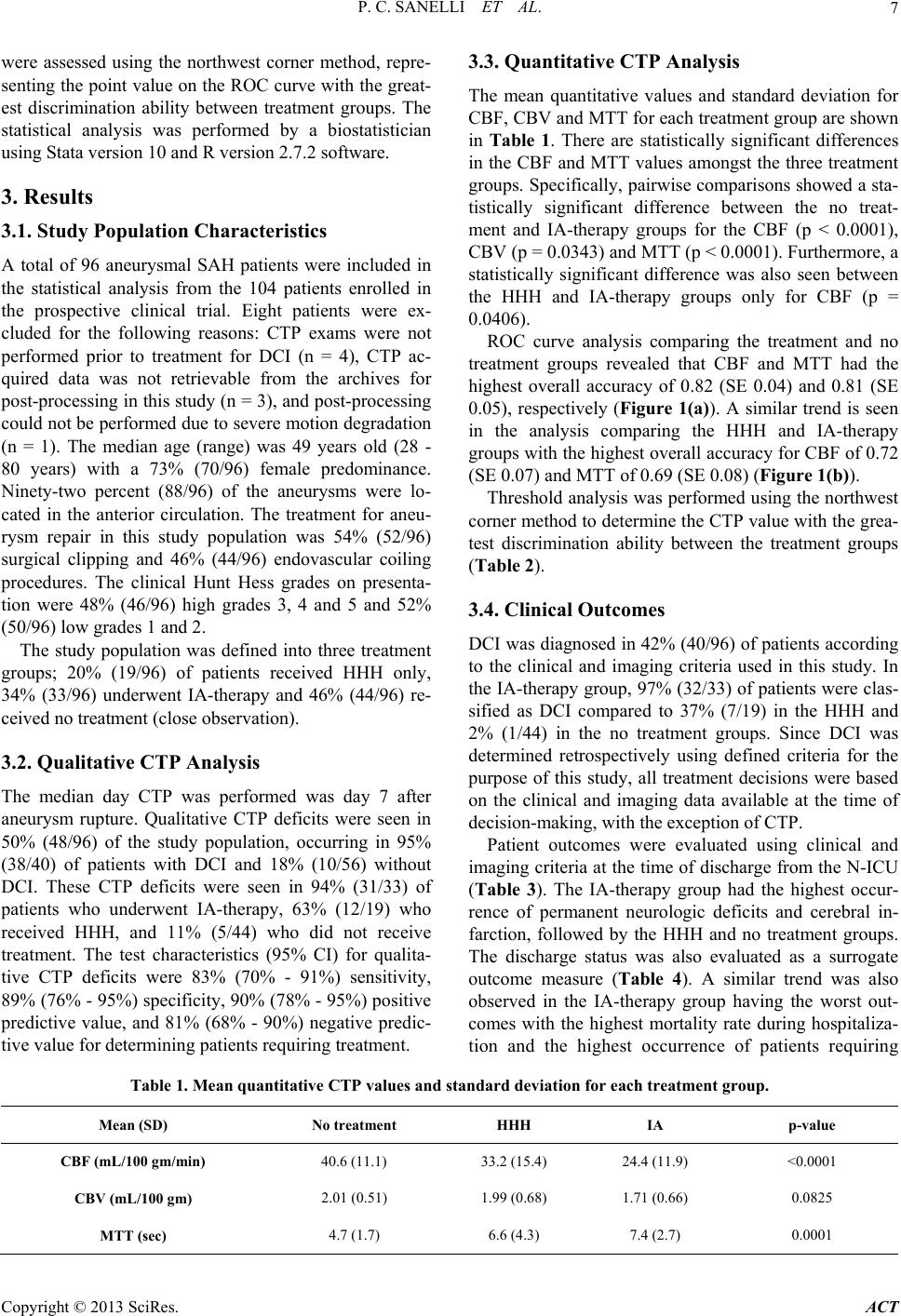 P. C. SANELLI ET AL. Copyright © 2013 SciRes. ACT 7 were assessed using the northwest corner method, repre- senting the point va lue on the ROC curve with the great- est discrimination ability between treatment groups. The statistical analysis was performed by a biostatistician using Stata version 10 and R version 2.7.2 software. 3. Results 3.1. Study Population Characteristics A total of 96 aneurysmal SAH patients were included in the statistical analysis from the 104 patients enrolled in the prospective clinical trial. Eight patients were ex- cluded for the following reasons: CTP exams were not performed prior to treatment for DCI (n = 4), CTP ac- quired data was not retrievable from the archives for post-processing in this study (n = 3), and post-processing could not be performed due to severe motion degradation (n = 1). The median age (range) was 49 years old (28 - 80 years) with a 73% (70/96) female predominance. Ninety-two percent (88/96) of the aneurysms were lo- cated in the anterior circulation. The treatment for aneu- rysm repair in this study population was 54% (52/96) surgical clipping and 46% (44/96) endovascular coiling procedures. The clinical Hunt Hess grades on presenta- tion were 48% (46/96) high grades 3, 4 and 5 and 52% (50/96) l o w g r a des 1 and 2. The study population was defined into three treatment groups; 20% (19/96) of patients received HHH only, 34% (33/96) underwent IA-therapy and 46% (44/96) re- ceived no treatment (close observation). 3.2. Qualitative CTP Analysis The median day CTP was performed was day 7 after aneurysm rupture. Qualitative CTP deficits were seen in 50% (48/96) of the study population, occurring in 95% (38/40) of patients with DCI and 18% (10/56) without DCI. These CTP deficits were seen in 94% (31/33) of patients who underwent IA-therapy, 63% (12/19) who received HHH, and 11% (5/44) who did not receive treatment. The test characteristics (95% CI) for qualita- tive CTP deficits were 83% (70% - 91%) sensitivity, 89% (76% - 95%) specificity, 90% (7 8% - 95%) positive predictive value, and 81% (68% - 90%) negative predic- tive value for determining patients requiring treatment. 3.3. Quantitative CTP Analysis The mean quantitative values and standard deviation for CBF, CBV and MTT for each treatment group are shown in Table 1. There are statistically significant differences in the CBF and MTT values amongst the three treatment groups. Specifically, pairwise comparisons showed a sta- tistically significant difference between the no treat- ment and IA-therapy groups for the CBF (p < 0.0001), CBV (p = 0.0343) an d MTT (p < 0.000 1) . Fur ther more, a statistically significant difference was also seen between the HHH and IA-therapy groups only for CBF (p = 0.0406). ROC curve analysis comparing the treatment and no treatment groups revealed that CBF and MTT had the highest overall accuracy of 0.82 (SE 0.04) and 0.81 (SE 0.05), respectively (Figure 1(a)). A similar trend is seen in the analysis comparing the HHH and IA-therapy groups with the highest overall accuracy for CBF of 0.72 (SE 0.07) an d MTT of 0.69 ( SE 0 .08) (Figure 1(b)). Threshold analysis was performed using the northwest corner method to determine the CTP value with the grea- test discrimination ability between the treatment groups (Table 2). 3.4. Clinical Outcomes DCI was diagnosed in 42% (40/96) of patients according to the clinical and imaging criteria used in this study. In the IA-therapy group, 97% (32/33) of patients were clas- sified as DCI compared to 37% (7/19) in the HHH and 2% (1/44) in the no treatment groups. Since DCI was determined retrospectively using defined criteria for the purpose of this study, all treatment decisions were based on the clinical and imaging data available at the time of decision-making, with the exception of CTP. Patient outcomes were evaluated using clinical and imaging criteria at the time of discharge from the N-ICU (Table 3). The IA-therapy group had the highest occur- rence of permanent neurologic deficits and cerebral in- farction, followed by the HHH and no treatment groups. The discharge status was also evaluated as a surrogate outcome measure (Table 4). A similar trend was also observed in the IA-therapy group having the worst out- comes with the highest mortality rate during hospitaliza- tion and the highest occurrence of patients requiring Table 1. Mean quantitative CTP values and standard deviation for each treatment group. Mean (SD) No treatment HHH IA p-value CBF (mL/100 gm/min) 40.6 (11.1) 33.2 (15.4) 24.4 (11.9) <0.0001 CBV (mL/100 gm) 2.01 (0.51) 1.99 (0.68) 1.71 (0.66) 0.0825 MTT (sec) 4.7 (1.7) 6.6 (4.3) 7.4 (2.7) 0.0001  P. C. SANELLI ET AL. 8 (a) (b) Figure 1. Receiver operator characteristic (ROC) curves of CBF, CBV and MTT for comparison of treatment groups. Table 2. Threshold analysi s for treatment options; (a) Treat- ment vs. no treatment; (b) HHH vs. IA-therapy. (a) Threshold Specificity/Sensitivity CBF 30 %71%/89 MTT 5.1 %71%/75 CBV 1.7 %52%/75 (b) Threshold Specificity/Sensitivity CBF 26 %79%/74 MTT 6.4 %61%/74 CBV 1.5 %52%/84 Table 3. Clinical outcomes determined by clinical and im- aging criteria. Total (n = 96) No treatment (n = 44) HHH (n = 19) IA (n = 33) Functional deficit/disability 28% (27/96) 11% (5/44) 26% (5/19) 52% (17/33) Cerebral infarction 14% (13/96) 0% 11% (2/19) 33% (11/33) rehabilitative care following discharg e. 4. Discussion DCI related to vasospasm is a complex entity involving Table 4. Discharge status for treatment gr oups. Discharge status Total (n = 96) No treatment (n = 44) HHH (n = 19) IA (n = 33) Home 52% (50/96)68% (30/44) 53% (10/19)30% (10/33) Rehabilitative care 43 % (41/96)32% (14/44) 42% (8/19)58% (19/33) Acute rehabilitation 33% (32/96)23% (10/44) 26% (5/19)52% (17/33) Long-term skilled 10% (9/96)9% (4/44) 16% (3/19)6% (2/33) Death 5% (5/96)0% 5% (1/19) 12% (4/33) delayed narrowing of the intracranial arteries that may lead to clinical deterioration, cerebral infarction and death. Current treatment options include medical man- agement with HHH as the initial treatment to increase systemic blood pressure and flow in order to globally increase cerebral perfusion. Typically, if patients do not adequately respond to HHH then more invasive tech- niques, such as intra-arterial therapy with vasodilatory medications and/or angioplasty, are used to improve re- gional blood flow by selectively dilating individual cere- bral arteries. Finally, close observation in the N-ICU may also be sufficient in some patients given the life-threaten- ing complications associated with both HHH and IA- therapy. Therefore, more accurate patient selection for treatment of DCI is critical in order to provide maximal treatment benefit while minimizing exposure of patients to serious risks. Copyright © 2013 SciRes. ACT  P. C. SANELLI ET AL. 9 In current practice, patient selection for treatment of D limited due to few studies pe if there are qualitative CI is based on clinical signs and symptoms coupled with imaging information of arterial narrowing for vaso- spasm. In recent years, CTP has also been used as a complimentary diagnostic tool for evaluation of DCI related to vasospasm. Specifically, qualitative CTP defi- cits have a high sensitivity and specificity [1-3,5,13-15] for vasospasm reported in 97% of patients with vaso- spasm compared to 24% without vasospasm [5]. Fur- thermore, Aralasmak et al. correlated CTP deficits and the degree of vessel narrowing with CTP deficits occur- ring in 83% of patients with severe vasospasm compared to 26% with mild-moderate and 15% without vasospasm [2]. Dankbaar et al. also demonstrated that the flow terri- tory of the vessel with the most severe vasospasm corre- sponded with the least perfused region on CTP [16]. More recently, investigations have focused on using quan- titative CTP and determining a threshold value. Several studies have reported CBF and MTT with the highest diagnostic accuracy for DCI [3,5,16]. A CBF threshold of 35 mL/100 gm/min (90% sensitivity, 68% specificity) and MTT threshold of 5.5 sec (73% sensitivity, 79% specificity) have been reported [5]. Similarly, Dankbaar et al. described an MTT threshold of 5.9 sec for DCI [16]. Wintermark et al. also described the utility of CTP in the diagnosis and management of vasospasm suggesting that a CBF threshold of 39 .3 mL/100 gm/min repr esented the most accurate (94.8%) indicator for endovascular therapy [3]. These results support the possibility that CTP-based criteria may be able to assist in differentiating patients according to the severity of DCI in attempts to guide pa- tient selection for treatment. A review of the literature is rformed in evaluating imaging based criteria for pa- tient selection for treatment of DCI. In recent years, it is becoming more apparent that image-based criteria may have a role in patient selection for treatment of acute stroke. Turk et al. reported similar rates of good func- tional outcome and low rates of intracranial hemorrhage when endovascular treatment was performed based on CTP rather than time-guided selection [17,18]. This po- tential for image-based criteria may also have a role in aneurysmal SAH, particularly in patients with limited clinical examination, uncertain clinical findings, or dis- crepant clinical and imaging data. In the prospective ma- nagement of patients, which perfusion deficits require treatment with HHH and/or IA-therapy still remains un- certain because not all perfusion deficits correspond to symptoms of DCI or arterial narrowing on imaging stud- ies indicating that not all perfusion deficits require treat- ment (Figure 2). Thereby, further quantitative analysis of CTP deficits is needed to differentiate treatment groups in order to guide patient selection. Our study focused on determining (a) (b) (c) Figure 2. 57-year-old status coil embolization of a right infarction on follow-up CT imaging. post posterior communicating artery aneurysm. CT perfusion was performed on day 7 for the purposes of the study pro- tocol and shows a focal perfusion deficit with elevated MTT (a) and reduced CBF (b) in the right MCAterritory in the parietal region. Quantitative evaluation of the perfusion deficit reveals a CBF of 37 mL/100 gm/min. The patient was asymptomatic and noncontrast CT (c) reveals no corre- sponding abnormality in this region. The patient did not receive treatment and was discharged home with resump- tion of normal daily activities. There was no evidence of a permanent neurologic deficit on clinical exam or cerebral Copyright © 2013 SciRes. ACT  P. C. SANELLI ET AL. 10 and/or quantitative differences in CTP deficits for dif- ferent treatment groups of DCI. The presence of a CTP deficit has a 90% positive predictive value for treatment. CTP deficits occurred in 94% (31/33) of the IA-therapy and 63% (12/19) of HHH groups compared to 11% (5/44) of the no treatment group. Further quantitative analysis revealed that CBF was the only perfusion parameter that showed a statistically significant difference in the pair- wise comparisons of all three treatment groups. In addi- tion, ROC curve analyses supported that CBF has the highest accuracy for determining the treatment group (Figure 1). Using this data, threshold analysis can be performed to help guide patien t selection by determining the CBF value on the ROC curve with the greatest dis- crimination ability between treatment groups. In this study, a CBF threshold of 30 mL/100 gm/min can be used as a guideline for a patient that should be carefully considered for treatment of DCI. Furthermore, a CBF threshold of 26 mL/100 gm/min can be used as a rough guide for possibly considering patients who may also require more invasive techniques with IA-therapy. Given the proximity of these two threshold values, caution is emphasized when using absolu te quantitative CTP values for the diagnosis and management of patients in clinical practice. The use of quantitative threshold values is lim- ited in clinical practice due to differences in scanner equipment, techniques and post-processing methods em- ployed. Importantly, standardization and validation of CTP methodology and post-processing techniques are necessary for its widespread implementation in SAH pa- tients. In this study, CTP deficits in the IA-therapy group had the most severely reduced CBF and prolonged MTT (Figure 3). The IA-therapy group had 97% of patients diagnosed with DCI, whereas the no treatment group had only 2% of patients with DCI. As expected, the IA- the- rapy group also had the worst outcomes with the highest mortality rate (12%) and greatest percentage of patients with functional disability (52%) and cerebral infarction (33%). Whereas patients who had no treatment with close observation, the mean CBF and MTT values were within normal ranges. Improved outcomes were seen in the no treatment group with the highest rate of patients discharged home (68%), lowest incidence of functional disability (11%) and no cerebral infarction on follow-up CT/MR imaging. These findings support that there was appropriate classification of aneurysmal SAH patients in the different treatment groups. We acknowledge several limitations in this study; in- cluding the retrospective study design limiting the out- comes data collection, specifically for clinical informa- tion. In addition, CTP provides limited brain coverage for evaluation of perfusion deficits. However, emerging CT scanner technology will continue to improve and (a) (b) (c) Figure 3. 35 year-old status post surgical clipping of a right posterior communicating a aneurysm and developed rtery new left-sided upper extremity hemiparesis and pronator drift on day 6. Noncontrast CT (a) demonstrates no new hemorrhage or hydrocephalus to explain the patient’s sym- ptoms. However, there is early hypodensity involving the right basal ganglia. CT perfusion performed the same day shows a large perfusion deficit in the right MCA territory in the frontal and parietal regions and focal perfusion defi- cit in the left ACA territory with elevated MTT (b) and reduced CBF (c). Quantitative evaluation reveals a CBF of 18 mL/100 gm/ min in the right MCA and CBF of 17 mL/ 100 gm/min in the left ACA perfusion deficits. The patient received HHH therapy and also underw ent IA-therapy wi th verapamil and angioplasty of the right MCA and left ACA. Patient developed cerebral infarction in the right anterior temporal lobe and basal ganglia. Copyright © 2013 SciRes. ACT  P. C. SANELLI ET AL. 11 provide broader coverage for which this data may be applicable. Another limitation is that the region of CTP imaging was not coordinated with the location of the arterial narrowing on DSA or region referable to symp- toms. Therefore, it is conceivable that this region may not have been imaged on the CTP or possibly averaged with a larger region of normal perfusion, resulting in in- creased false negatives and lower accuracy in our study. This method was not used in order to reduce work-up and observer bias by not having knowledge of the clini- cal exam and DSA results prior to CTP scanning and in- terpretation. 5. Conclusion There is a clinical need to more accurately select aneu- nts for treatment of DCI in order to This publication was made possible by Grant Num ational Institute of [1] J. W. Dankbaar, N. K. de Rooij, B. K. Velthius, C. J. Frijns, G. J. Rchaaf, “Diagnosing Delayed Cerebrent CT Modalit rysmal SAH patie provide maximal treatment benefit while minimizing patient exposure to serious complications. Critically ill and comatose patients with limited clinical examinations, uncertain clinical findings, and discrepant clinical and imaging data remain challenging to assess for treatment. CTP provides additional information regarding hemody- namic disturbance in the brain. Qualitative CTP deficits have a 90% positive predictive value for determining patients who require treatment. Since it remains uncer- tain which perfusion deficits require HHH and/or IA- therapy, further evaluation with quantitative analysis is needed to differentiate treatment groups. In this study, CBF showed statistically significant differences for the different treatment groups. CBF also had the highest ac- curacy and discrimination ability on the ROC curve for determining treatment groups using threshold analysis. These preliminary findings support continued work in this field with larger prospective clinical trials as CTP may have a role in guiding patient selection for treatment of DCI by its quantitative evaluation of perfusion defi- cits. 6. Acknowledgements ber Neu- 5K23NS058387-02 from the N rological Disorders and Stroke (NINDS), a component of the National Institutes of Health (NIH). Its contents are solely the responsibility of the authors and do not neces- sarily represent the official view of NINDS or NIH. REFERENCES inkel and I. C. van der S ral Ischemia with Diffe ies in Patients with Subarachnoid Hemorrhage with Clinical Deterioration,” Stroke, Vol. 40, No. 11, 2009, pp. 3493- 3498. doi:10.1161/STROKEAHA.109.559013 [2] A. Aralasmak, M. Akyuz, C. Ozkaynak, T. Sindel and R. Tuncer, “CT Angiography and Perfusion Imaging in Pa- tients with Subarachnoid Hemorrhage: Correlation of Va- sospasm to Perfusion Abnormality,” Neurora diology, Vol. 51, No. 2, 2009, pp. 85-93. doi:10.1007/s00234-008-0466-7 [3] M. Wintermark, N. U. Ko, W. S. Smith, S. Liu, R. T. Higashida and W. P. Dillon chnoid Hemorrhage: Utility of Pe , “Vasospasm after Subara- rfusion CT and CT An- Subarachnoid Haemorrhage,” Lancet Neurology, giography on Diagnosis and Management,” AJNR Ameri- can Journal of Neuroradiology, Vol. 27, No. 1, 2006, pp. 26-34. [4] J. Sen, A. Belli, H. Albon, L. Morgan, A. Petzold and N. Kitchen, “Triple-H Therapy in the Management of Aneu- rysmal Vol. 2, No. 10, 2003, pp. 614-621. doi:10.1016/S1474-4422(03)00531-3 [5] P. C. Sanelli, I. Ugorec, C. E. Johnson, J. Tan, A. Z. Se- gal, M. Fink, et al., “Using Quantita Evaluation of Delayed Cerebral Is tive CT Perfusion for chemia Following Aneurysmal Subarachnoid Hemorrhage,” AJNR American Journal of Neuroradiology, Vol. 32, No. 11, 2011, pp. 2047-2053. doi:10.3174/ajnr.A2693 [6] M. D. I. Vergouwen, M. Vermeulen, J. van Gijn, G. J. Rinkel, E. F. Wijdicks, J. P. Muizelaar, et al., “Definition of Delayed Cerebral Ischemia After Aneurysmal Suba- rachnoid Hemorrhage as an Outcome Event in Clinical Trials and Observational Studies: Proposal of a Multidis- ciplinary Research Group,” Stroke, Vol. 41, No. 10, 2010, pp. 2391-2395. doi:10.1161/STROKEAHA.110.589275 [7] M. B. Reichman, E. D. Greenberg, R. L. Gold and P. C. Sanelli, “Developing Patient-Centered Outcome Meas- ures for Evaluating Vasospasm in Aneurysmal Subara- chnoid Hemorrhage,” Academic Radiology, Vol. 16, No. 5, 2009, pp. 541-545. doi:10.1016/j.acra.2009.01.018 [8] J. A. Frontera, A. Fernandez, J. M. Schmidt, J. Claassen, K. E. Wartenberg, N. Badjatia, et al., “Defining Vaso- spasm after Subarachnoid Hemorrhage: What Is the Most Clinically Relevant Definition?” Stroke, Vol. 40, No. 6, 2009, pp. 1963-1968. doi:10.1161/STROKEAHA.108.544700 [9] M. Wintermark, P. Maeder, J. P. Thiran, P. Schnyder and R. Meuli, “Quantitative Flows by Perfusion CT Studies at Low In Assessment of Regional Blood jection Rates: A Critical Review of the Underlying Theoretical Models,” European Radiology, Vol. 11, No. 7, 2001, pp. 1220- 1230. doi:10.1007/s003300000707 [10] P. C. Sanelli, M. H. Lev, J. D. Eastwood, R. G. Gonzalez and T. Y. Lee, “The Effect of Varying User-Selected In- put Parameters on Quantitative Values in CT Perfusion Maps,” Academic Radiology, Vol. 11, No. 10, 2004, pp. 1085-1092. doi:10.1016/j.acra.2004.07.002 [11] M. Wintermark, B. C. Lau, J. Chien and S. Arora, “The Anterior Cerebral Artery Is an Appropriate Arterial Input Function for Perfusion-CT Processing in Patients with Acute Stroke,” Neuroradiology, Vol. 50, No. 3, 2008, pp. 227-236. doi:10.1007/s00234-007-0336-8 [12] B. D. Murphy, A. J. Fox, D. H. Lee, D. J. Sahlas, S. E. Black, M. J. Hogan, et al., “Identification of Penumbra Copyright © 2013 SciRes. ACT  P. C. SANELLI ET AL. Copyright © 2013 SciRes. ACT 12 ing Computedand Infarct in Acute Ischemic Stroke Us Tomography Perfusion-Derived Blood Flow and Blood Volume Measurements,” Stroke, Vol. 37, No. 7, 2006, pp. 1771-1777. doi:10.1161/01.STR.0000227243.96808.53 [13] S. Binaghi, M. L. Colleoni, P. Maeder, A. Uske, L. Regli, A. R. Dehdashti, et al., “CT Angiography and Perfusion CT in Cerebral Vasospasm after Subarachnoid Hemor- on CT Imaging: Comparisons rhage,” AJNR American Journal Neuroradiology, Vol. 28, No. 4, 2007, pp. 750-758. [14] M. Wintermark, W. P. Dillon, W. S. Smith, B. C. Lau, S. Chaudhary, S. Liu, et al., “Visual Grading System for Va- sospasm Based on Perfusi with Conventional Angiography and Quantitative Perfu- sion CT,” Cerebrovascular Disease, Vol. 26, No. 2, 2008, pp. 163-170. doi:10.1159/000139664 [15] J. W. Dankbaar, M. Rijsdijk, I. C. van der Schaaf, B. K. Velthuis, M. J. Wermer and G. J. Rinkel, “Relationship between Vasospasm, Cerebral Perfusion, and Delayed Ce- rebral Ischemia after Aneurysmal Subarachnoid Hemor- rhage,” Neuroradiology, Vol. 51, No. 12, 2009, pp. 813- 819. doi:10.1007/s00234-009-0575-y [16] J. W. Dankbaar, N. K. de Rooij, M. Rijsdijk, B. K. 2 Velthuis, C. J. Frijns, G. J. Rinkel, et al., “Diagnostic Threshold Values of Cerebral Perfusion Measured with Computed Tomography for Delayed Cerebral Ischemia after Aneurysmal Subarachnoid Hemorrhage,” Stroke, Vol. 41, No. 9, 2010, pp. 1927-1932. doi:10.1161/STROKEAHA.109.57439 . Turner, J. Ni- bbreviations ography perfusion hemorrhage ion hyperv ol emia doppler ultrasound MR: magnetic resonance ACA: anterior cerebral artery ry nit tic val [17] A. Turk, J. A. Magarik, I. Chaudry, R. D cholas, C. A. Holmstedt, et al., “CT Perfusion-Guided Pa- tient Selection for Endovascular Treatment of Acute Is- chemic Stroke is Safe and Effective,” Journal of Neuro- interventional Surgery, Vol. 4, No. 4, 2012, pp. 261-265. [18] A. S. Turk, J. A. Magarik, D. Frei, K. M. Fargen, I. Chau- dry, C. A. Hol mstedt, et al., “CT Perfusion-Guided Patient Selection for Endovascular Recanalization in Acute Ische- mic Stroke: A Multicenter Study,” Journal of Neuroin- terventional Surgery, 2012, (Epub ahead of print) in PubMed. A CTP: computed tom SAH: subarachnoid DCI: delayed cerebral ischemia CBF: cerebral blood flow CBV: cerebral blood volume MTT: mean transit time HHH: hypertension hemodilut IA: intra-arterial DSA: digital subtraction angiography TCD: transcranial CTA: computed tomography angiography MCA: middle cerebral arte PCA: posterior cerebral ar tery N-ICU: ne ur o- i nt e nsi ve care u ROI: region of interest ROC: receiver operator characteris AUC: area under curve ANOVA: analysis of variance SE: standard error SD: standard deviation CI: confidence inter |